

An automated pheochromocytoma and paraganglioma lesion segmentation AI-model at whole-body 68Ga- DOTATATE PET/CT
- PMID: 39500789
- PMCID: PMC11538206
- DOI: 10.1186/s13550-024-01168-5
An automated pheochromocytoma and paraganglioma lesion segmentation AI-model at whole-body 68Ga- DOTATATE PET/CT
Abstract
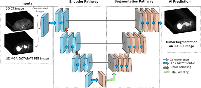
Background: Somatostatin receptor (SSR) targeting radiotracer 68Ga-DOTATATE is used for Positron Emission Tomography (PET)/Computed Tomography (CT) imaging to assess patients with Pheochromocytoma and paraganglioma (PPGL), rare types of Neuroendocrine tumor (NET) which can metastasize thereby becoming difficult to quantify. The goal of this study is to develop an artificial intelligence (AI) model for automated lesion segmentation on whole-body 3D DOTATATE-PET/CT and to automate the tumor burden calculation. 132 68Ga-DOTATATE PET/CT scans from 38 patients with metastatic and inoperable PPGL, were split into 70, and 62 scans, from 20, and 18 patients for training, and test sets, respectively. The training set was further divided into patient-stratified 5 folds for cross-validation. 3D-full resolution nnUNet configuration was trained with 5-fold cross-validation. The model's detection performance was evaluated at both scan and lesion levels for the PPGL test set and two other clinical cohorts with NET (n = 9) and olfactory neuroblastoma (ONB, n = 5). Additionally, quantitative statistical analysis of PET parameters including SUVmax, total lesion uptake (TLU), and total tumor volume (TTV), was conducted.
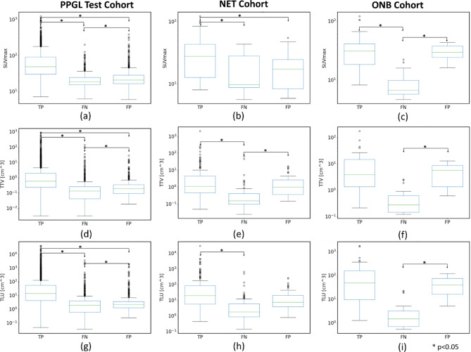
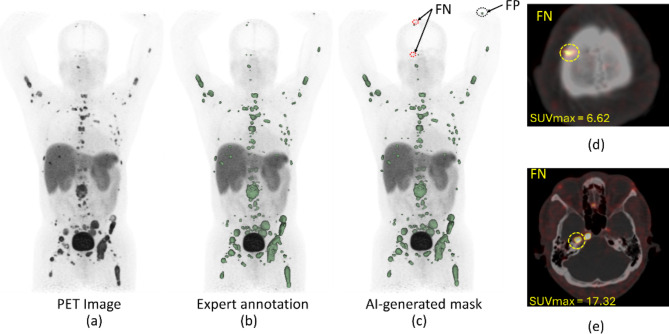
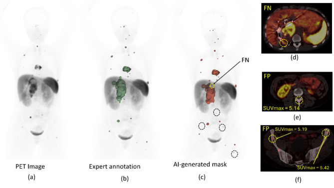
Results: The nnUNet AI model achieved an average 5-fold validation dice similarity coefficient of 0.84 at the scan level. The model achieved dice similarity coefficients (DSC) of 0.88, 0.6, and 0.67 at the scan level, the sensitivity of 86%, 61.13%, and 61.64%, and a positive predictive value of 89%, 74%, and 86.54% at the lesion level for the PPGL test, NET and ONB cohorts, respectively. For PPGL cohorts, smaller lesions with low uptake were missed by the AI model (p < 0.001). Anatomical region-based failure analysis showed most of the false negative and false positive lesions within the liver for all the cohorts, mainly due to the high physiologic liver background activity and image noise on 68Ga- DOTATATE PET scans.
Conclusions: The developed deep learning-based AI model showed reliable performance for automated segmentation of metastatic PPGL lesions on whole-body 68Ga-DOTATATE-PET/CT images, which may be beneficial for tumor burden estimation for objective evaluation during therapy follow-up. https://www.
Clinicaltrials: gov/study/NCT03206060 , https://www.
Clinicaltrials: gov/study/NCT04086485 , https://www.
Clinicaltrials: gov/study/NCT05012098 .
Keywords: Artificial intelligence; DOTATATE PET/CT; Deep learning; Image processing; Lesion segmentation; Machine learning; Neuroendocrine; Oncology; PPGL; Pheochromocytoma and paraganglioma.
© 2024. This is a U.S. Government work and not under copyright protection in the US; foreign copyright protection may apply.
Conflict of interest statement
The authors declare that they have no competing interests.
Figures

References
-
- Jha A, Patel M, Ling A, Shah R, Chen CC, Millo C, et al. Diagnostic performance of [(68)Ga]DOTATATE PET/CT, [(18)F]FDG PET/CT, MRI of the spine, and whole-body diagnostic CT and MRI in the detection of spinal bone metastases associated with pheochromocytoma and paraganglioma. Eur Radiol. 2024. 10.1007/s00330-024-10652-4. - PMC - PubMed
-
- Taieb D, Nolting S, Perrier ND, Fassnacht M, Carrasquillo JA, Grossman AB, et al. Management of phaeochromocytoma and paraganglioma in patients with germline SDHB pathogenic variants: an international expert Consensus statement. Nat Rev Endocrinol. 2024;20(3):168–84. 10.1038/s41574-023-00926-0. - PubMed
Associated data
LinkOut - more resources
Full Text Sources
Medical
