. 2024 Nov 5;15(11):790.
doi: 10.1038/s41419-024-07117-3.

USP3 promotes DNA damage response and chemotherapy resistance through stabilizing and deubiquitinating SMARCA5 in prostate cancer

Affiliations

USP3 promotes DNA damage response and chemotherapy resistance through stabilizing and deubiquitinating SMARCA5 in prostate cancer

Sheng Li et al. Cell Death Dis. .

Abstract

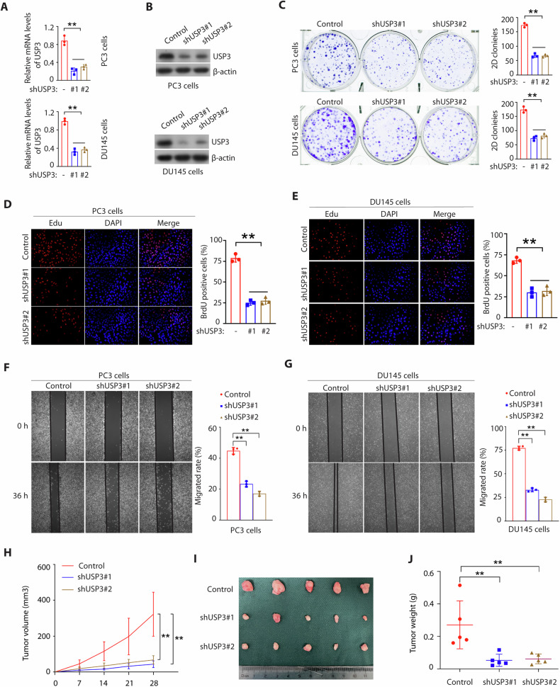
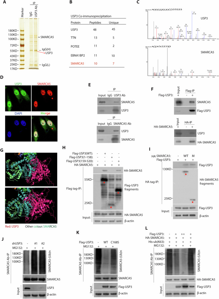
The chromatin-remodeling enzyme SMARCA5 plays a key role in DNA-templated events including transcription, DNA replication, and DNA repair. Loss of function of the SMARCA5 can cause neurodevelopmental disorder and Williams syndrome. However, the molecular mechanism underlying the regulation of SMARCA5 in prostate cancer remains largely elusive. Here, we report that the deubiquitinating enzyme USP3 directly interacts with SMARCA5 and removes K63-linked polyubiquitination of SMARCA5 to maintain its stability, which promotes DNA damage repair and chemotherapy resistance. Depletion of USP3 or SMARCA5 promoted PCa cells sensitive to docetaxel and overexpression of USP3 restored the cells resistance to docetaxel treatment in SMARCA5 silenced cells in vitro and vivo. Clinically, USP3 was significantly up-regulated in prostate cancer tissues and positively associated with SMARCA5 expression. Collectively, our findings uncover a novel molecular mechanism for the USP3-SMARCA5 axis in regulating DSB repair with an important role in chemotherapy response in human prostate cancers, highlighting that targeting USP3-SMARCA5 axis could be a valuable strategy to treat USP3/SMARCA5-overexpressing chemotherapy-resistant patients and improve drug treatment.


Conflict of interest statement

The authors declare no competing interests.

Figures

Fig. 1
Fig. 1. USP3 is elevated in PCa tissues and correlates with prostate cancer progression.
A, B USP3 mRNA was overexpression in primary prostate cancer tissues compared with normal prostate tissues (GSE69223 and GSE70768). C Expression profile of USP3 mRNA in paired primary prostate cancer tissues (n = 53) and matched normal prostate tissues (n = 53). ***p < 0.001; TCGA. D, E USP3 protein expression in adjacent non-tumor tissues and matched PCa tissues was detected by western blot. USP3 protein expression was quantified using Image J software. F qPCR was used to detect the levels of USP3 mRNA in the above tissues. G Representative staining of USP3 in PCa tissues and adjacent non-tumor tissues. H USP3 protein expression was upregulated in adjacent non-tumor tissues (n = 99) and PCa tissues (n = 99). **p < 0.01. I USP3 protein expression was upregulated in paired adjacent non-tumor tissues (n = 99) and matched PCa tissues (n = 99). **p < 0.01. JM A high USP3 expression was not related with age (p = 0.987), but related with a higher preoperative clinical T3b/T4 stage (p = 0.019), Tissue grade III-IV (p = 0.018) and Gleason score (p = 0.008). Relationship between USP3 expression and clinicopathological features. Chi-square tests were used to assess associations, with statistical significance indicated.
Fig. 2
Fig. 2. USP3 knockdown suppresses PCa cell proliferation in vitro and vivo.
A, B USP3 was knocked down in PC3 and DU145 cells by lentivirus control plasmid or shUSP3 (#1 and #2), and were detected by qPCR and Western blot. **p < 0.01. C The cells were generated as in A and colony-formation assays were performed (**p < 0.01). D, E Representative micrographs (left panel) and quantification (right panel) of Edu labeling in PC3 cells or DU145 cells stably expressing lentivirus control plasmid or shUSP3 (#1 and #2). **p < 0.01. F, G The indicated cells as described in A were examined by wound healing assays. Representative micrographs (left panel) and quantification (right panel) of migrated cells. H PC3 shNC cells or shUSP3 cells were subcutaneously injected into Balb/c nude mice. Tumor volume growth curves from the indicated days and tumor growth was measured every 7 days. I After 28 days, mice were sacrificed and representative tumor images at the end of the experiment are presented. J Tumor weights were examined in the two groups. The statistical analyses were performed with the ANOVA. **p < 0.01.
Fig. 3
Fig. 3. USP3 directly interacts with and deubiquitinates SMARCA5.
A The PC3 cells were lysed and subjected to immunoprecipitation using anti-USP3 antibody and followed by protein A/G agarose beads. The complexes were then separated, and the gels were stained with silver. B List of USP3-associated proteins identified by mass spectrometric analysis. C Representative best unique peptides of USP3 and SMARCA5 were identified by mass spectrometry assays. D Immunostaining of USP3 (green) and SMARCA5 (red) were detected by their antibody in PC3 cells. Nuclear 4’, 6-diamidino-2-phenylindole (DAPI; blue). E, F The interaction between USP3 and SMARCA5 in PC3 cells was detected by co-immunoprecipitation assay. G The both binding interface between USP3 and SMARCA5 was based on the molecular docking model. H HEK293T cells were cotransfected with HA-SMARCA5 and Flag-tagged full-length USP3 or its deletion mutants, and cell lysates were subjected to IP and detected with the indicated antibodies. I HEK293T cells were co-transfected with Flag-USP3 and HA-tagged full-length SMARCA5 or its deletion mutant (487–638) and cell lysates were subjected to IP and detected with the indicated antibodies. J The PC3 cells stably expressing control or USP3 shRNA#1 and or #2 were subjected to deubiquitination assay and the polyubiquitylated SMARCA5 protein was detected by the anti-Ub antibody. K The PC3 cells were transfected with Flag-USP3 and Flag-USP3 (C168S) as indicated. The polyubiquitylated SMARCA5 protein was detected by the anti-Ub antibody. L HEK293T cells were transfected with Flag-USP3 (+means 2 μg, ++ means 4 μg), HA-SMARCA5, and His-ub (K63). The polyubiquitylated SMARCA5 protein was detected by the anti-Ub antibody.
Fig. 4
Fig. 4. USP3 is a bona fide DUB targeting SMARCA5 protein for deubiquitination and stabilization.
A The PCa cells transfected with the indicated constructs were treated with or without MG132 for 4 h before harvest. USP3 and SMARCA5 protein were detected by the indicated antibodies. B PC3 cells (up) and DU145 cells (down) were transduced with USP3 shRNA#1 and or #2, treated with 50 mg/mL cycloheximide, harvested at different time points, and then immunoblotted with antibodies to USP3, SMARCA5 and β-actin. Right, quantification of SMARCA5 protein levels (normalized to β-actin). C The cells were transfected with Flag-USP3 (WT) or Flag-USP3 (Mut) for 48 h, then analyzed and quantified as described as in B. D USP3 protein were upregulated in 10 freshly collected paired human prostate tumor tissues (T) and matched adjacent non-tumor tissues (NT), and positively related with SMARCA5 expression. E Representative staining of USP3 and SMARCA5 in paired adjacent non-tumor tissues (n = 99) and matched PCa tissues. F The USP3 protein levels were positively correlated with SMARCA5 protein levels in human prostate tumor tissues. G USP3 and SMARCA5 protein expression status in adjacent nontumoral prostate tissue and prostate carcinoma specimens, and the correlation study of USP3 and SMARCA5 expression level in PCa tissues. Statistical analyses were undertaken with the χ2 test, P < 0.001. R, Pearson’s correlation coefficient.
Fig. 5
Fig. 5. USP3 regulates DNA damage response (DDR) through SMARCA5.
A The PC3 cells with or without USP3 silencing were treated with 2.5 nM docetaxel (Dox) at the indicated time points before fixing and processed for γ-H2AX immunofluorescence. N.S means no significance. **p < 0.01. B The PC3 cells with or without SMARCA5 silencing were treated with 2.5 nM docetaxel (Dox) at the indicated time points before fixing and processed for γ-H2AX immunofluorescence. N.S means no significance. **p < 0.01. C Flag-USP3 rescued SMARCA5 knockdown-mediated high levels of γ-H2AX post-docetaxel treatment at the indicated time points. Cells as described in Supplementary Fig. S5G were fixing and processed for γ-H2AX immunofluorescence. D Representative micrographs (left panel) and quantification (right panel) of Edu labeling in PC3 cells stably expressing lentivirus control plasmids or other indicated plasmids (Flag-USP3, shSMARCA5 or Flag-USP3+shSMARCA5). **p < 0.01. EG The indicated PC3 cells (Control, shUSP3 or shUSP3+Flag-SMARCA5) were subcutaneously injected into Balb/c nude mice. Tumor volume growth curves from the indicated days and tumor growth was measured every 7 days (E). F After 28 days, mice were sacrificed and representative tumor images at the end of the experiment are presented. G Tumor weights were examined in the two groups. The statistical analyses were performed with the ANOVA. **p < 0.01.
Fig. 6
Fig. 6. USP3 regulates chemotherapy resistance via SMARCA5.
A, B The PC3 cells and DU145 cells stably expressing USP3 shRNA were transfected with or without SMARCA5 shRNA and cells were analyzed by Western blotting for indicated proteins. Surviving cell percentage was counted after treated 2.5 nM docetaxel with for 2 days (mean ± SD, n = 3). C, D The PC3 cells and DU145 cells stably expressing Flag-USP3 were transfected with or without SMARCA5 shRNA and cells were analyzed by Western blotting for indicated proteins. Surviving cell percentage was counted after treated 2.5 nM docetaxel for 2 days. E, F The PC3 cells and DU145 cells stably expressing USP3 shRNA were transfected with Flag-USP3 (WT) or Flag-USP3 (CA) and cells were analyzed by Western blotting for indicated proteins. Surviving cell percentage was counted after treated 2.5 nM docetaxel for 2 days. GJ Tumor xenograft assays were performed by subcutaneous injection of PC3 cells stably expressing USP3 shRNA or control. Tumor growth rate in nude mice treated every other day with docetaxel (20 mg/kg) is shown G. Tumors were dissected and recorded after euthanizing the mice (H). Mice were sacrificed after 28 days. Tumor weights were measured as shown in I. Representative data are shown from five mice each group by two-sided unpaired t-test. J Representative staining of Ki67 on the tumor sections derived from above treatment mice. The staining was developed by DAB (brown) and counterstained by hematoxylin (blue). Statistical analyses were performed with the ANOVA, *p < 0.05; **p < 0.01. K Schematic model showing that USP3 deubiquitinates and stabilizes SMARCA5 by K63-linked deubiquitin, which promotes DNA damage response.

References

    1. Jiang X, Guo S, Wang S, Zhang Y, Chen H, Wang Y, et al. EIF4A3-Induced circARHGAP29 Promotes Aerobic Glycolysis in Docetaxel-Resistant Prostate Cancer through IGF2BP2/c-Myc/LDHA Signaling. Cancer Res. 2022;82:831–45. - PubMed
    1. Pritchard CC, Mateo J, Walsh MF, De Sarkar N, Abida W, Beltran H, et al. Inherited DNA-Repair Gene Mutations in Men with Metastatic Prostate Cancer. N Engl J Med. 2016;375:443–53. - PMC - PubMed
    1. Robinson D, Van Allen EM, Wu YM, Schultz N, Lonigro RJ, Mosquera JM, et al. Integrative clinical genomics of advanced prostate cancer. Cell. 2015;161:1215–28. - PMC - PubMed
    1. Li L, Chang W, Yang G, Ren C, Park S, Karantanos T, et al. Targeting poly(ADP-ribose) polymerase and the c-Myb-regulated DNA damage response pathway in castration-resistant prostate cancer. Sci Signal. 2014;7:ra47. - PMC - PubMed
    1. Goodwin JF, Schiewer MJ, Dean JL, Schrecengost RS, de Leeuw R, Han S, et al. A hormone-DNA repair circuit governs the response to genotoxic insult. Cancer Discov. 2013;3:1254–71. - PMC - PubMed

MeSH terms