. 2024 Nov 5;10(1):119.
doi: 10.1038/s41522-024-00593-7.

Combating biofilm-associated Klebsiella pneumoniae infections using a bovine microbial enzyme

Affiliations

Combating biofilm-associated Klebsiella pneumoniae infections using a bovine microbial enzyme

Reshma Ramakrishnan et al. NPJ Biofilms Microbiomes. .

Abstract

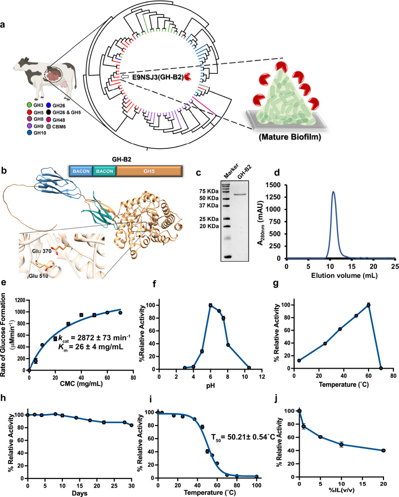
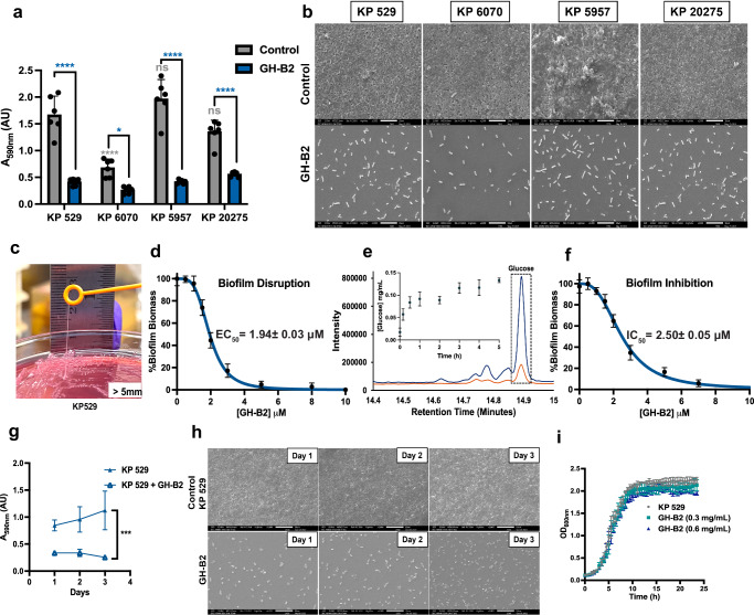
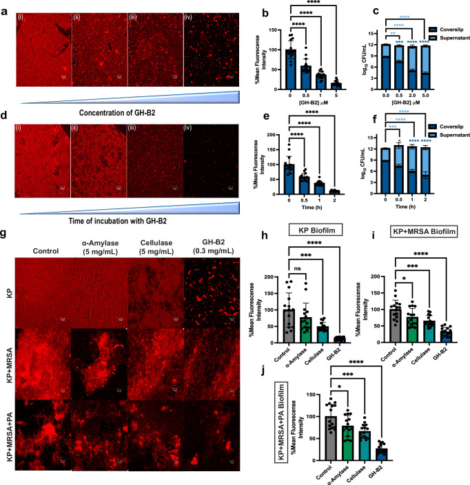
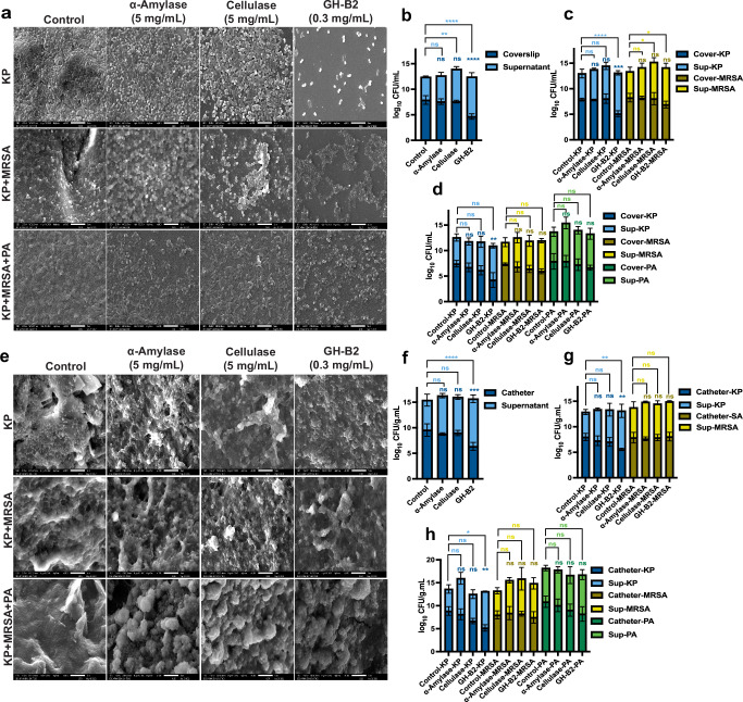
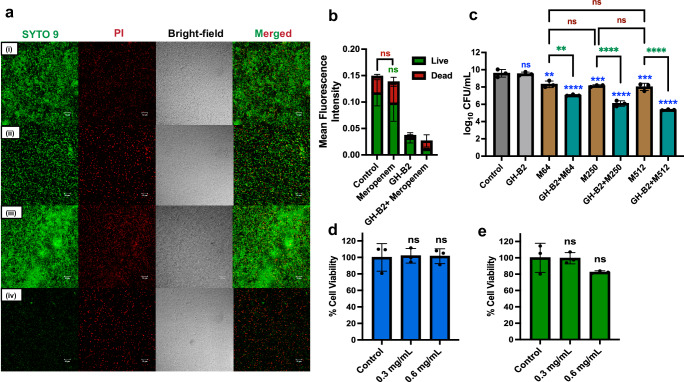
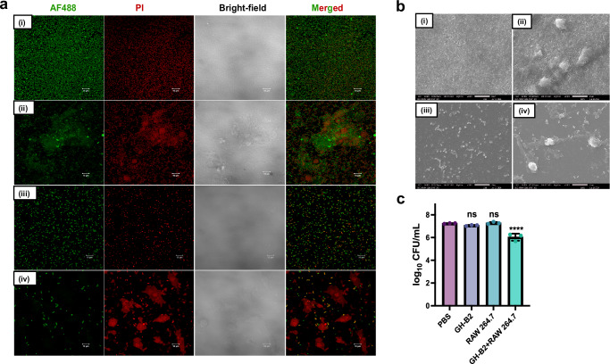
The emergence of multidrug-resistant Klebsiella pneumoniae poses significant clinical challenges with limited treatment options. Biofilm is an important virulence factor of K. pneumoniae, serving as a protective barrier against antibiotics and the immune system. Here, we present the remarkable ability of a bovine microbial enzyme to prevent biofilm formation (IC50 2.50 μM) and degrade pre-formed K. pneumoniae biofilms (EC50 1.94 μM) by degrading the matrix polysaccharides. The treatment was effective against four different clinical K. pneumoniae isolates tested. Moreover, the enzyme significantly improved the biofilm sensitivity of a poorly performing broad-spectrum antibiotic, meropenem, and immune cells, resulting in facile biofilm clearance from the mouse wound infection. Notably, well-known powerful enzymes of the same class, cellulase, and α-amylase, were nearly inactive against the K. pneumoniae biofilms. The enzyme exhibited antibiofilm activity without showing toxicity to the mammalian and microbial cells, highlighting the potential of the enzyme for in vivo applications.


Conflict of interest statement

The authors declare a conflict of interest in the work and filed a patent application to protect the findings. Applicant: Indian Institute of Science, Bangalore, India. Name of inventors: D.D., D.C., R.R., A.V.N., and K.P. Application No. 202341053814, which has been granted (Indian Patent No. 524536).

Figures

Fig. 1
Fig. 1. Characterization of GH-B2.
a Phylogenetic tree of the 90 predicted GHs found in the cow rumen microbiome using maximum parsimony, created using Clustal Omega and analyzed with iTOL (Interactive Tree Of Life software, version 6.9.1) tool. Each clade color indicates a particular GH family. Our selected enzyme GH-B2 (UniProt ID: E9NSJ3) is marked with an arrow. b The AlphaFold2-predicted structure of GH-B2. The zoomed-in view of the active site of GH-B2 displays the conserved catalytic residues, Glu370 and Glu510. c SDS–PAGE analysis of the purified GH-B2. d Size exclusion chromatography (SEC) elution profile of GH-B2. e Michaelis–Menten kinetics of GH-B2 with CMC. f Relative activity of GH-B2 at different pHs. g Relative activity of GH-B2 at different temperatures. h Relative activity of GH-B2 as a function of time monitored till 30 days at 37 °C. i Thermal stability measurement of GH-B2. j Relative activity of GH-B2 at different concentrations of the ionic liquid 1-ethyl-3-methylimidazolium acetate. The observed maximum activity is defined as 100%. All experiments were performed in triplicate (n = 3). Error bars correspond to mean ± standard deviation (SD).
Fig. 2
Fig. 2. GH-B2 prevents biofilm formation and degrades mature K. pneumoniae biofilms.
a Quantification of biomass of 3-day-old mature biofilm formed by various clinical isolates of K. pneumoniae in the absence of GH-B2 (control) and after incubation with GH-B2 (0.3 mg/mL) for 2 h at 37 °C. b Scanning electron microscopic (SEM) images of 3-day-old mature biofilms formed by various clinical isolates of K. pneumoniae (control) and the corresponding images after incubation with GH-B2 (0.3 mg/mL) for 2 h at 37 °C, scale bar: 10 μm. c Overnight incubated K. pneumoniae (KP 529) on MacConkey agar plate exhibiting hypermucoviscous thick biofilm. d GH-B2 dose-dependent disruption of the 3-day-old mature KP 529 biofilm in 2 h at 37 °C. e Sugar release from EPS of K. pneumoniae biofilm by GH-B2 at 37 °C. Overlaid gas chromatographs of derivatized samples of EPS with (blue) and without (orange) GH-B2. Silylated glucose O-methyl oxime peak (retention time: 14.9 min) is highlighted in a box. The inset shows the amount of glucose released with time. f GH-B2 dose-dependent inhibition of KP 529 biofilm formation. Biofilm biomass was quantified after 3 days post-incubation. EC50 and IC50 values were calculated using nonlinear least-squares fitting to a dose-response model in GraphPad Prism (version 10.1.1) software. g KP biofilm formation on a glass coverslip in the absence (control, KP 529) and presence of GH-B2 over 3 days. The biomass quantification was done using the crystal violet (CV) staining assay, and the error bars indicate the standard deviation of n = 6 data. Statistical significance was evaluated relative to control using one-way ANOVA with Dunnet’s post-hoc test to analyze the data; p values ****<0.0001, ***<0.001, *<0.05. h SEM images of KP biofilm formation on a glass coverslip in the absence (control) and presence of GH-B2 over 3 days, scale bar: 10 μm. i The growth curve of K. pneumoniae in the presence of GH-B2 (0.3 mg/mL and 0.6 mg/mL). The absorbance of the culture (OD600) was measured every 30 min over 24 h. Error bars indicate the standard deviation of triplicate (n = 3) data. Representative SEM images were shown.
Fig. 3
Fig. 3. CLSM analysis of GH-B2 mediated degradation of polysaccharides of the mature K. pneumoniae biofilms.
a Dispersion of K. pneumoniae biofilm as a function of the concentration of GH-B2. (i) Untreated (ii) 0.03 mg/mL (iii) 0.06 mg/mL, and (iv) 0.3 mg/mL GH-B2-treated 3-day-old biofilm. The enzyme treatment was carried out for 2 h at 37 °C. Corresponding b confocal image analysis and c CFU in the supernatant and the remaining biofilm on the glass coverslip, n = 3. d dispersion of K. pneumoniae biofilm as a function of time. (i) Untreated (ii) 30 min (iii) 1 h, and (iv) 2 h GH-B2 (0.3 mg/mL)-treated 3-day-old biofilm at 37 °C. Corresponding e confocal image analysis f CFU in the supernatant and the remaining biofilm on the glass coverslip g comparison of the biofilm dispersal activity of α-amylase (5 mg/mL), cellulase (5 mg/mL), and GH-B2 (0.3 mg/mL) on K. pneumoniae (KP), KP+ methicillin-resistant S. aureus (MRSA), KP + MRSA + P. aeruginosa (PA) biofilms, respectively. Corresponding confocal images analysis h KP i KP + MRSA j KP + MRSA + PA. Biofilm was stained with the Texas red conjugated with Concanavalin A, and representative confocal images of maximum intensity projection of Z-stacks are shown, scale bar: 10 μm. For confocal image analysis, each bar indicates mean ± standard deviation. Five confocal images were analyzed from each of three biological replicates, N = 3. Statistical significance was evaluated relative to control using one-way ANOVA with Dunnet’s post-hoc test to analyze the data; p values ****<0.0001, ***<0.001, **<0.01, *<0.05, ns no significant difference.
Fig. 4
Fig. 4. SEM analysis of GH-B2-mediated degradation of polysaccharides of the mature K. pneumoniae biofilms.
SEM images of the biofilm dispersal activity of cellulase (5 mg/mL), α-amylase (5 mg/mL), and GH-B2 (0.3 mg/mL) on K. pneumoniae (KP), KP + methicillin-resistant S. aureus (MRSA), KP + MRSA + P. aeruginosa (PA) biofilms, respectively, grown a on glass coverslips, bd corresponding number of CFU in the supernatant and number of CFU remaining in the biofilm; e on Foley catheter, fh corresponding number of CFU in the supernatant and number of CFU remaining in the biofilm. All experiments were performed in triplicate (n = 3), and representative images were shown, scale bar: 5 μm. Statistical significance was evaluated using two-way ANOVA with Tukey’s post-hoc test to analyze the grouped data; p values ****<0.0001, ***<0.001, **<0.01, *<0.05, ns no significant difference.
Fig. 5
Fig. 5. GH-B2 enhances the antibiotic efficacy against biofilm-associated K. pneumoniae.
a The biofilm was grown for 3 days and subjected to the combined treatment with GH-B2 and meropenem. CLSM images of (i) phosphate-buffered saline (PBS)-treated, (ii) GH-B2 (0.3 mg/mL)-treated, (iii) meropenem (250 µg/mL)-treated, and (iv) GH-B2 (0.3 mg/mL) + meropenem (250 µg/mL)-treated KP biofilm. The biofilm was subjected to 2 h GH-B2 treatment followed by 24 h meropenem treatment at 37 °C. Green (Syto 9-labeled) cells denote the live cells and red (propidium iodide-labeled) cells with damaged membranes. b Corresponding confocal images analysis. c CFU analysis of post 2 h GH-B2 treatment followed by 24 h meropenem treatment at 37 °C. M meropenem, and the number following M represents the antibiotic concentration in µg/mL. Statistical significance was evaluated relative to control (blue), meropenem (red), and GH-B2 + meropenem (green) using two-way ANOVA with Tukey’s post-hoc to analyze the grouped data; p values ****<0.0001, ***<0.001, **<0.01, ns no significant difference. Cytotoxicity analysis of GH-B2 on d HeLa cells and e macrophage (RAW264.6) upon treatment with GH-B2 (0.3 and 0.6 mg/mL) for 12 h at 37 °C. All experiments were performed in triplicate (n = 3), and representative confocal images were shown, scale bar: 10 μm. For confocal image analysis, each bar indicates mean ± standard deviation. Five confocal images were analyzed from each of the two biological replicates, N = 2. Statistical significance was evaluated relative to PBS control using one-way ANOVA with Dunnet’s post-hoc test to analyze the data; p values ****<0.0001, ***<0.001, **<0.01, *<0.05, ns no significant difference.
Fig. 6
Fig. 6. GH-B2 enhances the sensitivity of biofilm-encased KP cells to macrophages.
The biofilm was grown for 3 days and subjected to the treatment with GH-B2 along with macrophage. CLSM images showing a (i) PBS-treated, (ii) macrophage (RAW264.7 cells)-treated, (iii) GH-B2-treated, and (iv) GH-B2 + macrophage-treated KP biofilm, scale bar: 10 μm, AF488 Alexa flour-488, PI propidium iodide. b Corresponding SEM images, scale bar: 10 µm. Both the enzyme and macrophage treatments were carried out for 2 h at 37 °C. c CFU analysis of post 2 h GH-B2 treatment followed by 2 h macrophage treatment at 37 °C. Experiments were performed in triplicate (n = 3), and representative images were shown. Statistical significance was evaluated relative to PBS control using one-way ANOVA with Dunnet’s post-hoc test to analyze the data; p values ****<0.0001, ns no significant difference.
Fig. 7
Fig. 7. GH-B2-mediated treatment of biofilm-associated K. pneumoniae infection in mice.
a Representative images of different treatment groups—K. pneumoniae infected wound with PBS (control), meropenem (512 μg/mL), GH-B2 (0.6 mg/mL), and GH-B2 (0.6 mg/mL) + meropenem (512 μg/mL). b Body weight data represent mean ± SD, n = 5 mice/group. c CFU of K. pneumoniae on day 7 after different treatments, n = 5. d Histopathological analysis of wound tissue using H&E staining at day 7, uninfected (control) wound, PBS-treated, GH-B2 (0.6 mg/mL)-treated, meropenem (512 μg)-treated, and GH-B2 (0.6 mg/mL) + meropenem (512 μg)-treated infected wounds. Scale bar: 50 μm, E epidermis, D dermis, CF collagen fibers, IF inflammatory infiltrates, N necrosis, A angiogenesis, HF hair follicles. Statistical significance was evaluated relative to PBS control using one-way ANOVA with Dunnet’s post-hoc to analyze the data; p values ****<0.0001, **<0.01, *<0.05, ns no significant difference.

References

    1. Kochan, T. J. et al. Klebsiella pneumoniae clinical isolates with features of both multidrug-resistance and hypervirulence have unexpectedly low virulence. Nat. Commun.14, 7962 (2023). - PMC - PubMed
    1. Verani, J. R. et al. Child deaths caused by Klebsiella pneumoniae in sub-Saharan Africa and South Asia: a secondary analysis of Child Health and Mortality Prevention Surveillance (CHAMPS) data. Lancet Microbe5, e131–e141 (2024). - PMC - PubMed
    1. Cillóniz, C., Dominedò, C. & Torres, A. Multidrug resistant gram-negative bacteria in community-acquired pneumonia. Crit. Care34, 459–475 (2019). - PMC - PubMed
    1. David, S. et al. Epidemic of carbapenem-resistant Klebsiella pneumoniae in Europe is driven by nosocomial spread. Nat. Microbiol.4, 1919–1929 (2019). - PMC - PubMed
    1. Munoz-Price, L. S. et al. Clinical epidemiology of the global expansion of Klebsiella pneumoniae carbapenemases. Lancet Infect. Dis.13, 785–796 (2013). - PMC - PubMed

LinkOut - more resources