. 2024 Nov 5;14(1):26795.
doi: 10.1038/s41598-024-74661-5.

A novel therapeutic pathway to the human cochlear nerve

Affiliations

A novel therapeutic pathway to the human cochlear nerve

Hao Li et al. Sci Rep. .

Abstract

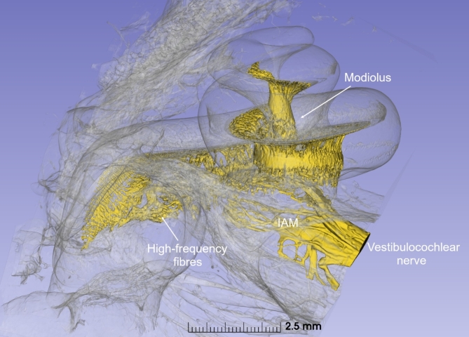
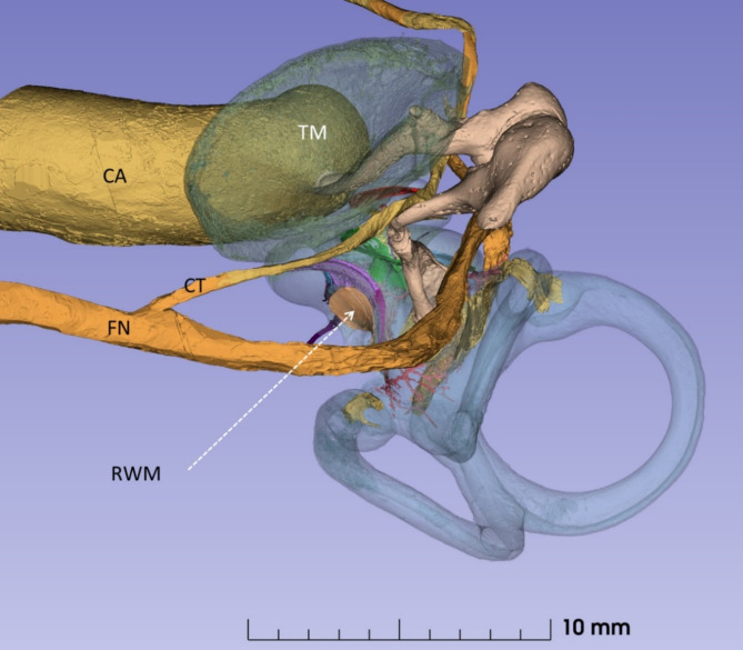
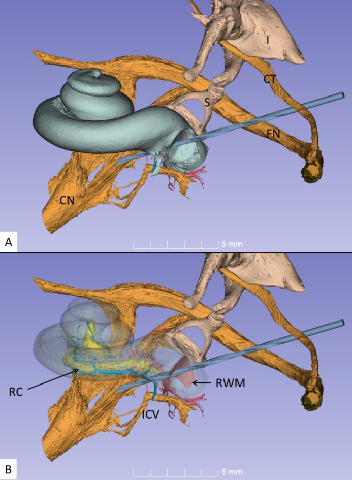
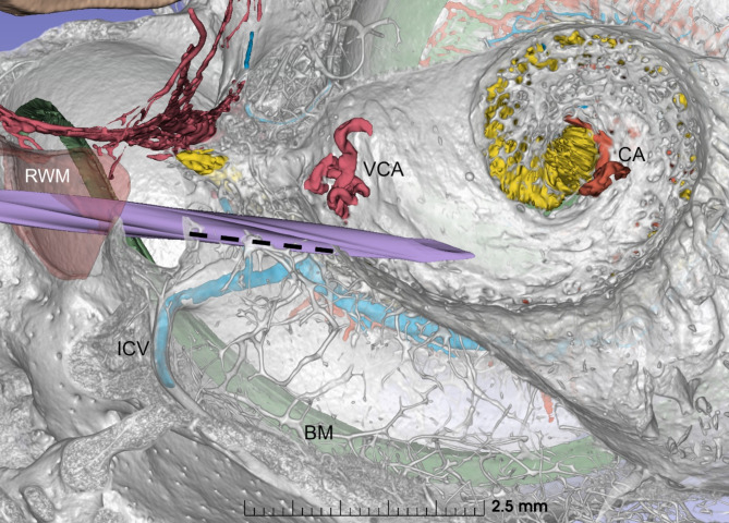
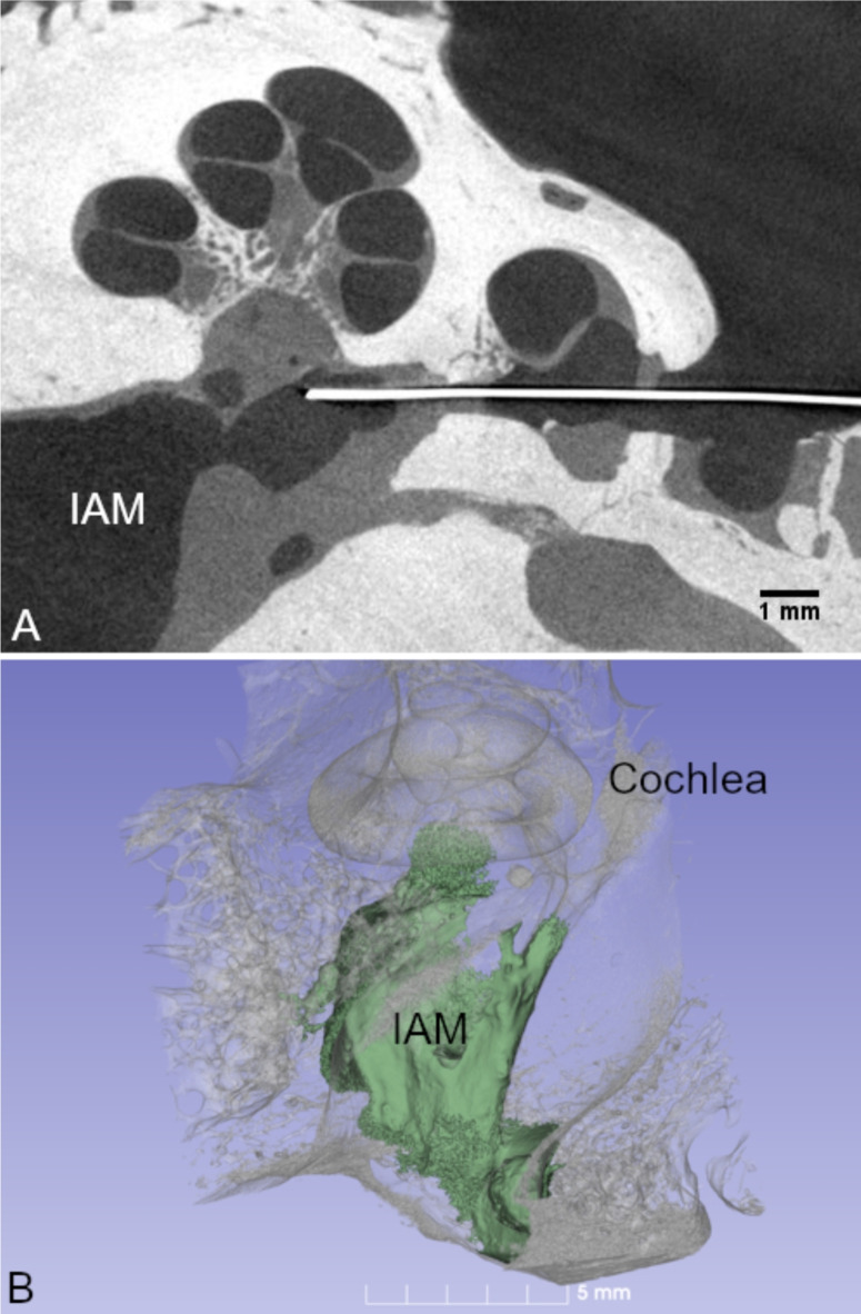
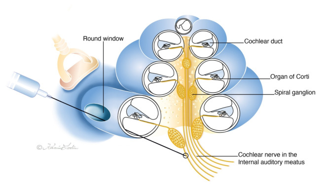
Traditional approaches to the human cochlear nerve have been impeded by its bony encasement deep inside the skull base. We present an innovative, minimally invasive, therapeutic pathway for direct access to the nerve to deliver novel regenerative therapies. Neuroanatomical studies on 10 cadaveric human temporal bones were undertaken to identify a potentially safe therapeutic pathway to the cochlear nerve. Simulations based on three-dimensional delineation of anatomical structures obtained from synchrotron phase-contrast imaging were analyzed. This enabled the identification of an approach to the nerve in the fundus of the internal auditory meatus by trephining the medial modiolar wall of the cochlea via the round window for a median depth of 1.48 mm (range 1.21-1.91 mm). The anatomical access was validated on 9 additional human temporal bones using radio-opaque markers and contrast injection with micro-computed tomography surveillance. We thus created an effective conduit for the delivery of therapeutic agents to the cochlear nerve.

Keywords: Cochlear nerve; Deafness; Gene Therapy; Human; Micro-CT; Stem cell therapy; Synchrotron Imaging.


Conflict of interest statement

Prof. Marcelo Rivolta is the Founder Director and Chief Scientific Officer of Rinri Therapeutics. Prof. Douglas Hartley is Rinri Therapeutics Chief Medical Officer. Other authors do not have any competing interest.

Figures

Fig. 1
Fig. 1
SR-PCI 3D reconstruction delineating the sensory nerve elements in a left human cochlea. The otic capsule is made semi-transparent to visualize the innervation of the cochlea and how the central axonal processes of the cochlear nerve are an accessible target for therapeutic intervention. The vestibulocochlear nerve within the IAM is visualized. IAM, internal acoustic meatus.
Fig. 2
Fig. 2
SR-PCI of a left human ear. It demonstrates the exposure obtained of the round window (arrow) through a routine trans-mastoid approach. FN facial nerve, CT chorda tympani, RWM round window membrane, TM tympanic membrane, CA carotid artery.
Fig. 3
Fig. 3
SR-PCI section and 3D reconstruction of a left human ear. The proximity of the RWM to the IAM is shown. The distance across the modiolar bone to reach the IAM is typically just over 1 mm. Broken arrow shows the desired trajectory to reach the IAM from the posterior tympanotomy. IAM internal acoustic meatus, RWM round window membrane.
Fig. 4
Fig. 4
(A) Inferior view of the left cochlea showing the RW trajectory to reach the high-frequency cochlear nerve fibres in the fundus of the IAM. The shaft of the drill is positioned on the bony rim of the round window (crista fenestra) and is angled anteriorly towards the chorda tympani (CT). FN, facial nerve; CN, cochlear nerve; S, stapes; I, incus. (B). Same exposure as in A with the cochlea rendered transparent. Note the position of Rosenthal’s canal (RC) housing the cell bodies of the cochlear neurons. ICV inferior cochlear vein, RWM round window membrane.
Fig. 5
Fig. 5
Vertical cross-section of a left human cochlea displays the relationship between the RWM, basal turn of the cochlea, and the spirally shaped cribriform area in the fundus of the IAM housing the fibres of the cochlear nerve. The drill has penetrated the modiolus creating a bony tunnel allowing delivery of therapeutic agents directly to the cochlear nerve. The average length of the tunnel created from the crista fenestra to the entry point in the IAM is 1.43 mm (interrupted line). Note the position of the blood vessels in the IAM fundus. The cochlear artery (CA) enters the cochlea through the central cochlear aperture; being more distant from the entry point of the drill, it is less at risk. However, the vestibulo-cochlear artery (VCA) enters the cochlea through one of the minor cochlear foramina in the cribriform area and is thus more vulnerable. ICV inferior cochlear vein, BM basilar membrane, RWM round window membrane, VCA vestibulo-cochlear artery, CA cochlear artery.
Fig. 6
Fig. 6
Micrograph showing the completed bony access tunnel from the round window membrane, through the modiolus, to reach the IAM and containing a radio-opaque marker needle (A). Cell suspension and contrast medium (green, 20uL, otic neuroprogenitors/potassium iodide) was injected through the tunnel and was distributed throughout the IAM (B). IAM, internal acoustic meatus.
Fig. 7
Fig. 7
Drawing showing the trajectory for injection to the cochlear nerve in the internal auditory meatus via the round window. Image by Karin Lodin.

References

    1. WHO World Report on Hearing 2021.
    1. Hudspeth, A. J. Integrating the active process of hair cells with cochlear function. Nat. Rev. Neurosci. 2014 159 15, 600–614 (2014). - PubMed
    1. Kujawa, S. G. & Liberman, M. C. Adding insult to Injury: cochlear nerve degeneration after temporary noise-Induced hearing loss. J. Neurosci. 29, 14077 (2009). - PMC - PubMed
    1. Liberman, M. C. & Kujawa, S. G. Cochlear synaptopathy in acquired sensorineural hearing loss: manifestations and mechanisms. Hear. Res. 349, 138–147 (2017). - PMC - PubMed
    1. Viana, L. M. et al. Cochlear neuropathy in human presbycusis: confocal analysis of hidden hearing loss in post-mortem tissue. Hear. Res. 327, 78 (2015). - PMC - PubMed