. 2024 Nov 5;14(1):26747.
doi: 10.1038/s41598-024-77995-2.

Injectable alginate chitosan hydrogel as a promising bioengineered therapy for acute spinal cord injury

Affiliations

Injectable alginate chitosan hydrogel as a promising bioengineered therapy for acute spinal cord injury

Fatemeh Saadinam et al. Sci Rep. .

Abstract

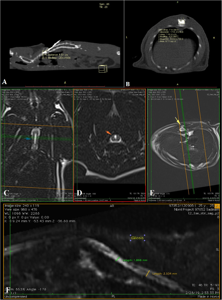
Dealing with spinal cord injuries presents problematic due to multiple secondary mechanisms. Beyond primary concerns like paralysis and disability, complications including urinary, gastrointestinal, cardiac, and respiratory disorders, along with substantial economic burdens may occur. Limited research focuses on modeling and treating contusion and compression injuries. Tissue engineering emerges as an innovative treatment, targeting lesion pathophysiology. This study was evaluated implanting injectable biomaterials into injury-induced cavity before glial scar formation, avoiding tissue incisions and minimizing further damage. The efficacy of injectable alginate/thiolated chitosan hydrogel was investigated for acute spinal cord injury induced by Vanický method in Wistar rats. Three days post-injury, hydrogel was administrated through microinjection after laminectomy. After 60 days, the hydrogel group demonstrated notable motor function enhancement compared to the control by the BBB locomotor test (P < 0.05). However, no statistically significant differences were observed in MRI assessment concerning lesion severity. Stereological and histopathological evaluations revealed a reduction in vacuole volume and the presence of axon profiles within the scaffold (P < 0.05), alongside reduced infiltration of inflammatory and Gitter cells in the hydrogel group, although the latter was not statistically significant compared to the control. Thiolated chitosan/ alginate hydrogel implantation may be regarded as a promising treatment to enhance motor function by restraining destructive processes post-acute spinal cord injury.

Keywords: Alginates; Chitosan; Contusions; Rats; Spinal cord injuries.


Conflict of interest statement

The authors declare no competing interests.

Figures

Fig. 1
Fig. 1
SEM image of thiolated chitosan/alginate hydrogel structure. 400x magnification.
Fig. 2
Fig. 2
(A) Rheometric properties evaluation diagram of thiolated chitosan/alginate hydrogel. (B) Assessment of cell viability through Alamar Blue staining (Mean ± SEM). The graph illustrates the growth in cell population after 1, 3, 5 and 7 days of culture. (C) FTIR spectra of alginate, thiolated chitosan and hydrogel are displaced.
Fig. 3
Fig. 3
The Basso–Beattie–Bresnahan (BBB) locomotor rating scale was evaluated post-operation weekly. Repeated measures ANOVA test demonstrated a significant difference between the control group (n = 5) and the scaffold group (n = 10) (P < 0.05). Data shows in Median ± SD.
Fig. 4
Fig. 4
Longitudinal (A) and transverse sections (B) obtained from CT scan images of Fogarty’s balloon and vertebral column. (C & D) Some findings resulting from SCI are visible in MRI images: Intraspinal necrosis evident in longitudinal (blue arrow) and transverse (red arrow) views. (E) Dura mater adhesion highlighted by the yellow arrow. (F) Reduced diameter of the spinal cord, hyperintensity and the presence of glial scar are demonstrated.
Fig. 5
Fig. 5
(A & B) The statistical evaluation diagrams present a comparison of data between the scaffold and control groups based on MRI images. (C & D) Histopathological parameters including axon counting in 5 HPF (high power field; 400X) and the percentage of cavity area in the histopathology section, were assessed. The cavity area in the control group is significantly greater than in the scaffold sections. There is a statistically significant increase in the number of axons observed in the scaffold group compared to the control defects. Data are presented as Mean ± Std Deviation. ns: Not significant (P > 0.05); *, **: Significant (P < 0.05).
Fig. 6
Fig. 6
Microscopic image of the transverse section in the region of SCI and glial scar, stained with Hematoxylin-eosin. (A) control group. 100x magnification. (B) The black arrows indicate Wallerian degeneration and axonopathy, while gitter cells (macrophages with foamy cytoplasm) are marked with blue arrowheads. control group. 400x magnification. (C) scaffold group. 100x magnification. (D) the repair tissue formed in the scaffold group reveals a low density of mononuclear inflammatory cells, along with fibroblasts and delicate collagen fibers. Notably, normal axonal structures (black arrows) are visible, indicating the communication of nerve fibers within the repair tissue. scaffold group. 400x magnification.
Fig. 7
Fig. 7
(A) The distance between the laminotomy site (blue arrow) and the location of the Fogarty catheter balloon was determined through a CT scan, involving a laminectomy performed above the spinal cord lesion site (black arrow). (B) Subsequently, scaffold injection was carried out using a microinjection device.

References

    1. Verma, R., Virdi, J. K., Singh, N. & Jaggi, A. S. Animals models of spinal cord contusion injury. Korean J. Pain. 32, 12–21 (2019). - PMC - PubMed
    1. Ryan, F. et al. Tlr4 deletion modulates cytokine and extracellular matrix expression in chronic spinal cord injury, leading to improved secondary damage and functional recovery. J. Neurosci.44, e0778232023. 10.1523/JNEUROSCI.0778-23.2023 (2024). - PMC - PubMed
    1. Yuan, H. et al. Microarray analysis of lncRNAs and mRNAs in spinal cord contusion rats with iPSC-derived A2B5 + oligodendrocyte precursor cells transplantation. Heliyon. 10, e22808. 10.1016/j.heliyon.2023.e22808 (2024). - PMC - PubMed
    1. Sharif-Alhoseini, M. et al. Animal models of spinal cord injury: a systematic review. Spinal cord. 55, 714–721 (2017). - PubMed
    1. Slater, P. G., Domínguez-Romero, M. E., Villarreal, M., Eisner, V. & Larraín, J. Mitochondrial function in spinal cord injury and regeneration. Cell. Mol. Life Sci.79, 239 (2022). - PMC - PubMed

LinkOut - more resources