. 2024 Nov 5;14(1):26819.
doi: 10.1038/s41598-024-77823-7.

The power of 810 nm near-infrared photobiomodulation therapy for human asthenozoospermia

Affiliations

The power of 810 nm near-infrared photobiomodulation therapy for human asthenozoospermia

Sara Stigliani et al. Sci Rep. .

Abstract

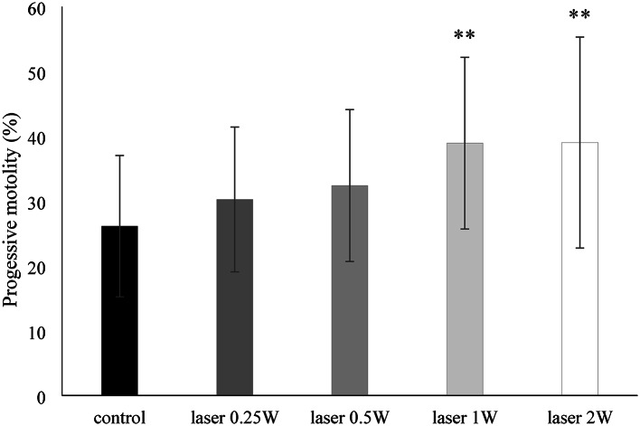
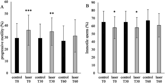
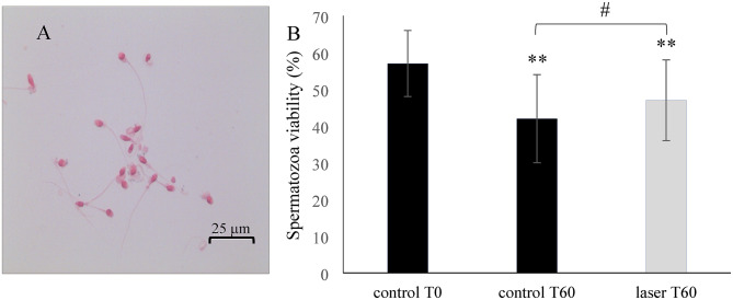
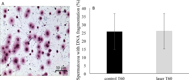
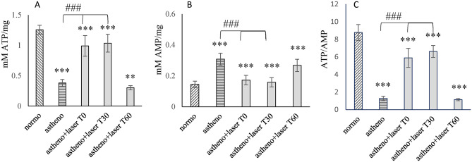
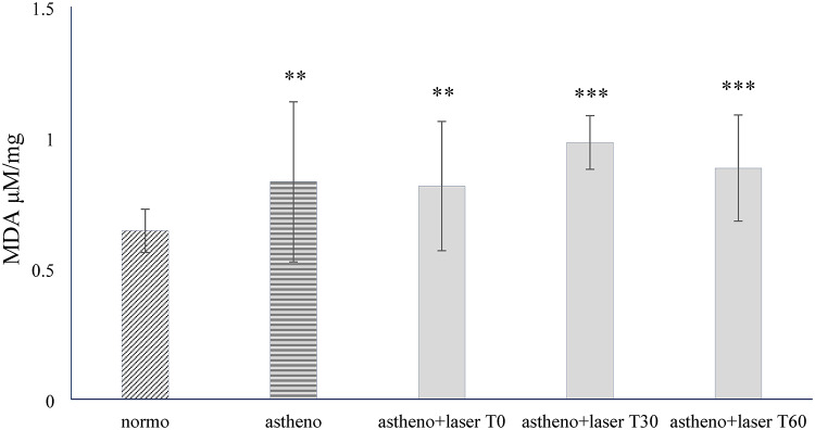
Sperm motility is a crucial factor in male fertility. Photobiomodulation (PBM) has been reported to increase sperm motility, but a consistent approach suitable for identifying standardizable protocols is lacking. We collected asthenozoospermic (n = 70) and normozoospermic (n = 20) semen. The asthenozoospermic samples were irradiated with an 810 nm diode laser, in continuous wave mode, at 0.25 W, 0.5 W, 1 W and 2 W for 60 s on a circular area of 1 cm2 through a novel handpiece with an innovative flat-top profile. Sperm motility was assessed immediately, after 30 and 60 min. A sample size calculator, unpaired t-test and one-way ANOVA with post-hoc Tukey HSD tests were used for statistics. One and 2 W were the most effective outputs in increasing progressive motility compared to control (p < 0.001). The maximum effect was immediately after 1 W-PBM (p < 0.001) and decreased after 60 min (p < 0.001). Time physiologically decreased vitality (p < 0.001), but less in the 1 W-PBM samples (p < 0.05). 1 W-PBM did not affect chromatin condensation. Asthenozoospermic samples displayed an impairment of 80% in oxygen consumption and ATP production and a slight inefficiency of oxidative phosphorylation compared to normozoospermic samples (p < 0.001). 1 W-PBM partially restored the functionality of aerobic metabolism (p < 0.001) by recovery of oxidative phosphorylation efficiency. PBM did not affect lactate dehydrogenase (glycolysis pathway). No irradiated samples increased accumulated malondialdehyde, a marker of lipidic peroxidation. In conclusion, PBM improves progressive motility in asthenozoospermia through increased mitochondrial energetic metabolism without harmful oxidative stress.

Keywords: Asthenozoospermia; DNA fragmentation; Low level laser therapy; Male fertility; Mitochondrial metabolism; Sperm motility; Sperm vitality.


Conflict of interest statement

The authors declare no competing interests.

Figures

Fig. 1
Fig. 1
Study design. The patients were recruited at the SS Physiopathology of Human Reproduction, IRCCS Ospedale Policlinico San Martino, Genova, Italy (A). Semen samples were analyzed (B), and according to the inclusion and exclusion criteria, 70 astenozoospermic semen and 20 normozoospermic semen samples were included in our study (C). Astenozoospermic samples were divided into aliquots and irradiated with 810 nm photobiomodulation therapies (D). Samples irradiated with laser switch-off were considered controls. To assess the beneficial or detrimental effects of the treatment, the asthenozoospermic samples were then analyzed in a blinded fashion (E). The normozoospermic samples’ energy metabolism and oxidative stress were also examined and considered (E).
Fig. 2
Fig. 2
Graph showing results of progressive motility in samples of 20 asthenozoospermic patients immediately after irradiation at different powers. The progressive motility rate was calculated as the percentage of a + b, where a corresponds to rapid progressive motility and b to slow progressive motility. Data are expressed as mean ± SD and compared with one-way ANOVA followed by Tukey’s multiple comparison test. ** indicates a significant difference (p < 0.01) compared to the control.
Fig. 3
Fig. 3
Bar graphs showing PBM effects on sperm motility of 70 asthenozoospermic patients. (A) percentage of progressive motility, and (B) percentage of immotile sperm in control and treated samples over time (T0: immediately after irradiation; T30: 30 min after irradiation; T60: 60 min after irradiation). Data are expressed as mean ± SD. Comparisons were made by one-way ANOVA followed by Tukey’s multiple comparison test. *, **, and *** indicate a significant difference for p < 0.05, p < 0.01, and p < 0.001, respectively, between the control and the treated sample at the same time point.
Fig. 4
Fig. 4
Sperm viability test using eosin staining.  (A) A representative image of sperm viability test. Live sperm heads were unstained, and dead sperm heads were stained red-pink (bar: 25 μm). (B) Graph showing the viability percentage at the baseline (control T0) and after 60 min from irradiation in controls (control T60) and treated samples (laser T60) of 25 asthenozoospermic patients. Data were expressed as mean ± SD and compared with one-way ANOVA followed by Tukey’s multiple comparison test. *** indicates a significant (p < 0.001) difference between control T0 vs. control T60 and laser T60, respectively. # indicates a significant (p < 0.05) difference between control T60 vs. laser T60.
Fig. 5
Fig. 5
Sperm DNA integrity test using Sperm Chromatin Dispersion (SCD) technique. (A) A representative image of sperm DNA integrity test. Sperm without DNA fragmentation shows a dispersion halo, sperm with fragmented DNA do not show a dispersion halo, or the halo is minimal (bar: 50 μm). (B) The percentage of sperm with fragmented DNA after 60 min from irradiation in controls (control T60) and treated samples (laser T60) of 25 asthenozoospermic patients. Data reported in Panel B were expressed as mean ± SD and compared with the unpaired t-test.
Fig. 6
Fig. 6
PBM effect on OxPhos activity in samples from 20 asthenozoospermic and 20 normozoospermic patients. (A) ATP synthase activity; (B) oxygen consumption rate; (C) P/O ratio as a marker of OxPhos efficiency in normozoospermic samples (normo), asthenozoospermic samples (astheno) and asthenozoospermic samples lasered and immediately evaluated (astheno + laser T0) or evaluated after 30 min (astheno + laser T 30). Data are expressed as mean  ±  SD. Statistical analysis was performed using one-way ANOVA followed by Tukey’s multiple comparison test. *** indicates a significant difference (p < 0.001) between normo and astheno, astheno + laser T0), or astheno + laser T 30. ### indicates a significant difference (p < 0.001) between astheno versus astheno + laser T0 or astheno + laser T30.
Fig. 7
Fig. 7
PBM-induced changes of cellular energy status in samples from 20 asthenozoospermic and 20 normozoospermic patients. (A) ATP intracellular concentration; (B); AMP intracellular concentration; (C) ATP/AMP ratio as a marker of cellular energy status in normozoospermic samples (normo), asthenozoospermic samples (astheno) and asthenozoospermic samples lasered and immediately evaluated (astheno+laser T0) or evaluated after 30 minutes (astheno+laser T 30). Data are expressed as mean + SD. Statistical analysis was performed using one-way ANOVA followed by Tukey’s multiple comparison test. *** indicates a significant difference (< 0.001) between normo and astheno, astheno+laser T0), or astheno+laser T 30. ### indicates a significant difference (p<0.001) between astheno versus astheno+laser T0 or astheno+laser T30.
Fig. 8
Fig. 8
PBM effect on LDH activity in samples from 20 asthenozoospermic and 20 normozoospermic patients. Graph reports the LDH activity in normozoospermic samples (normo), asthenozoospermic samples (astheno), and asthenozoospermic samples lasered and immediately evaluated (astheno+laser T0) or assessed after 30 minutes (astheno+laser T 30). Data are expressed as mean + SD. Statistical analysis was performed using one-way ANOVA followed by Tukey’s multiple comparison test, and no significant differences were observed.
Fig. 9
Fig. 9
PBM effects on lipid peroxidation accumulation were evaluated in samples from 20 asthenozoospermic and 20 normozoospermic patients. Graph reports the MDA intracellular concentration as a marker of oxidative damage in normozoospermic samples (normo), asthenozoospermic samples (astheno) and asthenozoospermic samples lasered and immediately evaluated (astheno + laser T0) or assessed after 30 min (astheno + laser T 30). Data are expressed as mean ± SD. Statistical analysis was performed using one-way ANOVA followed by Tukey’s multiple comparison test. ** and *** indicate a significant difference (p < 0.01 and 0.001, respectively) between normo and astheno, astheno + laser T0), or astheno + laser T 30. No significant differences have been observed between astheno versus astheno + laser T0 or astheno + laser T30.

Similar articles

Cited by

References

    1. Agarwal, A., Mulgund, A., Hamada, A. & Chyatte, M. <ArticleTitle Language=“En”>R. A unique view on male infertility around the globe. Reproductive Biology Endocrinol.13, 1–9 (2015). - PMC - PubMed
    1. Levine, H. et al. Temporal trends in sperm count: a systematic review and meta-regression analysis of samples collected globally in the 20th and 21st centuries. Hum. Reprod. Update. 29, 157–176 (2023). - PubMed
    1. Amaral, A., Lourenço, B., Marques, M. & Ramalho-Santos, J. Mitochondria functionality and sperm quality. Reproduction. 146, R163–R174 (2013). - PubMed
    1. Boguenet, M., Bouet, P. E., Spiers, A. & Reynier, P. May-Panloup, P. Mitochondria: their role in spermatozoa and in male infertility. Hum. Reprod. Update. 27, 697–719 (2021). - PubMed
    1. Gu, N. H., Zhao, W. L., Wang, G. S. & Sun, F. Comparative analysis of mammalian sperm ultrastructure reveals relationships between sperm morphology, mitochondrial functions and motility. Reprod. Biol. Endocrinol.17, (2019). - PMC - PubMed

LinkOut - more resources