. 2025 Feb;70(2):87-97.
doi: 10.1038/s10038-024-01301-1. Epub 2024 Nov 6.

Homozygous synonymous FAM111A variant underlies an autosomal recessive form of Kenny-Caffey syndrome

Affiliations

Homozygous synonymous FAM111A variant underlies an autosomal recessive form of Kenny-Caffey syndrome

Loisa Dana Bonde et al. J Hum Genet. 2025 Feb.

Abstract

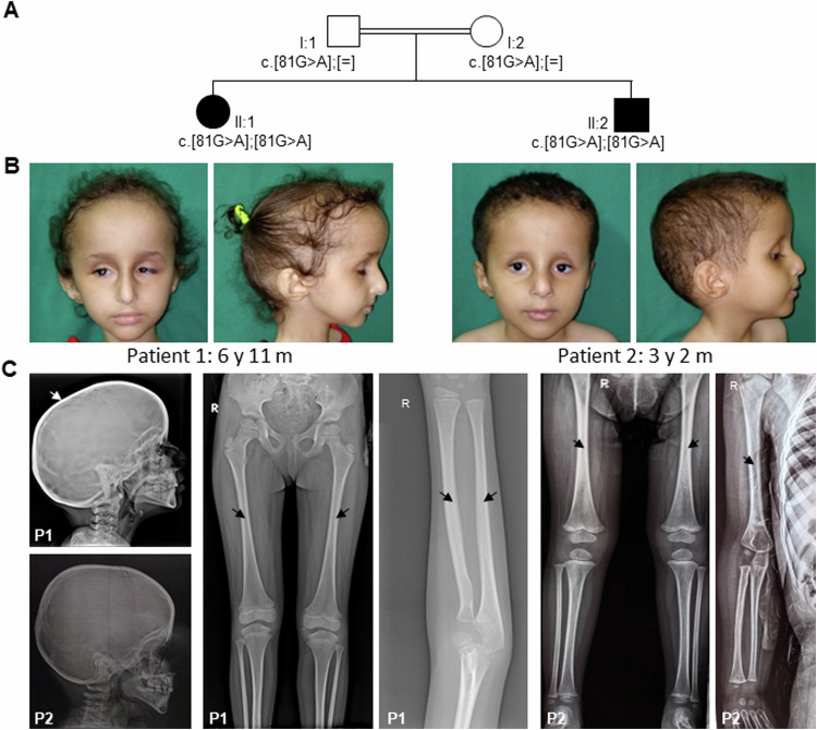
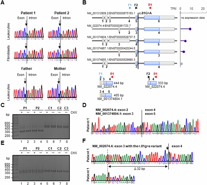
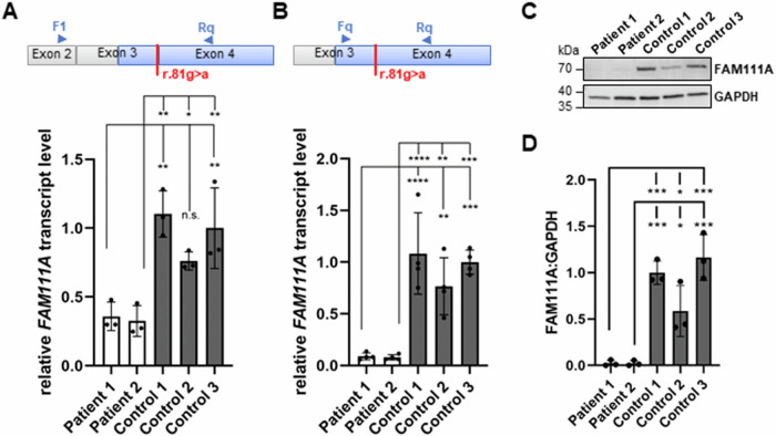
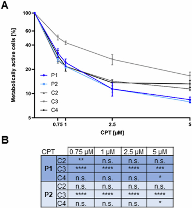
FAM111A (family with sequence similarity 111 member A) is a serine protease and removes covalent DNA-protein cross-links during DNA replication. Heterozygous gain-of-function variants in FAM111A cause skeletal dysplasias, such as the perinatal lethal osteocraniostenosis and the milder Kenny-Caffey syndrome (KCS). We report two siblings born to consanguineous parents with dysmorphic craniofacial features, postnatal growth retardation, ophthalmologic manifestations, hair and nail anomalies, and skeletal abnormalities such as thickened cortex and stenosis of the medullary cavity of the long bones suggestive of KCS. Using exome sequencing, a homozygous synonymous FAM111A variant, NM_001312909.2:c.81 G > A; p.Pro27=, that affects the last base of the exon and is predicted to alter FAM111A pre-mRNA splicing, was identified in both siblings. We identified aberrantly spliced FAM111A transcripts, reduced FAM111A mRNA levels, and near-complete absence of FAM111A protein in fibroblasts of both patients. After treatment of patient and control fibroblasts with different concentrations of camptothecin that induces covalent DNA-protein cross-links, we observed a tendency towards a reduced proportion of metabolically active cells in patient compared to control fibroblasts. However, under these culture conditions, we did not find consistent and statistically significant differences in cell cycle progression and apoptotic cell death between patient and control cells. Our findings show that FAM111A deficiency underlies an autosomal recessive form of FAM111A-related KCS. Based on our results and published data, we hypothesize that loss of FAM111A and FAM111A protease hyperactivity, as observed for gain-of-function patient-variant proteins, may converge on a similar pathomechanism underlying skeletal dysplasias.


Conflict of interest statement

Competing interests: The authors declare no competing interests.

Figures

Fig. 1
Fig. 1
Pedigree, facial photographs, and radiographs of both affected siblings with the homozygous FAM111A variant c.81 G > A. A Pedigree of the patients’ family. The healthy father (I:1) and the healthy mother (I:2) are first-degree cousins and heterozygous carriers of the FAM111A c.81 G > A variant. Both affected siblings, patient 1 (II:1) and patient 2 (II:2), carry the FAM111A c.81 G > A variant in the homozygous state. B Facial photographs of patient 1 at the age of 6 years and 11 months (left) and of patient 2 at the age of 3 years and 2 months (right) show prominent forehead, triangular face, sparse eyebrows, deeply set eyes, bilateral microphthalmia, a long and narrow nose with a prominent nasal bridge, and posteriorly rotated ears with a prominent antihelix. C Radiographs of patient 1 (P1) at the age of 6 years and 10 months and of patient 2 (P2) at the age of 2 years (right upper limb) and 3 years and 2 months (skull and lower limbs). Skull radiographs show mild cortical thickening of the skull with narrowed diploic space indicated by a white arrow. Upper and lower limb radiographs show thickened cortex of long bones and stenosis of the medullary cavity of the long bones in both patients indicated by black arrows. m months, y years
Fig. 2
Fig. 2
Segregation analysis of the FAM111A c.81 G > A variant in the family, FAM111A transcript variants, and FAM111A transcript analysis in patient and control fibroblasts. A Partial sequence electropherograms demonstrating the FAM111A NM_001312909.2:c.81 G > A variant in the homozygous state in leukocyte- and fibroblast-derived DNA of patients 1 and 2, and in the heterozygous state in leukocyte-derived DNA of healthy parents (mother and father). The exon-intron boundary is indicated by a black line. Arrows point to the G-to-A change at the last exon position. B The MANE FAM111A transcript (top) and the four FAM111A transcript variants (TVs) expressed in cultured fibroblasts are shown, all encoding the same FAM111A protein. Exons are given by boxes and are numbered. The coding region is indicated in blue, untranslated regions in gray. Expression levels of FAM111A TVs in cultured fibroblasts according to the GTEx database (last accessed 08/2024) are shown on the right. Length of the bars represents the rate of expression (violet, strong expression; gray, no expression). Primers used for RT-PCRs are indicated by arrows above the transcript variants. The expected PCR amplicon sizes using the primer combination F1 and R1 (444 bp and 405 bp) and F2 and R1 (333 bp) are depicted in the lower left and the lower right panel, respectively. C 2% agarose gel showing RT-PCR amplicons generated with primers F1 and R1 using fibroblast-derived cDNA from patients (P1, P2) and three controls (C1-C3). Fibroblasts were either treated with cycloheximide (CHX, +) or DMSO (─) prior to RNA isolation. In control cells, the expected RT-PCR products of 444 bp and 405 bp were amplified. In contrast, a major amplicon of ~300 bp was obtained from cDNA of patient-derived cells. D Partial sequence electropherogram of an aberrantly spliced FAM111A transcript in patient 1. Direct sequencing of the 287-bp RT-PCR amplicon obtained with primers F1 and R1 revealed skipping of the r.81 g > a change containing exon 3 (NM_022074.4) or exon 4 (NM_001374804.1) in FAM111A transcripts. E 2% agarose gel showing RT-PCR amplicons generated with primers F2 and R1 using fibroblast-derived cDNA from patients (P1, P2) and three controls (C1-C3). Fibroblasts were either treated with CHX (+) or DMSO (─) prior to RNA isolation. The expected RT-PCR product of 333 bp was amplified from cDNA of control and patient cells. A second amplicon of ~300 bp was obtained from patient-derived cDNAs. F Cloning of patient 1-derived RT-PCR amplicons followed by colony PCR and Sanger sequencing of individual amplicons identified the larger amplicon (333 bp) as transcripts in which exon 3 with the r.81 g > a variant (indicated by an arrow) was correctly spliced to exon 4 (upper electropherogram). The smaller amplicon (301 bp) corresponds to aberrantly spliced FAM111A transcripts lacking the last 32 bp of exon 3 (Δ32 bp; lower electropherogram). bp base pairs, F forward primer, R reverse primer, TPM transcripts per million
Fig. 3
Fig. 3
Determination of FAM111A transcript and FAM111A protein levels in patient and control fibroblasts. A, B Relative quantification of FAM111A mRNA levels by RT-qPCR using fibroblast-derived cDNA from the two patients and three healthy controls. The schematics above the RT-qPCR data show the location of the F1 primer in exon 2 (A), the Fq primer in exon 3 (B), and the Rq primer in exon 4 of FAM111A (according to NM_022074.4) (A, B). The coding region is indicated in blue and untranslated region in gray. For quantification, FAM111A mRNA levels were normalized to GAPDH mRNA levels. The bars and errors represent the mean ± SD of three independent experiments (n  =  3), each performed in triplicate. Individual data points are shown. One-way ANOVA followed by Dunnett’s post hoc test was used for statistical analysis to compare relative FAM111A mRNA levels in fibroblasts from patients 1 and 2 separately with controls 1–3. C Representative immunoblot of whole-cell lysates obtained from fibroblasts of patients 1 and 2 and controls 1–3. The amount of FAM111A was monitored by using an anti-FAM111A antibody. Anti-GAPDH antibody was used to control for equal loading. D Band intensities of fluorescence signals were quantified using the ChemiDoc imaging system. Levels of FAM111A were normalized to GAPDH. The bars and errors represent the mean ± SD of three independent experiments (n  =  3). Individual data points are shown. One-way ANOVA followed by Dunnett’s post hoc test was used for statistical analysis to compare the relative FAM111A protein levels in fibroblasts from patients 1 and 2 separately with controls 1–3. *p ≤ 0.05; **p ≤ 0.01; ***p ≤ 0.001; ****p ≤ 0.0001. F1 and Fq forward primers for RT-qPCR, kDa kilodalton, n.s. not significant, Rq reverse primer for RT-qPCR
Fig. 4
Fig. 4
Determination of the proportion of metabolically active cells in patient and control fibroblasts after camptothecin treatment. A Patient- and control-derived fibroblasts were treated with camptothecin (CPT) at a concentration of 0.75 µM, 1 µM, 2.5 µM, or 5 µM or with an equal volume of DMSO for 72 h. The graph shows the proportion of metabolically active cells in patient (P1, P2) and control (C2-C4) fibroblasts following treatment with the indicated concentrations of CPT for 72 h relative to DMSO-treated cells. The mean ± SD of three individual experiments is shown (n = 3), each performed in triplicate. B Two-way ANOVA followed by Dunnett’s post hoc test was used for statistical analysis to compare the proportion of metabolically active fibroblasts from patients 1 and 2 separately with that of fibroblasts from controls 2–4. *p ≤ 0.05; **p ≤ 0.01; ***p ≤ 0.001; ****p ≤ 0.0001. n.s. not significant

Similar articles

Cited by

References

    1. Welter AL, Machida YJ. Functions and evolution of FAM111 serine proteases. Front Mol Biosci. 2022;9:1081166. - PMC - PubMed
    1. Naicker D, Rhoda C, Sunda F, Arowolo A. Unravelling the intricate roles of FAM111A and FAM111B: from protease-mediated cellular processes to disease implications. Int J Mol Sci. 2024;25:2845. - PMC - PubMed
    1. Alabert C, Bukowski-Wills JC, Lee SB, Kustatscher G, Nakamura K, de Lima Alves F, et al. Nascent chromatin capture proteomics determines chromatin dynamics during DNA replication and identifies unknown fork components. Nat Cell Biol. 2014;16:281–93. - PMC - PubMed
    1. Hoffmann S, Pentakota S, Mund A, Haahr P, Coscia F, Gallo M, et al. FAM111 protease activity undermines cellular fitness and is amplified by gain-of-function mutations in human disease. EMBO Rep. 2020;21:e50662. - PMC - PubMed
    1. Nie M, Oravcova M, Jami-Alahmadi Y, Wohlschlegel JA, Lazzerini-Denchi E, Boddy MN. FAM111A induces nuclear dysfunction in disease and viral restriction. EMBO Rep. 2021;22:e50803. - PMC - PubMed

Substances

LinkOut - more resources