. 2024 Nov 6;22(1):513.
doi: 10.1186/s12916-024-03738-9.

Western diet promotes endometriotic lesion growth in mice and induces depletion of Akkermansia muciniphila in intestinal microbiota

Affiliations

Western diet promotes endometriotic lesion growth in mice and induces depletion of Akkermansia muciniphila in intestinal microbiota

Guillaume Parpex et al. BMC Med. .

Abstract

Background: Endometriosis, affecting 10% of women in their reproductive years, remains poorly understood. Both individual and environmental unexplained factors are implicated in this heterogenous condition. This study aims to examine the influence of a Western diet on endometriosis lesion development in mice and to uncover the mechanisms involved.

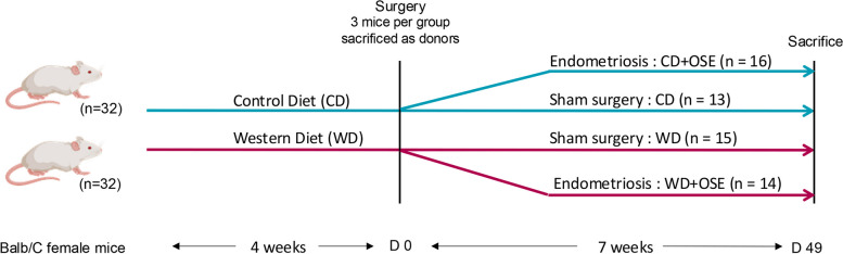
Methods: Mice were fed either a control diet or a Western diet (high in fatty acids and low in fiber) for 4 weeks. Endometriosis was then surgically induced, and lesion development was monitored by ultrasound. After 7 weeks, the mice were sacrificed for analysis of lesion characteristics through RT-qPCR, immunohistochemistry, and flow cytometry. Additionally, the intestinal microbiota was assessed using 16S rRNA gene sequencing.

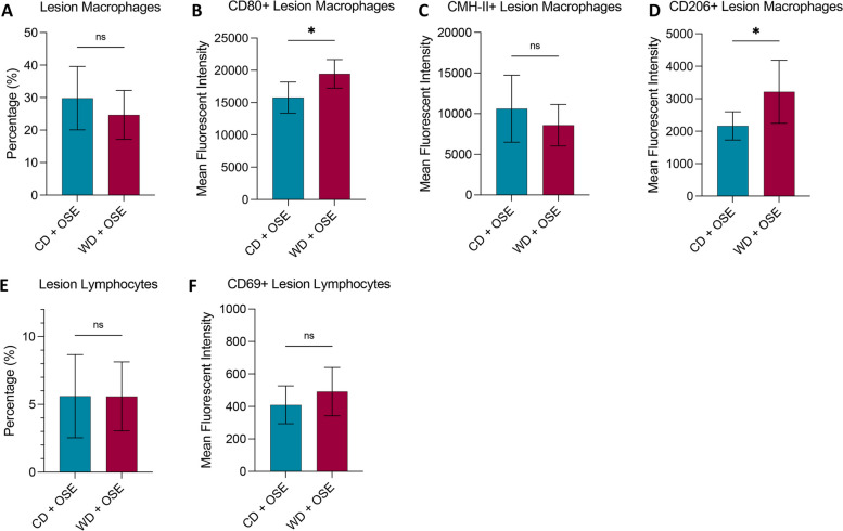
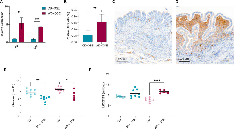
Results: Mice on the Western diet developed lesions that were significantly twice as large compared to those on the control diet. These lesions exhibited greater fibrosis and proliferation, alongside enhanced macrophage activity and leptin pathway expression. Changes in the intestinal microbiota were significantly noted after endometriosis induction, regardless of diet. Notably, mice on the Western diet with the most substantial lesions showed a loss of Akkermansia Muciniphila in their intestinal microbiota.

Conclusions: A Western diet significantly exacerbates lesion size in a mouse model of endometriosis, accompanied by metabolic and immune alterations. The onset of endometriosis also leads to substantial shifts in intestinal microbiota, suggesting a potential link between diet, intestinal health, and endometriosis development.

Keywords: Akkermansia muciniphila; Diet; Endometriosis; Intestinal Microbiota.


Conflict of interest statement

The authors declare no competing interests.

Figures

Fig. 1
Fig. 1
Experimental design
Fig. 2
Fig. 2
Promotion of endometriosis lesions by Western diet in vivo. A (a) Macroscopic view of the implants on day 49. (b) Ultrasonography images of peritoneal implants in mice on day 40. (c) Staining with hematoxylin and eosin of the lesion on day 49 (G: glandular cells; L: lumen; Sc: stromal cells). Original magnification × 100. B Implants weight per mice before surgical implantation to induce endometriosis in CD group (n = 16) and WD group (n = 15) (mg). C Volume evolution of the implants between day 0 (the day of the surgery) and day 40 evaluated by ultrasound (mm3) in the WD + OSE (n = 14) and CD + OSE groups (n = 16). D Weight gain of mice between day 0 and day 49 (g) in the CD (n = 13), CD + OSE (n = 16), WD (n = 15), and WD + OSE groups (n = 14). Unpaired t-test (B, C), one-way ANOVA (D). ns, non-significant. *p < 0.05, **p < 0.01, ***p < 0.001. Data represent the mean and SEM from at least n = 7 mice per group
Fig. 3
Fig. 3
Macrophage activation by Western diet in lesions. AF Flow cytometry analysis on macrophage within lesions (CD11b + F4/80 +) and lymphocytes (CD45 + CD3e +). Macrophage activation markers CD206, CD80, and CMH-II, as well as lymphocyte activation marker CD69, were assessed by mean fluorescent activity and SEM. CD + OSE group (n = 6), WD + OSE group (n = 6). T-test. ns, non-significant. *p % 0.05, **p % 0.01, ***p % 0.001
Fig. 4
Fig. 4
Impact of Western diet on lesion characteristics. A qRT-PCR assessment of Ptgs2 and Col1a1 mRNA expression in lesions. Representative sections of ectopic lesions stained with Masson’s trichrome in the CD + OSE (B) and WD + OSE groups (C). D The area of fibrosis was assessed based on the blue coloration in the CD + OSE and WD + OSE lesions. E Percentage of stromal cells positive for Ki-67 immunostaining. Representative sections of ectopic lesions showing Ki-67-positive stromal cells in CD + OSE (F) and WD + OSE (G) lesions. CD group (n = 7), WD group (n = 6), CD + OSE group (n = 8), WD + OSE group (n = 7). Unpaired t-test. ns, non-significant. *p < 0.05, **p < 0.01, ***p < 0.001
Fig. 5
Fig. 5
Impact of Western diet on metabolic changes. A qRT-PCR assessment of Ob and Obr mRNA expression in lesions. B Percentage of stromal cells positive for Obr immunostaining. Representative sections of ectopic lesions stained with Obr antibody in the CD + OSE (C) and WD + OSE groups (D). Levels of glucose (E) and lactate (F) in mouse serum. CD group (n = 8), WD group (n = 7), CD + OSE group (n = 8), WD + OSE group (n = 5). ANOVA test with Bonferroni correction. *p < 0.05, **p < 0.01, ***p < 0.0001
Fig. 6
Fig. 6
Impact of endometriosis on intestinal microbiota. AC Principal coordinate analysis (PCoA) plots were used to assess the variation between the groups (beta diversity), analyzed by permutational multivariate analysis of variance. Representative visualizations of variations observed by the induction of endometriosis in both diet groups (A and B) or by the diet changes (C). D Shannon diversity index. E Relative abundance of A. muciniphila. F Firmicutes/Bacteroidetes ratio. G The 10 most abundant taxa (at genus level) among different study groups. CD group (n = 8), WD group (n = 7), CD + OSE group (n = 7), WD + OSE group (n = 5). ANOVA test with Bonferroni correction. NS, non-significant. *p % 0.05, **p % 0.01, ***p % 0.001

References

    1. Chapron C, Marcellin L, Borghese B, Santulli P. Rethinking mechanisms, diagnosis and management of endometriosis. Nat Rev Endocrinol. 2019;15:666–82. - PubMed
    1. Chapron C, Chopin N, Borghese B, Foulot H, Dousset B, Vacher-Lavenu MC, et al. Deeply infiltrating endometriosis: pathogenetic implications of the anatomical distribution. Hum Reprod Oxf Engl. 2006;21:1839–45. - PubMed
    1. Sampson JA. The development of the implantation theory for the origin of peritoneal endometriosis. Am J Obstet Gynecol. 1940;40:549–57.
    1. Christ JP, Yu O, Schulze-Rath R, Grafton J, Hansen K, Reed SD. Incidence, prevalence, and trends in endometriosis diagnosis: a United States population-based study from 2006 to 2015. Am J Obstet Gynecol. 2021;225:500.e1-500.e9. - PubMed
    1. Vallée A, Ceccaldi PF, Carbonnel M, Feki A, Ayoubi JM. Pollution and endometriosis: a deep dive into the environmental impacts on women’s health. BJOG Int J Obstet Gynaecol. 2024;131:401–14. - PubMed

Publication types

Substances

Supplementary concepts

LinkOut - more resources