. 2024 Nov 5;23(1):397.
doi: 10.1186/s12933-024-02487-6.

Healthy longevity-associated protein improves cardiac function in murine models of cardiomyopathy with preserved ejection fraction

Affiliations

Healthy longevity-associated protein improves cardiac function in murine models of cardiomyopathy with preserved ejection fraction

Valeria Vincenza Alvino et al. Cardiovasc Diabetol. .

Abstract

Aims: Aging is influenced by genetic determinants and comorbidities, among which diabetes increases the risk for heart failure with preserved ejection fraction. There is no therapy to prevent heart dysfunction in aging and diabetic individuals. In previous studies, a single administration of the longevity-associated variant (LAV) of the human BPIFB4 gene halted heart decline in older and type 2 diabetic mice. Here, we asked whether orally administered LAV-BPIFB4 protein replicates these benefits.

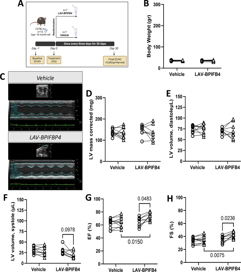
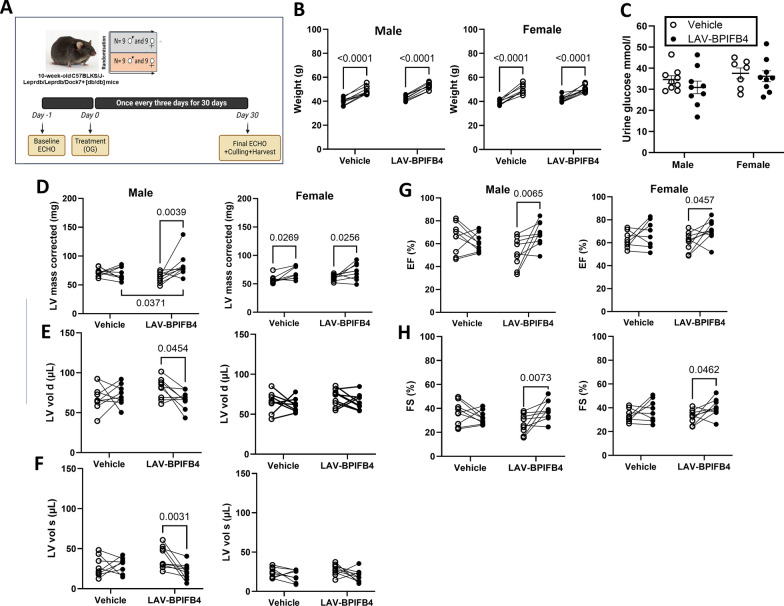
Materials and methods: In two controlled, randomized studies, 18-month-old male C57BL/6 J mice and 9-week-old C57BLKS/J-Leprdb/Leprdb/Dock7 + [db/db] mice of both sexes underwent baseline echocardiography. They then received a recombinant purified LAV-BPIFB4 protein (3 µg/animal, every three days) or vehicle by gavage. After 30 days, the animals underwent echocardiography, and the hearts were collected post-termination for histology.

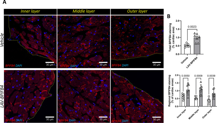
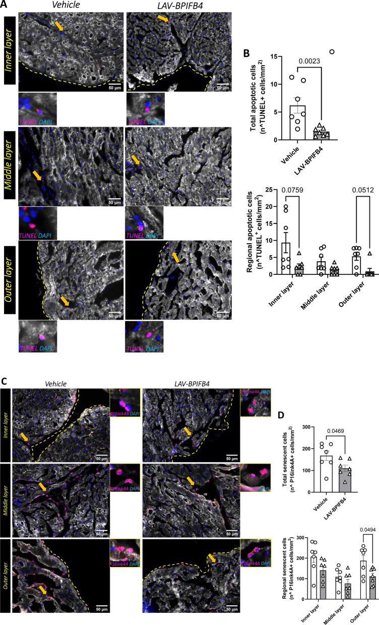
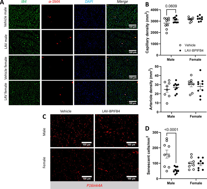
Results: All the animals completed the study except one female diabetic mouse, which was culled prematurely because tooth malocclusion caused eating problems. There was no effect of the LAV-BPIFB4 protein on body weight in the two studies or glycosuria in the diabetic study. In aging mice, LAV-BPIFB4 increased myocardial Bpifb4 expression, improving heart contractility and capillarity while reducing perivascular fibrosis and senesce. In male diabetic mice, LAV-BPIFB4 therapy improved systolic function, microvascular density, and senescence, whereas the benefit was limited to systolic function in females.

Conclusions: This study shows the feasibility and efficacy of a variant protein associated with human longevity in contrasting pivotal risk factors for heart failure in animal models. The diabetic study revealed that sex influences the treatment efficacy.


Conflict of interest statement

A.A.P. owns shares of LGV1 Inc. and has filed a patent. All the other authors declare that there is no conflict of interest.

Figures

Fig. 1
Fig. 1
Primary endpoints of the study in older mice. A Experimental protocol. B Body weight at baseline and the end of the follow-up. C Representative echocardiography traces. DH Echocardiography data: left ventricular mass (D), left ventricular volume at the end of diastole (E) and systole (F), left ventricular ejection fraction (G), and left ventricular fractional shortening (H). Each point represents a single mouse, with a line connecting the baseline and final measurements. The mean values and standard deviations are reported in Supplementary Table 2
Fig. 2
Fig. 2
Effect of treatment on the myocardial expression of BPIFB4. A Representative images with inserts. B Bar graphs with individual values
Fig. 3
Fig. 3
Histological endpoints of the study in older mice. A Representative images illustrating the abundance of capillaries and arterioles. The inserts show specific staining. B Bar graphs with individual values of capillary and arteriole density in the whole left ventricular wall and single regions of the heart. C, D Quantification of vessels covered by PDGFRβ PCs. Representative images with inserts (C) and bar graphs with individual values for capillary and arteriole coverage in the whole left ventricular wall and single regions of the heart (D). E, F Quantification of fibrosis via Azan-Mallory staining. Representative images (E) and bar graphs with individual values of perivascular and interstitial fibrosis (F)
Fig. 3
Fig. 3
Histological endpoints of the study in older mice. A Representative images illustrating the abundance of capillaries and arterioles. The inserts show specific staining. B Bar graphs with individual values of capillary and arteriole density in the whole left ventricular wall and single regions of the heart. C, D Quantification of vessels covered by PDGFRβ PCs. Representative images with inserts (C) and bar graphs with individual values for capillary and arteriole coverage in the whole left ventricular wall and single regions of the heart (D). E, F Quantification of fibrosis via Azan-Mallory staining. Representative images (E) and bar graphs with individual values of perivascular and interstitial fibrosis (F)
Fig. 4
Fig. 4
Effect of treatment on markers of viability and senescence. A, B TUNEL staining for visualization and quantification of apoptotic cells. Representative images with inserts (A) and bar graphs with individual values (B). C, D Staining with P16ink4A for visualization and quantification of senescent cells. Representative images with inserts (C) and bar graphs with individual values (D) are shown. E, F Staining with SA-β-Gal for visualization and quantification of senescent cells. Representative images with inserts E and bar graphs with individual values F are shown
Fig. 5
Fig. 5
Main endpoints of the study in male and female diabetic mice. A Experimental protocol. B Body weight at baseline and at the end of the follow-up. C Urinary glucose levels. D-H) Echocardiography data: left ventricular mass (D), left ventricular volume at the end of diastole (E) and systole (F), left ventricular ejection fraction (G), and left ventricular fractional shortening (H). Each point represents a single mouse, with a line connecting the baseline and final measurements. The mean values and standard deviations are reported in Supplementary Table 3
Fig. 6
Fig. 6
Vascular endpoints of the study in diabetic mice. A Representative images illustrating the abundance of capillaries and arterioles. B Graphs with individual values of capillary density (B) and arteriole density (B). C, D P16inkA + senescent cells (illustrative images (C) and graphs (D))

References

    1. Roger VL. Epidemiology of heart failure: a contemporary perspective. Circ Res. 2021;128:1421–34. 10.1161/CIRCRESAHA.121.318172. - PubMed
    1. Tsao CW, Lyass A, Enserro D, Larson MG, Ho JE, Kizer JR, Gottdiener JS, Psaty BM, Vasan RS. Temporal trends in the incidence of and mortality associated with heart failure with preserved and reduced ejection fraction. JACC Heart Fail. 2018;6:678–85. 10.1016/j.jchf.2018.03.006. - PMC - PubMed
    1. Redfield MM, Borlaug BA. Heart failure with preserved ejection fraction: a review. JAMA. 2023;329:827–38. 10.1001/jama.2023.2020. - PubMed
    1. Heidenreich PA, Bozkurt B, Aguilar D, Allen LA, Byun JJ, Colvin MM, Deswal A, Drazner MH, Dunlay SM, Evers LR, et al. 2022 AHA/ACC/HFSA guideline for the management of heart failure: a report of the American college of cardiology/American heart association joint committee on clinical practice guidelines. Circulation. 2022;145:e895–1032. 10.1161/CIR.0000000000001063. - PubMed
    1. McDonagh TA, Metra M, Adamo M, Gardner RS, Baumbach A, Bohm M, Burri H, Butler J, Celutkiene J, Chioncel O, et al. Focused update of the 2021 ESC Guidelines for the diagnosis and treatment of acute and chronic heart failure. Eur Heart J. 2023;44:3627–39. 10.1093/eurheartj/ehad195. - PubMed

Publication types

MeSH terms

LinkOut - more resources