Review
. 2024 Nov 5;22(1):676.
doi: 10.1186/s12951-024-02906-6.

Recent advances of photoresponsive nanomaterials for diagnosis and treatment of acute kidney injury

Affiliations
Review

Recent advances of photoresponsive nanomaterials for diagnosis and treatment of acute kidney injury

Shijie Yao et al. J Nanobiotechnology. .

Abstract

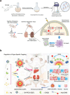
Non-invasive imaging in the near-infrared region (NIR) offers enhanced tissue penetration, reduced spontaneous fluorescence of biological tissues, and improved signal-to-noise ratio (SNR), rendering it more suitable for in vivo deep tissue imaging. In recent years, a plethora of NIR photoresponsive materials have been employed for disease diagnosis, particularly acute kidney injury (AKI). These encompass inorganic nonmetallic materials such as carbon (C), silicon (Si), phosphorus (P), and upconversion nanoparticles (UCNPs); precious metal nanoparticles like gold and silver; as well as small molecule and organic semiconductor polymer nanoparticles with near infrared responsiveness. These materials enable effective therapy triggered by NIR light and serve as valuable tools for monitoring AKI in living systems. The review provides a concise overview of the current state and pathological characteristics of AKI, followed by an exploration of the application of nanomaterials and photoresponsive nanomaterials in AKI. Finally, it presents the design challenges and prospects associated with NIR photoresponsive materials in AKI.

Keywords: Acute kidney injury; Diagnosis and treatment; Fluorescent probe; Nanomaterials; Near-infrared.


Conflict of interest statement

The authors declare they have no competing interests.

Figures

Fig. 1
Fig. 1
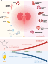
a Relationship between AKI, AKD, CKD, and NKD. b The development of AKI over time and the outcomes that would result
Fig. 2
Fig. 2
a Etiology of AKI and distal multi-organ dysfunction caused by AKI. b Staging of AKI according to current KDIGO
Fig. 3
Fig. 3
Etiology of AKI, including pre-renal, intrinsic renal, and post-renal
Fig. 4
Fig. 4
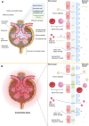
a Basic structure of the renal corpuscle and schematic diagram of the glomerular filtration barrier. b Schematic representation of the glomerular filtration barrier after injury
Fig. 5
Fig. 5
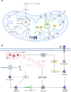
a Basic structure of mitochondria. b Endogenous and exogenous apoptotic pathways in mitochondria
Fig. 6
Fig. 6
Application of various nanomaterials in AKI
Fig. 7
Fig. 7
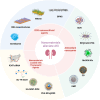
Different types of NIR Photoresponsive nanomaterials
Fig. 8
Fig. 8
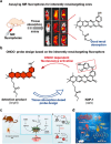
a Early diagnosis of AKI by designing inherently kidney-targeted NIR fluorescent probes and detecting ONOO rise during AKI. b The probe’s mechanism for detecting ONOO. Reproduced from ref [219] with permission. Copyright (2020) Springer. c The mechanism of ONOO production in ferroptosis-mediated AKI. Reproduced from ref [220] with permission. Copyright (2023) Springer. d The NRN probe attenuates fluorescence by responding to ONOO, while reduced glutathione reduces it to restore fluorescence. The probe enables diagnosis and treatment of DIAKI by simultaneously detecting two redox biomarkers in the kidney. Reproduced from ref [221] with permission. Copyright (2022) Springer
Fig. 9
Fig. 9
a FDOCl-22 reacts with HOCl and provides early diagnosis of AKI by NIR and PA imaging. b Renal PA imaging maps of mice at different times after intravenous injection of FDOCl-22 in a cisplatin-induced AKI mouse model. (200 μL × 0.5 mM); the histogram is the PA intensity of kidneys at different time points (PA2: PA intensity at different time points; PA1: PA intensity at 0 min; n = 3 per group). Reproduced from ref [222] with permission. Copyright (2020) Springer
Fig. 10
Fig. 10
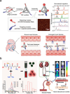
a Design and characterization of the engineering TDF nanodevice (Kim-TDF). The stepwise assembly of Kim-TDF was verified by polyacrylamide gel electrophoresis (PAGE). In vivo schematic diagrams of Kim-TDF in normal kidney and renal injury conditions. Reproduced from ref [223] with permission. Copyright (2022) Springer. b An illustrative diagram detailing the process of synthesizing NIR-CDs with both NIRF and ultra-small dimensions. These NIR-CDs are subsequently employed in imaging studies to track the renal clearance pathway and assess kidney function effectively. Reproduced from ref [158] with permission. Copyright (2022) Springer
Fig. 11
Fig. 11
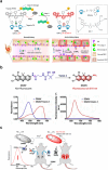
a Schematic design of caspase-3 activatable NIR fluorescent probe. In the early stage of AKI, phosphatidylserine (PS) is flipped from the inner membrane to the outer membrane, and 1-DPA2 is retained in the renal tubule by binding to the flipped PS. At the same time, caspase-3 can cleave 1-DPA2 and then intense NIR fluorescence appears. Reproduced from ref [224] with permission. Copyright (2021) Springer. b A depiction illustrating the process of DDAV mediated by Vanin-1, accompanied by the absorption and fluorescence spectra depicting the response of DDAV towards Vanin-1. Reproduced from ref [225] with permission. Copyright (2022) Springer. c In the drug-induced AKI mouse model, DSMN can diagnose AKI by detecting SO2. Reproduced from ref [226] with permission. Copyright (2023) Springer
Fig. 12
Fig. 12
a The ONOO-activated AUCN is applied for efficient NIR-II fluorescence imaging of AKI. Reproduced from ref [227] with permission. Copyright (2023) Springer. b TPPTS-AuNPs can be activated by GSH in the liver, while early imaging can be performed in metabolic acidosis-induced kidney injury. Reproduced from ref [228] with permission. Copyright (2023) Springer. c Schematic Diagram of the Dual-Ligands Stabilized Au NCs and NIR-II window fluorescence imaging in animal models of renal ischemia–reperfusion (RIR) and unilateral ureteral obstruction (UUO). Reproduced from ref [229] with permission. Copyright (2023) Springer. d Illustrations presenting the metabolic pathways of various dye-KTP conjugates in vivo and their application in noninvasive kidney monitoring within the NIR-II window. Reproduced from ref [230] with permission. Copyright (2021) Springer
Fig. 13
Fig. 13
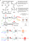
a In previous studies, Heptamethine Cyanine Dyes with hydrophilic groups located at one or both ends have been reported. Despite the introduction of these hydrophilic groups, these dyes maintain hepatic clearance. Chemical Structure and Features of the Probe PEG3-HC-PB and the Chromophore PEG3-HC-POH (the Activated Probe) Resulting from the Probe’s Response to the Biomarker H2O2, as well as the Probe’s Utilization in Detecting Contrast-Agent- and Ischemia/Reperfusion-Induced AKI by NIR-II Fluorescent and Optoacoustic Dual-Mode Imaging. Reproduced from ref [231] with permission. Copyright (2023) Springer. b Mechanism of NAG detection by BOD-I-NAG-NP and BOD-II-NAG-NP in vivo. Reproduced from ref [232] with permission. Copyright (2021) Springer
Fig. 14
Fig. 14
a Preparation of a novel liposome-mediated biomimetic delivery system with NIR-II triggered release and NIR light irradiation to kidney resulted in an excellent effect for reduced immune cell infiltration and renal inflammation. Mechanistically, inhibiting PLK3 suppressed the degradation of HIF-1α and ROS-induced OS. This protection shielded renal tubular epithelial cells from apoptosis and halted macrophage activation, thereby mitigating renal inflammation. Reproduced from ref [233] with permission. Copyright (2022) Springer. b A schematic diagram depicting the use of CB[7]-mediated ultrasmall luminescent Au nanocarriers for targeted delivery to specific organs, along with the therapeutic application of DXM-02AuNPs in mice with cisplatin-induced AKI. Reproduced from ref [234] with permission. Copyright (2023) Springer
Fig. 15
Fig. 15
a Structural schematic, TEM image, UV–Vis absorption spectrum, and XPS image of Au24Cd1 clusters. Reproduced from ref [235] with permission. Copyright (2023) Springer. b An illustrative representation of the NIR-II fluorescence emission of BPQDs upon excitation with an 808 nm laser. BPQDs undergo degradation into phosphate, phosphite, and other phosphorus oxide compounds triggered by water, oxygen, or ROS, resulting in decreased NIR-II fluorescence intensity. Reproduced from ref [236] with permission. Copyright (2021) Springer. c An illustrated depiction of a DNA origami plasmonic nanoantenna designed for the early detection and targeted treatment of AKI. Following intravenous administration, the rDONs@AuNR dimer exhibited preferential accumulation in the kidneys. Reproduced from ref [237] with permission. Copyright (2022) Springer. d Schematic diagram to indicate the design of a NIR fluorescent/photoacoustic probe, CDIA, for monitoring •OH in AKI and demonstration of the strategy for HTS of antioxidant natural products to attenuate AKI. Reproduced from ref [238] with permission. Copyright (2023) Springer

Similar articles

Cited by

References

    1. Vanmassenhove J, Kielstein J, Jörres A, Biesen WV. Management of patients at risk of acute kidney injury. Lancet. 2017;389:2139–51. - PubMed
    1. Vaara ST, Bhatraju PK, Stanski NL, McMahon BA, Liu K, Joannidis M, Bagshaw SM. Subphenotypes in acute kidney injury: a narrative review. Critical Care. 2022. 10.1186/s13054-022-04121-x. - PMC - PubMed
    1. Summary of recommendation statements. Kidney international supplements 2012, 2:8-12. - PMC - PubMed
    1. Kellum JA, Lameire N. Diagnosis, evaluation, and management of acute kidney injury: a KDIGO summary (part 1). Critical Care. 2013;17:204. - PMC - PubMed
    1. Kellum JA, Romagnani P, Ashuntantang G, Ronco C, Zarbock A, Anders HJ. Acute kidney injury. Nat Rev Dis Primers. 2021;7:52. - PubMed

LinkOut - more resources