. 2024 Nov 5;15(1):400.
doi: 10.1186/s13287-024-03972-1.

Harnessing the regenerative effects of human amniotic stem cells (hAFSCs) on restoring erectile function in a bilateral cavernous nerve crush (BCNC) injury rat model

Affiliations

Harnessing the regenerative effects of human amniotic stem cells (hAFSCs) on restoring erectile function in a bilateral cavernous nerve crush (BCNC) injury rat model

Chun-Hou Liao et al. Stem Cell Res Ther. .

Abstract

Background: Intracavernous (IC) injections of stem cells has been shown to ameliorate cavernous nerve (CN)-induced erectile dysfunction (ED). However, the regenerative effects underlying the recovery of erectile function (EF) in human amniotic fluid-derived stem cells (hAFSCs) remain unclear. In the bilateral cavernous nerve crushing (BCNC) injury rat paradigm, we sought to ascertain the effects of hAFSC treatment on EF recovery during the incipient phase.

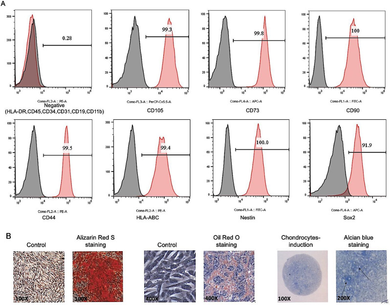
Methods: Three groups of 45 male rats were used in this study: sham (Group 1), saline IC injection after BCNC (Group 2), and hAFSC intracavernous injection (ICI) after BCNC (Group 3). hAFSCs from the fourth passage showed potential to differentiate into trilineage cells. All animals were subjected to EF analysis on the 28th day post-injection and tissues were retrieved for histopathological and immunohistochemical analyses.

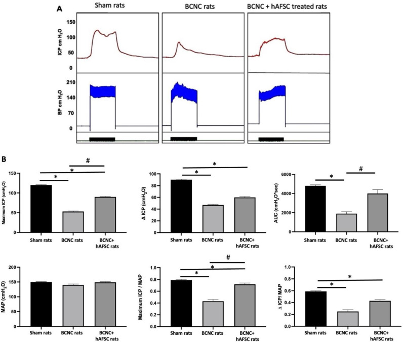
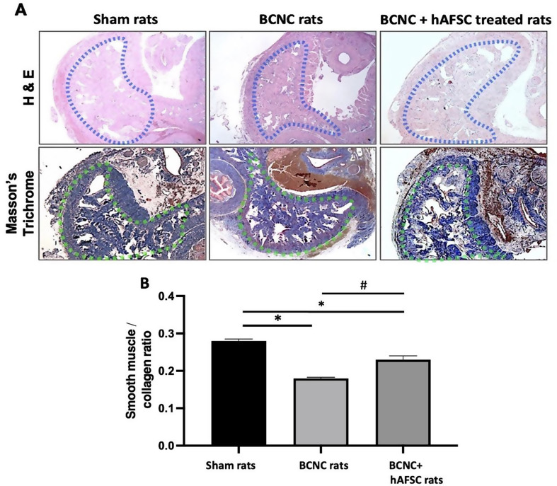
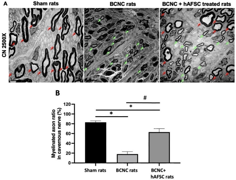
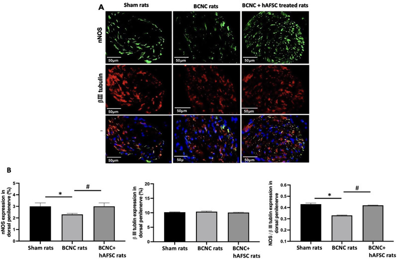
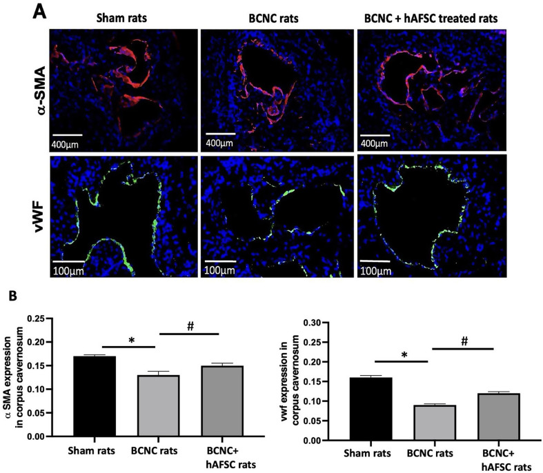
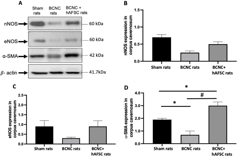
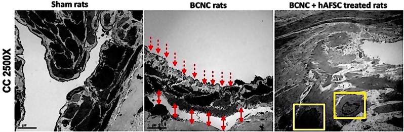
Results: IC injections of hAFSC significantly improved EF parameters in BCNC-ED rats at 28 days post-injury. AFSC treatment enhanced the smooth muscle condition and increased the smooth muscle/collagen ratio, as evidenced by histological analysis. Immunohistology revealed increased expression of 𝛼-SMA andvWf in the corpus cavernosum and enhanced expression of nNOS in the dorsal penile nerve in BCNC-ED rats (p < 0.05). Western blotting showed that hAFSC treatment significantly increased α-SMA expression in the hAFSC group compared with that in the BCNC group. Electron microscopy revealed significantly elevated myelination in the CN (p < 0.05), maintenance of smooth muscle structures, and restoration of EF in BCNC-ED rats treated with hAFSC.

Discussion and conclusions: hAFSC treatment increased EF in BCNC-ED rats at a single dose. As BCNC-ED resembles ED caused by radical prostatectomy (RP), this therapy has high potential for ED patients after RP surgery.

Keywords: BCNC injury; Electron microscopy; Erectile dysfunction; Stem cells; hAFSC.


Conflict of interest statement

The authors declare no conflicts of interest.

Figures

Fig. 1
Fig. 1
Immunophenotyping and trilineage differentiation capability of hAFSCs. A) Flow cytometry results depicting the pluripotent and mesenchymal markers of hAFSCs. (B)hAFSC profiles exhibiting typical MSC characteristics of various origins, including osteocytes (Alizarin Red staining), adipocytes (Oil Red staining), and chondrocytes (arrows indicate acidic polysaccharides). *Passage 4 cells were used for the analysis
Fig. 2
Fig. 2
2A and B: Comparison of EF among the sham, BCNC, and BCNC + hAFSC-treated groups. (A) Intracavernous pressure (ICP) (red) and blood pressure (BP) (blue) in sham, BCNC, and BCNC rats + hAFSC. Green bars indicate electrical stimulation for 60 s. (B) Quantitative results of EF parameters (MAP, ICP, and AUC) calculated for each group (sham rats, BCNC rats, and BCNC rats + hAFSC). (n = 15). * p < 0.05, compared to sham rats; # p < 0.05, compared to BCNC rats. (AUC, area under the curve; ICP, intracavernous pressure; MAP, mean arterial pressure)
Fig. 3
Fig. 3
(A) Micrograph showing hematoxylin and eosin and Masson’s trichrome staining of the corpus cavernosum (40× magnification). H&E staining revealed varying degrees of damage and poor tissue arrangements exclusively in the BCNC group (blue demarcated area), whereas no similar damage was observed in BCNC rats treated with hAFSC. The smooth muscle/collagen ratio was determined using Masson’s trichrome-stained corpus cavernosum (green demarcated area). (B) Quantification of the smooth muscle/collagen ratio in the sham, BCNC, and BCNC rats treated with hAFSC. BCNC refers to bilateral cavernous nerve crush injury and hAFSC denotes human amniotic fluid stem cells. * Indicates a significant difference from the sham rats (p < 0.05) and # indicates a significant difference from the BCNC rats (p < 0.05). For each group, 8 animals were used in this study (n = 8)
Fig. 4
Fig. 4
(A) Transmission electron microscopy of the cavernous nerve (CN) in sham rats, BCNC rats, and BCNC rats treated with hAFSC magnified 2500×. The image illustrates an increased number of myelinated sheaths (indicated by red arrows) and demyelinated nerve axons (indicated by green arrows) observed in BCNC rats treated with hAFSC compared to the vehicle-treated BCNC group. The scale shown in Fig. 3A is 5 μm. (B) Graph showing statistical results of the number of myelin sheaths in each group of rats. BCNC refers to bilateral cavernous nerve crush injury and hAFSC denotes human amniotic fluid stem cells. * Indicates a significant difference from the sham rats (p < 0.05) and # indicates a significant difference from the BCNC rats (p < 0.05). Six rats from each group were used for this study (n = 6)
Fig. 5
Fig. 5
(A) The illustration depicts Immunofluorescence staining of control, nNOS in the DPN of sham rats, BCNC rats, and BCNC rats treated with hAFSC 28 days post-BCNC. Representative images of the control and experimental groups are shown (nNOS in green, β-III tubulin in red) with an original magnification of 400x. nNOS-positive nerve fibers in DPN were quantified as the area of nNOS-positive nerve fibers/β-III tubulin. (B) Quantitative analysis revealed a decrease in the number of nNOS-positive nerve fibers in the BCNC vehicle-treated group compared to the sham and BCNC groups treated with hAFSC. * Indicates a significant difference from the sham rats (p < 0.05), and # indicates a significant difference from the BCNC rats (p < 0.05). For each group, eight rats were used in this study (n = 8)
Fig. 6
Fig. 6
(A) illustrates the expression of 𝛼-smooth muscle actin (𝛼-SMA) and Von Willebrand Factor (vWF) in assessing the endothelial contents in the corpus cavernosum by immuno- histological analysis in normal control, BCNC and the BCNC + hAFSC group of rats. The immuno-histopathological images indicate the expressions of 𝛼-SMA in red colour and the expression of vWF in green fluorescent colour. (B) Semi-quantification analysis of the expression of 𝛼-SMA and vWF in the corpus cavernosum of normal control, BCNC, and BCNC + hAFSC group of rats. *p < 0.05 – compared with normal sham rats; #p < 0.05 – compared with BCNC rats. In each group, eight rats were used in the study (n = 8)
Fig. 7
Fig. 7
(A) Western blot analysis of (the middle section of the corpus cavernosum tissue) was used to evaluate the protein expression of nNOS, eNOS, and α-SMA across different groups, with β-actin serving as the loading control. (B) Quantification of nNOS expression by Western blot analysis showed an increase in the BCNC + hAFSC group and a low level in the BCNC group. Compared with the sham group, both the BCNC + hAFSC and BCNC groups were lower. (C) A similar pattern was observed for eNOS expression, an increase in the BCNC + hAFSC group, a low level in the BCNC group, and low levels in both the BCNC + hAFSC and BCNC groups compared to the sham group. (D) A significantly increased level of α-SMA was observed in the BCNC + hAFSC group compared to the BCNC and sham groups. level of significance: *p < 0.05 - compared to sham rats; #p < 0.05 – compared to BCNC rats. n = 4 animals per group for Western blot analysis
Fig. 8
Fig. 8
Transmission electron microscopy image of the corporal smooth muscle in sham rats, BCNC rats, and BCNC rats treated with hAFSC at a magnification of 2500×. Sham control rats showed normal structures, characterized by intact adherent junctions and preserved smooth muscle cells. BCNC rats exhibited abnormal endothelial layer structures (indicated by red arrows) with disrupted adherent junctions and chasm between the smooth muscle layers (indicated by red double-sided arrows). In contrast, BCNC rats treated with hAFSC displayed tightly packed normal smooth muscle structures with well-defined adherent junctions. The yellow demarcated area represents the integration of hAFSC into the smooth muscle layer. Six rats were used for each group (n = 6)

Similar articles

References

    1. Liao CH, Chang CJ, Chen KC, Rajneesh CP, Tseng XW, Cheng JH, Chiang HS, Wu YN. Platelet-rich plasma glue placement at the prostatectomy site: effects on erectile function restoration and preservation of cavernous nerves in a rat model of nerve-sparing prostatectomy. Biomed Pharmacother. 2023;172:114499. 10.1016/j.biopha.2022.114499. - PubMed
    1. Irdam GA, Rasyid N, Taher A. Intracavernosal injection of mesenchymal stem cells for diabetic erectile dysfunction: a systematic review. Med J Indonesia. 2023;32(1):96–105. 10.13181/mji.v32i1.2549.
    1. Fandel TM, Albersen M, Lin G, Qiu X, Ning H, Banie L, Lue TF, Lin CS. Intracavernous injection of adipose-derived stem cells enhances erectile function in a rat model of cavernous nerve injury by recruiting cells to the major pelvic ganglion. Eur Urol. 2023;74(3):456–65. 10.1016/j.eururo.2022.08.036. - PMC - PubMed
    1. Mulhall JP, Smith AB. Penile rehabilitation following radical prostatectomy: a comprehensive review. Curr Opin Urol. 2006;16(6):410–4. 10.1016/j.biopha.2005.11.007. - PubMed
    1. Hatzimouratidis K, Salonia A. Pharmacotherapy for erectile dysfunction: recent advances and future prospects. J Sex Med. 2007;4(2):406–17. 10.1016/j.biopha.2006.07.004. - PubMed