

Bilateral Dermoid Ovarian Cysts in a Young Woman - A Case Report and Literature Review
- PMID: 39502610
- PMCID: PMC11534055
- DOI: 10.21873/cdp.10402
Bilateral Dermoid Ovarian Cysts in a Young Woman - A Case Report and Literature Review
Abstract
Background/aim: Ovarian tumors are a common type of neoplasm in women, with mature cystic teratomas being the most frequent variant. These tumors occur bilaterally in approximately 10% of cases. However, bilateral and multiple occurrences are rarely reported.
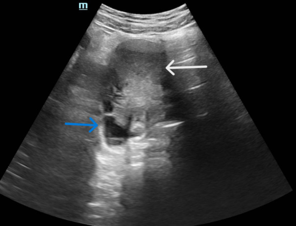
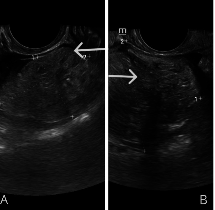
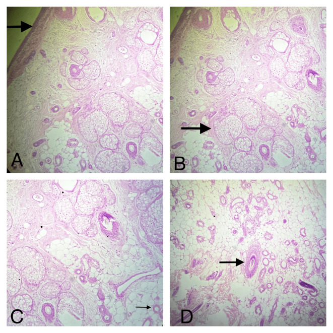
Case report: A 22-year-old nulliparous woman presented with amenorrhea and sudden, generalized, dull lower abdominal pain. Diagnostic imaging, including ultrasound and computed tomography (CT) scans, revealed large solid-cystic lesions in both ovaries, with internal hyperechoic foci consistent with fat and calcification, along with thin internal septations. A laparoscopic cystectomy was successfully performed, preserving ovarian function. Histopathological examination confirmed the presence of stratified keratinized squamous epithelium, sebaceous glands, hair follicles, mature adipose tissue, blood vessels, and lymphatic vessels within the resected cysts, with no evidence of malignancy.
Conclusion: This unique case provides valuable insights into the understanding and management of bilateral dermoid cysts, highlighting the importance of preserving ovarian function in young women.
Keywords: Mature cystic ovarian teratoma; bilateral dermoid cyst; dermoid cysts; histopathology; laparoscopic cystectomy.
©2024 The Author(s). Published by the International Institute of Anticancer Research.
Conflict of interest statement
The Authors declare no conflicts of interest in relation to this study.
Figures
References
-
- Ekici E, Soysal M, Kara S, Dogan M, Gokmen O. The efficiency of ultrasonography in the diagnosis of dermoid cysts. Zentralbl Gynakol. 1996;118(3):136–141. - PubMed
LinkOut - more resources
Full Text Sources