. 2024 Oct 22:15:1473133.
doi: 10.3389/fimmu.2024.1473133. eCollection 2024.

Leishmania major surface components and DKK1 signalling via LRP6 promote migration and longevity of neutrophils in the infection site

Affiliations

Leishmania major surface components and DKK1 signalling via LRP6 promote migration and longevity of neutrophils in the infection site

Olivia C Ihedioha et al. Front Immunol. .

Abstract

Background: Host-related factors highly regulate the increased circulation of neutrophils during Leishmania infection. Platelet-derived Dickkopf-1 (DKK1) is established as a high-affinity ligand to LRP6. Recently, we demonstrated that DKK1 upregulates leukocyte-platelet aggregation, infiltration of neutrophils to the draining lymph node and Th2 differentiation during Leishmania infection, suggesting the potential involvement of the DKK1-LRP6 signalling pathway in neutrophil migration in infectious diseases.

Results: In this study, we further explored the potential role of DKK1-LRP6 signalling in the migration and longevity of activated neutrophils in the infection site using BALB/c mice with PMNs deficient in LRP6 (LRP6NKO) or BALB/c mice deficient in both PMN LRP6 and platelet DKK1 (LRP6NKO DKK1PKO). Relative to the infected wild-type BALB/c mice, reduced neutrophil activation at the infection site of LRP6NKO or LRP6NKO DKK1PKO mice was noted. The neutrophils obtained from either infected LRP6NKO or LRP6NKO DKK1PKO mice additionally showed a high level of apoptosis. Notably, the level of LRP6 expressing neutrophils was elevated in infected BALB/c mice. Relative to infected BALB/c mice, a significant reduction in parasite load was observed in both LRP6NKO and LRP6NKO DKK1PKO infected mice. Notably, DKK1 levels were comparable in the LRP6NKO and BALB/c mice in response to infection, indicating that PMN activation is the major pathway for DKK1 in promoting parasitemia. Parasite-specific components also play a crucial role in modulating neutrophil circulation in Leishmania disease. Thus, we further determine the contribution of Leishmania membrane components in the migration of neutrophils to the infection site using null mutants deficient in LPG synthesis (Δlpg1- ) or lacking all ether phospholipids (plasmalogens, LPG, and GIPLs) synthesis (Δads1- ). Relative to the WT controls, Δads1- parasite-infected mice showed a sustained decrease in neutrophils and neutrophil-platelet aggregates (for at least 14 days PI), while neutrophils returned to normal in Δlpg1- parasite-infected mice after day 3 PI.

Conclusion: Our results suggest that DKK1 signalling and Leishmania pathogen-associated molecular patterns appear to regulate the migration and sustenance of viable activated neutrophils in the infection site resulting in chronic type 2 cell-mediated inflammation.

Keywords: apoptosis; innate response; leishmaniasis; neutrophils; platelet.


Conflict of interest statement

The authors declare that the research was conducted in the absence of any commercial or financial relationships that could be construed as a potential conflict of interest.

Figures

Figure 1
Figure 1
Decreased percentage of neutrophils and NPA in Δads1- and Δlpg1- parasite-infected mice. BALB/c mice were challenged with infective metacyclic promastigote (2 x 10⁶ parasites, n = 5) of (LV39c5 WT and Δlpg1-) and (Freidlin WT and Δads1- ) strains via the footpad. Control mice (n = 10/2 feet per mouse) were given 0.9% NaCl saline. Neutrophils were isolated from the footpads of all infected and non-infected mice at days 3, 7 and 14 PI. Isolated neutrophil samples were analyzed by flow cytometry for Ly6G-positive cells and NPA formation. Each dot indicates Ly6G+ cells (A–C) and NPA (G–I) obtained from Δads1 - parasite infected mice. Also, each dot indicates Ly6G+ cells (D–F) and NPA (J–L) obtained from Δlpg1 - parasite infected Mice. Representative flow cytometry dot plots showing the analyses of Ly6G + cells and NPA performed on day 3 PI, as well as the concatenated dot plot of each sample in all the experimental groups are presented in Supplementary Figure S2 . In all the experiments, WT-infected and non-infected mice served as positive and negative controls, respectively. Results are presented as mean (± SEM) and are representative of 3 independent experiments. One-way ANOVA with Bonferroni’s post hoc test was performed to analyze the data *p < 0.05, **p < 0.01, ***p < 0.001, ‘ns’ indicates not significant (p > 0.05).
Figure 2
Figure 2
Decreased CD11b and MHC class II positive neutrophils derived from LRP6NKO DKK1PKO and LRP6NKO infected mice. The WT-BALB/c, LRP6NKOand LRP6NKO DKK1PKO mice were challenged with infective metacyclic promastigote (2 x 10⁶ parasites, n = 5) of L. major via the footpad. Non-infected BALB/c mice (n = 10/2 feet per mouse) were given 0.9% NaCl saline. Neutrophils were isolated from the footpads of all infected and non-infected mice at day 3 PI. Isolated neutrophil samples were analyzed by flow cytometry for CD11b and MHC class II positive neutrophils. Each dot indicates MHC class II (A) and CD11b (B) positive cells. Representative flow cytometry dot plots showing the analyses of CD11b and MHC class II + neutrophils performed on day 3 PI, as well as the concatenated dot plot of each sample in all the experimental groups are presented in Supplementary Figure S3 . In all the experiments, WT-infected and non-infected mice served as positive and negative controls, respectively. Results are presented as mean (± SEM). One-way ANOVA with Bonferroni’s post hoc test was performed to analyze the data **p < 0.01, ***p < 0.001(p > 0.05).
Figure 3
Figure 3
Decreased myeloperoxidase positive neutrophils derived from LRP6NKO DKK1PKO and LRP6NKO infected mice on day 3 PI. The WT-BALB/c, LRP6NKO and LRP6NKO DKK1PKO mice were challenged with infective metacyclic promastigote (2 x 10⁶ parasites, n = 5) of L. major via the footpad. Non-infected BALB/c mice (n = 10/2 feet per mouse) were given 0.9% NaCl saline. Neutrophils were isolated from the footpads of all infected and non-infected mice at day 3 PI. Isolated neutrophil samples were analyzed by flow cytometry for myeloperoxidase + cells. Each dot indicates myeloperoxidase + neutrophils, and the percentage of myeloperoxidase+ cells in the different experimental groups is shown in column graphs. Representative flow cytometry dot plots showing the analyses of myeloperoxidase + neutrophils and IgG1 isotype control for non-specific antibody staining, as well as a dot plot of each sample in all the experimental groups are presented in Supplementary Figure S4 . In all the experiments, WT-infected and non-infected mice served as positive and negative controls, respectively. Results are presented as mean (± SEM). One-way ANOVA with Bonferroni’s post hoc test was performed to analyze the data **p < 0.01; ***p < 0.001; ns, non-significant (p > 0.05).
Figure 4
Figure 4
Infected LRP6NKO and LRP6NKO DKK1PKO mice show elevated neutrophil apoptosis. The WT-BALB/c, LRP6NKOand LRP6NKO DKK1PKO mice were challenged with infective metacyclic promastigote (2 x 10⁶ parasites, n = 5) of L. major via the footpad. Non-infected BALB/c mice (n = 10/2 feet per mouse) were given 0.9% NaCl saline. Neutrophils were isolated from the footpads of all infected and non-infected mice at day 3 PI. Apoptotic neutrophils were analyzed by flow cytometry and identified by the increase in fluorescence intensity of annexin V-FITC. Each dot indicates either viable or apoptotic Ly6G+ cells. Percentage of apoptotic cells (early apoptosis: Annexin+, late apoptosis: Annexin+ PI+) in the infected and non-infected mice are shown in column graph (A) & neutrophil viability is shown in column graph (B). Representative flow cytometry dot plots showing apoptotic and viable neutrophils after double staining with Annexin V-FITC and propidium iodide performed on day 3 PI. The first quadrant (Q1) represents necrotic neutrophils, the second quadrant (Q2) represents later apoptotic neutrophils, the third quadrant (Q3) represents early apoptotic neutrophils, and the fourth quadrant (Q4) represents normal neutrophils Supplementary Figure S5 . A concatenated dot plot of each sample in all the experimental groups is presented in Supplementary Figure S5 . In all the experiments, WT-infected and non-infected mice served as positive and negative controls, respectively. Results are presented as mean (± SEM). One-way ANOVA with Bonferroni’s post hoc test was performed to analyze the data *p < 0.05; **p < 0.01, ***p < 0.001.
Figure 5
Figure 5
Recombinant DKK1 delayed neutrophil apoptosis in a dose-dependent manner. Neutrophils isolated from naïve mice were incubated with various concentrations of rDKK1. Neutrophil samples harvested at 0-, 4-, and 24 hrs post incubation were used to determine neutrophil apoptosis by flow cytometry. The percentage of apoptotic cells in the different experimental conditions is shown in the bar graph. Representative flow cytometry contour plots generated 24 hrs post incubation showed total apoptotic neutrophils (Q3 & Q2) after double staining with Annexin V-FITC and propidium iodide, as presented in Supplementary Figure S6 . Also, a contour plot of each sample in all the experimental conditions is presented in Supplementary Figure S6 . In all the experiments, non-treated neutrophils served as controls. Results are presented as mean (± SEM). One-way ANOVA with Bonferroni’s post hoc test was performed to analyze the data (*p < 0.05, **p < 0.01). ns, non-significant.
Figure 6
Figure 6
Elevated LRP6 expression in infected BALB/c mice. The WT-BALB/c mice were challenged with infective metacyclic promastigote (2 x 10⁶ parasites, n = 5) of L. major via the footpad. Non-infected BALB/c mice (n = 10/2 feet per mouse) were given 0.9% NaCl saline. Neutrophils were isolated from the footpads of infected and non-infected mice at day 3 PI. Isolated neutrophil samples were analyzed by flow cytometry for LRP6 expression. The column graph indicates the percentage of LRP6 positive cells (A) and the expression of LRP6 by Ly6G+ cells in the different experimental groups (B). Representative flow cytometry dot plots showing the analyses of LRP6+ neutrophils and dot plots of each sample in all the experimental groups are presented in Supplementary Figure S1 . Results are presented as mean (± SEM). Student’s t-test was performed to analyze the data (*p < 0.05; ** p < 0.01).
Figure 7
Figure 7
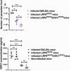
Parasite load in the infected LRP6NKO and LRP6NKO DKK1PKO mice were significantly reduced, while plasma DKK1 production in infected BALB/c and LRP6NKO mice were comparable. BALB/c, LRP6NKO and LRP6NKO DKK1PKO mice were challenged with infective metacyclic promastigote (2 x 10⁶ parasites, n = 5) of WT L. major strain via the footpad. Control mice (n = 10/2 feet per mouse) were given 0.9% NaCl Saline. The infected foot from each mouse was used to determine parasite load using a limiting dilution assay on day 14 PI. Parasite load in the infected BALB/c group was compared with the infected LRP6NKO and LRP6NKO DKK1PKO mice. To determine DKK1 production, blood was collected via the maxillary vein at day 3 PI. Plasma samples were analyzed by ELISA. In all experiments, Infected and non-infected BALB/c mice served as positive and negative controls, respectively. Results of parasite load are presented as mean +/- SEM, and data analysis was done using one-way ANOVA with Bonferroni’s post hoc test *p < 0.05, **p < 0.01, (p > 0.05). For DKK1 concentration, results are presented as mean +/- SEM of replicate wells. One-way ANOVA with Bonferroni’s post hoc test was performed to analyze the data **p < 0.01, ‘ns’ indicates not significant (p > 0.05).

Similar articles

References

    1. Lipoldová M, Svobodová M, Krulová M, Havelková H, Badalová J, Nohýnková E, et al. . Susceptibility to Leishmania major infection in mice: multiple loci and heterogeneity of immunopathological phenotypes. Genes Immun. (2000) 1:200–6. doi: 10.1038/sj.gene.6363660 - DOI - PubMed
    1. Reiner SL, Locksley RM. The regulation of immunity to Leishmania major. Annu Rev Immunol. (1995) 13:151–77. doi: 10.1146/annurev.iy.13.040195.001055 - DOI - PubMed
    1. Alexander J, Brombacher F. T helper1/t helper2 cells and resistance/susceptibility to leishmania infection: is this paradigm still relevant? Front Immunol. (2012) 3:80. doi: 10.3389/fimmu.2012.00080 - DOI - PMC - PubMed
    1. Sacks D, Noben-Trauth N. The immunology of susceptibility and resistance to Leishmania major in mice. Nat Rev Immunol. (2002) 2:845–58. doi: 10.1038/nri933 - DOI - PubMed
    1. Bennouna S, Bliss SK, Curiel TJ, Denkers EY. Cross-talk in the innate immune system: neutrophils instruct recruitment and activation of dendritic cells during microbial infection 1. J Immunol. (2003) 171:6052–8. doi: 10.4049/jimmunol.171.11.6052 - DOI - PubMed

MeSH terms

Substances

LinkOut - more resources