[Preprint]. 2024 Oct 21:rs.3.rs-5200876.
doi: 10.21203/rs.3.rs-5200876/v1.

Hemifusomes and Interacting Proteolipid Nanodroplets Mediate Multi-Vesicular Body Formation

Affiliations

Hemifusomes and Interacting Proteolipid Nanodroplets Mediate Multi-Vesicular Body Formation

Amirrasoul Tavakoli et al. Res Sq. .

Update in

Abstract

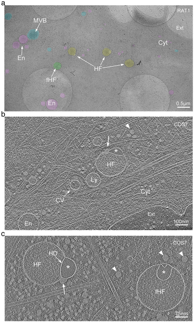
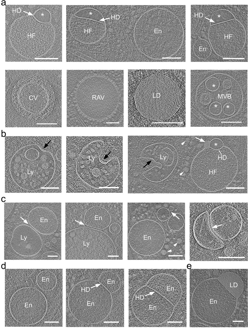
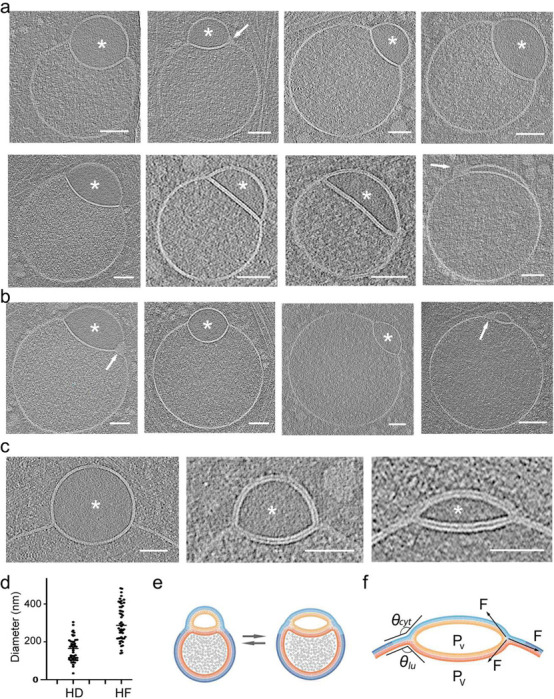
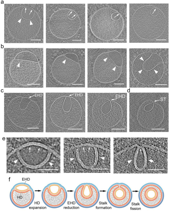
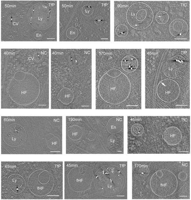
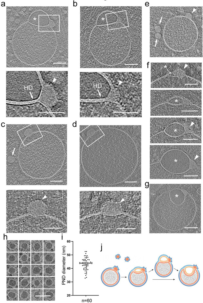
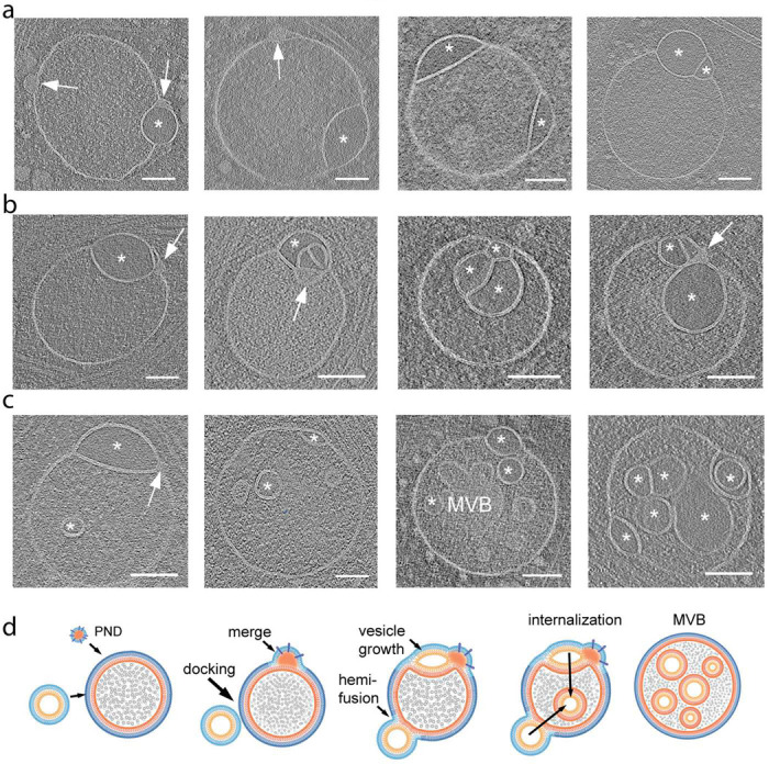
The complex, pleiomorphic membrane structure of the vesicular components within the endolysosomal system has been appreciated through decades of classical electron microscopy. However, due to the heavy fixation and staining required in these approaches, in situ visualization of fragile intermediates between early endosomes, late endosomes and ultimately multivesicular bodies (MVBs), remains elusive, raising the likelihood that other structures may have also been overlooked. Here, using in situ cryo-electron tomography in four mammalian cell lines, we discover heterotypic hemifused vesicles that share an extended hemifusion diaphragm, associated with a 42nm proteolipid nanodroplet (PND). We term this previously undescribed vesicular organelle-complex, "hemifusome". Hemifusomes make up approximately 10% of the organelle pool of the endolysosomal system, but do not participate directly in transferrin-mediated endocytosis. Hemifusomes exist in compound conformations and also contain intraluminal vesicles. Based on their range of morphologies, and the consistent presence of the PND at sites of compound hemifused vesicles, we propose that hemifusomes function as platforms for vesicular biogenesis mediated by the PND. These findings offer direct in situ evidence for a long-lived hemifusion diaphragm, and a new, ESCRT-independent model for the formation of late endosomes containing intraluminal vesicles and ultimately MVBs.


Conflict of interest statement

Additional Declarations: There is NO Competing Interest.

Figures

Figure 1.
Figure 1.. Cryo-electron tomography observation of hemifused vesicles at the leading edge of cultured cells
a. Representative cryo-electron microscopy image of the leading edge of a RAT1 cell cultured on cryo-EM grids. Lamellipodia and filopodia in the upper right corner delineate the cell border, separating the cytoplasm (Cyt) from the extracellular space (Ext). Vesicular organelles are highlighted in color: endosomes (En, pink), multivesicular bodies (MVB, blue), hemifusomes (HF, yellow), and flipped hemifusomes (fHF, green). Scale bar: 0.5 μm. b. Representative cryo-electron tomogram slice of the border of a COS7 cell highlighting cytoskeletal components, endosomes (En), lysosomes (Ly), a clathrin-coated vesicle (CV), and a hemifusome (HF). A single bilayer or hemifusion diaphragm separates the hemifused vesicles. The larger vesicle has a fine granular content, and the smaller hemifused vesicle has a smooth translucent lumen (*). The arrowhead points to the proteolipid particle and arrow points to the nanodroplet at the rim of the hemifusion diaphragm. Scale bar: 100 nm. c. Tomographic mid-cross-section through a direct (HF) and a flipped hemifusome (fHF) showing the well-defined bilayer outline of the vesicle membranes. A single bilayer or hemifusion diaphragm (HD) separates the hemifused vesicles. Arrowheads point to the proteolipid particle, as well as similar proteo-lipid particles seen free in the cytoplasm. Scale bar: 75 nm.
Figure 2.
Figure 2.. Hemifusome luminal content is distinct from other membrane-bound organelles of the endo-lysosomal system
a. Representative cryo-electron microscopy images of various membrane-bound organelles within the endo-lysosomal system. Endosomes (En), lysosomes (Ly), a clathrin-coated vesicle (CV), ribosome associated vesicles (RAV), multivesicular body (MVB), lipid droplet (LD), and hemifusomes (HF) are identified. The distinct luminal content of each organelle is visible, with the smaller hemifusome vesicles (*) consistently showing a unique light, smooth, and particle-free luminal content compared to other organelles. Similar smooth lumen vesicles were found only inside some MVBs (*). Hemifusion diaphragm (HD) is highlighted with white arrows. b. Series of lysosomes at various initial stages of inward budding of a vesicle obtained from tomographic slices. The last panel shows a side-by-side comparison of a lysosome and a hemifusome (HF). Black arrows point to a distinct surface protein complex, likely ESCRT, at the inwardly curved portion of the lysosomal membrane. White arrowhead points to proteolipid nanodroplets in the cytoplasm and white arrow points to the nanodroplet at the rim of the hemifusion diaphragm. c. Series of tomogram slices showing endosomal (En), and lysosomal (Ly) vesicles adhered or docked to each other (white arrows). d. Series of tomogram slices showing hemifused endosomes sharing an extended hemifusion diaphragm (HD) (white arrow). Right panel- a hemifused endosome (En) and lipid droplet (LD) Note the granular texture of the lumen of all vesicles. Scale bars: 100 nm.
Figure 3.
Figure 3.. Range of morphological appearances of direct hemifusomes
a. Cryo-electron tomography mid-cross-section slices of various direct hemifusomes highlighting the variability in sizes and shapes across and within each hemifusome pair. Note the smooth appearance of the smaller vesicle of the hemifusome (*). Hemifusomes also show variability in their hemifusion diaphragm diameter and curvature. Arrows point to proteolipid particles lodged in the hemifusome at the rim of the hemifusion diaphragm where it meets with membranes of the two vesicles. b. Hemifusomes showing deformation of the smaller vesicle with the expansion and curvature of the hemifusion diaphragm, resulting in a cross-sectional view that resembles a lens-shaped vesicle. In this specific configuration, the entire inner leaflet as well as the content of the smaller vesicle becomes fully embedded within the bilayer of the membrane containing the larger vesicle. c. Close-up views of the hemifusion diaphragm and degrees of flattening of the smaller vesicle (*) to form the lens-shaped structure embedded in the bilayer. d. Diameters of the hemifusion diaphragm (HD), mean= 158.4 +/− 60.9, and the larger vesicle of the hemifusome (HF), mean= 299.3 +/− 96.2. n = 50 hemifusomes. e. Diagram illustrating the hemifused configuration and possible interconversion between hemifusome conformations. f. Diagram illustrating some of the forces at play to modulate hemifusome angles between the membranes and overall shape. θ = angle of the cytoplasmic (cyt) and luminal (lu) leaflets of the bilayer at the point of junction with the hemifusion diaphragm. ; P, internal pressure of smaller (v) and larger vesicle (V), F, membrane tension vectors.
Figure 4.
Figure 4.. Range of morphological appearances of flipped hemifusomes and the proposed progression to form an intraluminal vesicle
a. Tomographic slices of various flipped hemifusomes (fHF) highlighting the variability in sizes and shapes across and within each hemifusome pair. b-d. Various degrees of curving and budding of the hemifusion diaphragm. The budding vesicle exhibits an elongated shape. In both scenarios, the intraluminal portion of the hemifused diaphragm expands, while the cytoplasmic side of the vesicle decreases in radius and forms an external segment of the bilayer shared by both vesicles. During this process, the cytoplasmic side of the lens-shaped structure transforms into an external hemifusion diaphragm (EHD), which reduces radially to form a stalk-like structure (ST). e. Close-up views of the curving and expansion of the hemifusion diaphragm (HD, arrowheads) and radial reduction of the EHD (arrows). f. Diagram depicting our proposed model, illustrating the progressive rounding and expansion of the hemifusion diaphragm (HD) and reduction of the EHD to form a stalk and ultimately scission to form an intraluminal vesicle. Scale bars: 200 nm.
Figure 5.
Figure 5.. Hemifusomes are not part of the uptake and cargo transfer of endocytosed nanogold particles
Tomographic slices of pulse-chase experiments showing the distribution nanogold particles of various surface-functionalization and size in clathrin-coated pits or vesicles (CV), endosomes (En), and lysosomes (Lys), but absent from either vesicle compartment of the hemifusomes (HF) or flipped hemifusomes (fHF). T = time of incubation with the gold nanoparticles. TP = gold nanoparticles with transferrin physiosorbed, TC = gold nanoparticles with transferrin covalently attached, and PC = gold nanoparticles with slightly positively charged non-reactive polymer. D = diameter of the gold nanoparticles. Scale bars: 200 nm.
Figure 6.
Figure 6.. Proteo-lipid particles associated with hemifusomes are located at the rim of the hemifusion diaphragm
and b. Tomographic slices and corresponding close-up views of hemifusomes (HF) with a prominent proteolipid nanodroplet (PND) at the rim of the hemifusion diaphragm (HD). Asterisk marks the smooth lumen of the smaller vesicle. Lower panels are close-up views of the area marked with a white rectangle showing the PND embedded in the hydrophobic core of the vesicle membrane and a series of particulate structures on its outer limit. These structural features reinforce the view that PNDs contain lipids and proteins. c and d. Tomographic slices and corresponding close-up views of PNDs fused to endosome-like vesicles. Arrows point to a PND free in the cytoplasm. Lower panels are high magnifications of the boxed regions, showing the PND encapsulated within the vesicle lipid bilayer. The outer leaflet enveloping the PND is decorated with protein particles (arrowheads). e. More examples of PNDs of uniform size and texture, within the cytoplasm (arrows) and fused to an endosome-like vesicle (arrowhead). f. Mid-cross-section of tomograms of a series of different hemifusomes showing a range of increasing size vesicles with a smooth lumen (asterisks) formed next to the PND site (arrowheads), suggesting that the smaller vesicle of the hemifusome may be forming by a de novo vesiculogenesis process. g. Tomogram slice of a flipped hemifusome showing the PND embedded at the three-way juncture of the hemifusion diaphragm and the membrane of the two vesicles. h. Panel of PNDs found free in the cytoplasm in the vicinity of endosomes and hemifusomes. Image contrast in this image is reversed to its original gray scale to show that the PNDs are electron dense or have a phase dark appearance. i. Plot of the diameter distribution of PNDs. The average diameter was calculated to be 42.4 ± 4.3 nm (n=60) j. Diagram illustrating the association of PNDs to endosomes to form a hemifusome and the progression to a flipped hemifusome.
Figure 7.
Figure 7.. Compound hemifusomes as hubs for the formation of late endosomes and multivesicular bodies
a. Tomographic slices illustrating compound hemifusomes with one or more additional vesicles in a hemifused conformation with either of the two vesicles of the initial hemifusome pair. Asterisks mark vesicles with clear luminal content. Hemifusomes with additional PNDs embedded in their membranes (arrows) suggest these may act as a hub for the formation of compound hemifusomes. b. Compound hemifusion with flipped hemifusomes. Multiple hemifusion events coalesce to form very complex compound structures. Asterisks mark vesicles with clear luminal content. c. Hemifusomes comprising hemifused vesicles as well as multiple intraluminal vesicles. Asterisks mark vesicles with clear luminal content. Arrow points to PND. d. Diagram illustrating the proposed path for the formation of compound hemifusomes, followed by the flipping of the vesicle into the luminal side of the larger vesicle and subsequent scission, providing an alternative path to the formation of late endosomes and multivesicular bodies (MVBs). Asterisks mark vesicles with clear luminal content. Scale bar: 200 nm.

Similar articles

References

    1. Knorr R. L., Mizushima N. & Dimova R. Fusion and scission of membranes: Ubiquitous topological transformations in cells. Traffic 18, 758–761 (2017). 10.1111/tra.12509 - DOI - PubMed
    1. Saffi G. T. & Botelho R. J. Lysosome Fission: Planning for an Exit. Trends Cell Biol 29, 635–646 (2019). 10.1016/j.tcb.2019.05.003 - DOI - PubMed
    1. Knorr R. L., Lipowsky R. & Dimova R. Autophagosome closure requires membrane scission. Autophagy 11, 2134–2137 (2015). 10.1080/15548627.2015.1091552 - DOI - PMC - PubMed
    1. Piper R. C. & Katzmann D. J. Biogenesis and function of multivesicular bodies. Annu Rev Cell Dev Biol 23, 519–547 (2007). 10.1146/annurev.cellbio.23.090506.123319 - DOI - PMC - PubMed
    1. Olmos Y. The ESCRT Machinery: Remodeling, Repairing, and Sealing Membranes. Membranes (Basel) 12 (2022). 10.3390/membranes12060633 - DOI - PMC - PubMed

Publication types

LinkOut - more resources