. 2025 Jan;12(1):e2405818.
doi: 10.1002/advs.202405818. Epub 2024 Nov 6.

Identifying Multiomic Signatures of X-Linked Retinoschisis-Derived Retinal Organoids and Mice Harboring Patient-Specific Mutation Using Spatiotemporal Single-Cell Transcriptomics

Affiliations

Identifying Multiomic Signatures of X-Linked Retinoschisis-Derived Retinal Organoids and Mice Harboring Patient-Specific Mutation Using Spatiotemporal Single-Cell Transcriptomics

Yueh Chien et al. Adv Sci (Weinh). 2025 Jan.

Abstract

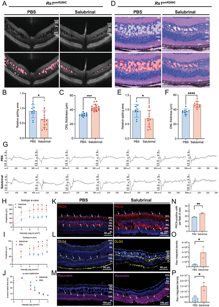
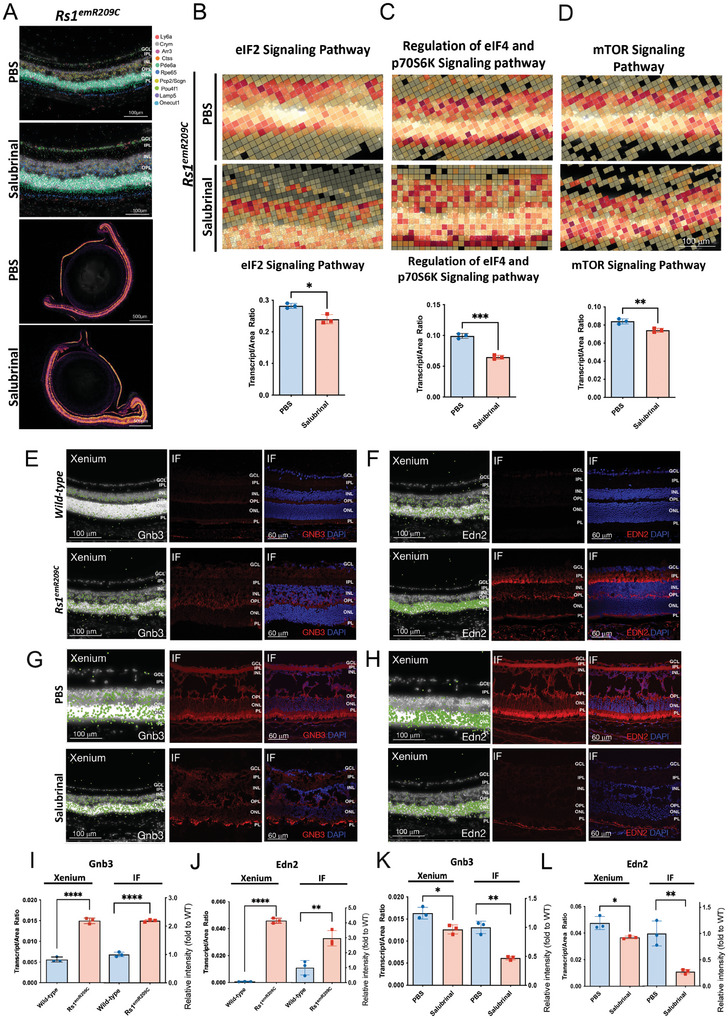
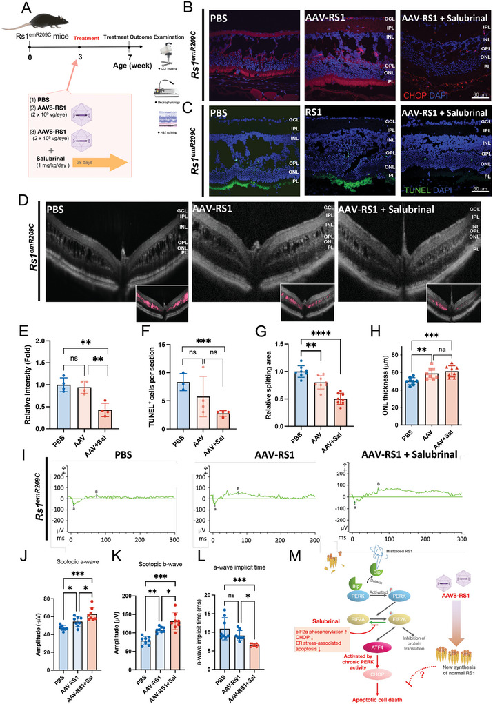
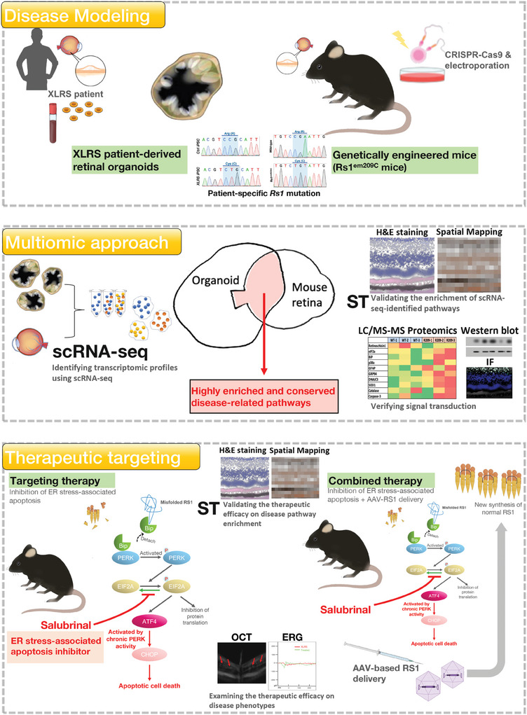
X-linked retinoschisis (XLRS) is an inherited retinal disorder with severe retinoschisis and visual impairments. Multiomics approaches integrate single-cell RNA-sequencing (scRNA-seq) and spatiotemporal transcriptomics (ST) offering potential for dissecting transcriptional networks and revealing cell-cell interactions involved in biomolecular pathomechanisms. Herein, a multimodal approach is demonstrated combining high-throughput scRNA-seq and ST to elucidate XLRS-specific transcriptomic signatures in two XLRS-like models with retinal splitting phenotypes, including genetically engineered (Rs1emR209C) mice and patient-derived retinal organoids harboring the same patient-specific p.R209C mutation. Through multiomics transcriptomic analysis, the endoplasmic reticulum (ER) stress/eukryotic initiation factor 2 (eIF2) signaling, mTOR pathway, and the regulation of eIF4 and p70S6K pathways are identified as chronically enriched and highly conserved disease pathways between two XLRS-like models. Western blots and proteomics analysis validate the occurrence of unfolded protein responses, chronic eIF2α signaling activation, and chronic ER stress-induced apoptosis. Furthermore, therapeutic targeting of the chronic ER stress/eIF2α pathway activation synergistically enhances the efficacy of AAV-mediated RS1 gene delivery, ultimately improving bipolar cell integrity, postsynaptic transmission, disorganized retinal architecture, and electrophysiological responses. Collectively, the complex transcriptomic signatures obtained from Rs1emR209C mice and patient-derived retinal organoids using the multiomics approach provide opportunities to unravel potential therapeutic targets for incurable retinal diseases, such as XLRS.

Keywords: X‐link retinoschisis (XLRS); chronic ER stress‐associated apoptosis; eIF2α signaling; genetically engineered mice; retinoschisin 1 (RS1); single‐cell RNA‐sequencing; spatiotemporal transcriptomics.


Conflict of interest statement

The authors declare no conflict of interest.

Figures

Figure 1
Figure 1
Characterizing XLRS patient‐derived retinal organoids and genetically engineered Rs1 knock‐in mice carrying patient‐specific point mutation p. R209C. A) Schematic illustration showing the disease modeling using XLRS patient‐derived retinal organoids and genetically engineered Rs1 knock‐in mice (Rs1 emR209C mice). Note that the patient‐derived retinal organoids and Rs1 emR209C mice carried the point mutation p. R209C identical to that of the enrolled XLRS patient. B) Clinical examination of XLRS patient with p. R209C point mutation by OCT imaging and color fundus photography. C) The design of the CRISPR/Cas9 genome editing system to generate Rs1 emR209C mice by introducing sgRNA to target the PAM sequence on the wild‐type allele. D) Confirmation of introducing point mutation p. R209C into Rs1 in Rs1 emR209C mice using Sanger sequencing. E) OCT imaging and F) H & E staining of Rs1 emR209C and age‐matched wild‐type mouse retinas at different ages. G) IF staining shows the expression of bipolar cell marker, PRKCA (green), and postsynaptic marker, DLG4 (red) in Rs1 emR209C and age‐matched wild‐type mouse retinas at different ages. Nuclei were stained with DAPI (blue). H) IF staining and quantification of GFAP stained müller glia (green) and in Rs1 emR209C and wild‐type retinas at different ages. I) Dark‐adapted ERG responses in Rs1 emR209C and age‐matched wild‐type mouse retinas at different ages. Scale bar = 60 µm.
Figure 2
Figure 2
ScRNAseq analysis of Rs1 emR209C mice and XLRS patient iPSC‐derived retinal organoids. A) Schematic illustration showing the multiomic approach integrating scRNA‐seq and ST to identify overlapping disease pathways on patient‐derived retinal organoids and Rs1 emR209C mice. The transcriptomic signatures were identified by deep scRNA‐seq analysis from both models. The overlapping transcriptomic signatures identified by scRNA‐seq delineated the crucial disease‐related pathways. ST was used for validating the disease‐related pathways identified by scRNA‐seq. B)The diagram shows the scRNA‐seq experimental procedures of XLRS patient‐derived retinal organoids and Rs1 emR209C mice. C) IF staining of RS1 (green) and bipolar cell marker PKCA (red) in Rs1 emR209C and wild‐type retinas at different ages. Nuclei were stained with DAPI (blue). D) UMAP visualization and cell clustering of Rs1 emR209C and age‐matched wild‐type mouse retinas. E) Relative proportions of cell clusters in Rs1 emR209C and wild‐type retinas at different ages. F) Up‐regulated and down‐regulated DEGs of scRNA‐seq dataset in bipolar cells from Rs1 emR209C and wild‐type mouse retinas at different ages. G) IPA results show the enriched pathways in the bipolar cells, cones, and rods of the Rs1emR209C retinas at indicated times. H) Venn diagrams show the number of overlapped pathways enriched in bipolar cells, cones, and rods between Rs1 emR209C retinas and XLRS patient‐specific retinal organoids. I) Overlapped pathways enriched in bipolar cells, cones, and rods of Rs1 emR209C retinas and XLRS patient‐specific retinal organoids. J) Venn diagram shows the number of overlapped enriched pathways across bipolar cells, cones, and rods in Rs1 emR209C retinas and XLRS patient‐specific retinal organoids. K) Gene interaction analysis reveals the interaction network among the genes involved in eIF2 signaling, mTOR and the regulation of the eIF4 and p70S6K signaling pathways.
Figure 3
Figure 3
Chronic enrichment of ER stress/eIF2α pathway in RS1‐expressing cells in Rs1 emR209C mouse retinas. A) Xenium analysis shows the visualization of selected marker genes to identify retinal ganglion cells, rod and cone photoreceptors, retinal pigment epithelium, microglia, and endothelial cells. The original H&E‐stained section is presented in the upper left corner. B) Xenium analysis shows the enrichment of (B) eIF2α pathway, C) regulation of eIF4 and p70S6K signaling pathway, and D) mTOR signaling pathway in Rs1 emR209C retinas compared to age‐matched wild‐type retinas. Transcripts per area were quantified and shown as the lower subpanel in panels B‐D (N = 3, mean ± s.e.). E) Using CytAssist Visium to match the gene expression captured spots in the spatial transcriptome (right) in the H&E‐stained images of 3‐week‐old wild‐type and Rs1 emR209C retinas (left). The captured spots were further divided into different retinal layers, including the retinal ganglion cell layer (blue), inner cell layer (orange), outer cell layer (green), and retinal pigment epithelium (red). F) Heatmap shows the expression of CytAssist Visium‐detected enriched genes in bulk RNAseq and the rod cell data from scRNA‐seq. CytAssist Visium‐captured spots shows the gene expression of (Panel G; Upper) Edn2 and (Panel H; Upper) Gnb3 in wild‐type and Rs1 emR209C retinas. scRNA‐seq and UMAP visualization show the distribution of Edn2 and Gnb3 expression in cell clusters (Panels G, H; Lower).
Figure 4
Figure 4
Misfolding of RS1 protein activates the ER stress/eIF2 pathway in Rs1 emR209C mouse retinas. A) Nonreducing SDS polyacrylamide gradient gels showing the failure of RS1 homo‐octameric complex in Rs1 emR209C mouse retinas (upper). In reducing gels, a large reduction of RS1 protein was observed in the lysates from Rs1 emR209C mouse retinas (lower). B) Quantification of RS1 protein of wild‐type and Rs1 emR209C mouse retinas in the reducing gel. Western blot shows increased C) BIP protein amount, D) the phosphorylation of eIF2α, E) ATF4 and CHOP protein amount in Rs1 emR209C retinas compared to wild‐type retinas. Quantification of F) BIP protein amount, G) eIF2α phosphorylation, H) ATF4, and I) CHOP protein amount (N = 3, mean ± s.e.). (J) Puromycin incorporation assay indicated failure of protein synthesis in Rs1 emR209C retinas (N = 2, mean ± s.e.). K) The quantification of protein synthesis through puromycin incorporation assay. L) TUNEL assay shows increased apoptosis in the Rs1 emR209C retinas. M) IF staining shows increased phosphorylation of PERK in Rs1 emR209C retinas at 6 months and 12 months, but no PERK phosphorylation was detected in wild‐type retinas at any given age. N) Mass spectrometry (LC/MS)‐based proteomic analysis showing the reduction in overall translation in the Rs1 emR209C retinas (N = 4). O) Comparison of the score sequest HT:sequest HT between wild‐type and Rs1 emR209C retinas at 3‐week ages (N = 3, mean ± s.e.). One‐way t‐test, *p‐value < 0.05. P) Heatmap comparing the expression of the candidate proteins involved in XLRS pathologies between wild‐type and Rs1 emR209C retinas at 3‐week ages. Q) Schematic illustration shows RS1 protein misfolding chronically enriched ER stress and eIF2α pathway, impaired protein synthesis, leading to increased ER stress‐induced apoptosis in Rs1 emR209C retinas.
Figure 5
Figure 5
Therapeutic targeting the chronic ER stress/ eIF2 pathway activation ameliorates retinoschisis and improves retinal electrophysiological functions. A) OCT imaging of Rs1 emR209C retinas with or without salubrinal treatment. B) Quantification of the schisis area based on the OCT imaging data of Rs1 emR209C retinas with or without salubrinal treatment (N = 10, mean ± s.e.). C) Quantification of the ONL thickness based on the OCT imaging of Rs1 emR209C retinas with or without salubrinal treatment (N = 10, mean ± s.e.). D) H & E staining of Rs1 emR209C retinas with or without salubrinal treatment. E) Quantification of the schisis area based on the H&E staining of Rs1 emR209C retinas with or without salubrinal treatment (N = 10, mean ± s.e.). F) Quantification of the ONL thickness based on the H&E staining data of Rs1 emR209C retinas with or without salubrinal treatment (N = 10, mean ± s.e.). G) The intensity‐response relation for the dark‐adapted ERG of Rs1 emR209C retinas with or without salubrinal treatment. H) The scotopic a‐waves, I) scotopic b‐waves, and J) the a‐wave implicit time of Rs1 emR209C retinas with and without salubrinal treatment. In panels H, I, and J, the results are mean ± s.e. of eight independent experiments. K) IF staining of bipolar cell marker PKCA (red), L) postsynaptic marker DLG4 (yellow), and M) photoreceptor marker recoverin (purple) in Rs1 emR209C retinas treated with salubrinal or PBS. Nuclei were stained with DAPI (blue). N) Quantification of the PKCA‐positive cells, and intensity of the area positive for O) DLG4, and P) recoverin in Rs1 emR209C retinas treated with salubrinal or PBS. In panels N, O, and P, the results are mean ± s.e. of three independent experiments.
Figure 6
Figure 6
In situ spatial transcriptomics of differential gene expression in Rs1 emR209C mouse retinas. A) Using the Xenium ST to visualize several cell types in Rs1 emR209C mouse retina receiving the administration of salubrinal or PBS. Xenium analysis and quantification of the transcripts per area of the Xenium data showing the treatment effect of salubrinal on the enrichment of B) eIF2α pathway, regulation of C) eIF4 and p70S6K signaling pathway, and D) mTOR signaling pathway in Rs1 emR209C retinas compared to PBS treated retinas (N = 3, mean ± s.e.). Xenium analysis and IF show the enrichment of E) Gnb3 and F) Edn2 at transcriptomic (left) and protein levels (middle and right) in Rs1 emR209C retinas compared to age‐matched wild‐type retinas. Xenium analysis and IF show the effect of salubrinal treatment on the enrichment of G) Gnb3 and H) Edn2 gene at transcriptomic (left) and protein levels (middle and right) in Rs1 emR209C retinas compared to PBS‐treated retina. The quantification of I) Gnb3 and J) Edn2 expression based on the Xenium analysis and IF data for Rs1emR209C retinas and wild‐type retinas (N = 3, mean ± s.e.). The quantification of K) Gnb3 and L) Edn2 expression based on the Xenium analysis and IF data for Rs1emR209C retinas treated with salubrinal or PBS (N = 3, mean ± s.e.).
Figure 7
Figure 7
Combination of salubrinal administration and AAV‐based RS1 gene delivery synergistically ameliorates retinoschisis and improves retinal electrophysiological functions in Rs1 emR209C mouse retinas. A) The experimental design shows the combination of salubrinal administration and AAV‐based RS1 gene delivery in Rs1 emR209C retinas. B) CHOP staining and C) TUNEL assay in PBS‐treated Rs1 emR209C retinas, AAV8‐based RS1 gene delivery, and the combination of salubrinal and AAV8‐based RS1 delivery. D) OCT imaging of Rs1 emR209C PBS‐treated retinas, AAV8‐based RS1 gene delivery, and the combination of salubrinal and AAV8‐based RS1 delivery. The schisis splitting area (pink) is presented in the lower right corner. The quantification of E) CHOP protein content (N = 4, mean ± s.e.), F) apoptotic signals (N = 4, mean ± s.e.), G) the area of schisis splitting cavities (N = 7, mean ± s.e.), and H) ONL thickness (N = 8, mean ± s.e.) in Rs1 emR209C retinas with indicated treatment are shown. I) The intensity‐response relation for the dark‐adapted full‐field ERG of PBS‐treated Rs1 emR209C mouse retinas, AAV8‐based RS1 gene delivery, and the combination of salubrinal and AAV8‐based RS1 delivery. J) Comparison of the scotopic a‐wave amplitudes, K) scotopic b‐wave amplitudes, and L) the a‐wave implicit time of the PBS‐treated Rs1 emR209C retinas, AAV8‐based RS1 gene delivery, and the combination of salubrinal and AAV8‐based RS1 delivery (N = 8, mean ± s.e.). M) A schematic diagram shows the enrichment of ER stress/eIF2 signaling pathways in XLRS pathogenesis. Misfolded RS1 protein increased ER stress, BIP, phosphorylated eIF2α, ATF4, and CHOP. This signaling cascade leads to the increase of apoptosis of retinal cells. Salubrinal that ameliorates ER stress and attenuates the eIF2 pathway exhibited a synergistic efficacy with AAV‐based RS1 gene delivery in the treatment of XLRS.
Figure 8
Figure 8
Schematic illustration showing the use of multiomic approaches to identify XLRS‐related disease pathways in in vitro and in vivo XLRS‐like models. For disease modeling (Upper), patient's peripheral blood was collected for the generation of patient iPSCs and patient iPSC‐derived retinal organoids. These patient‐derived retinal organoids carried patient's genetic background and unique splitting features. Meanwhile, using a CRISPR‐Cas9‐based technologies, we also generated the genetically engineered mice (Rs1 emR209C mice) that exhibited XLRS‐like retinoschisis phenotypes. Sanger sequencing confirmed that both XLRS‐like models harbored the same patient‐specific Rs1 point mutation. For the multiomic approach (Middle), we subjected the dissociated single cells from both patient‐derived retinal organoids and Rs1 emR209C mouse retina to scRNA‐seq. The transcriptomic signatures of both XLRS‐like models identified by scRNA‐seq were integrated and overlapped to obtain the highly enriched and conserved pathways between two XLRS‐like models. In situ spatial transcriptomics was used to validate the enrichment of identified disease‐related pathways. The signal transduction of the disease‐related pathways were verified using immunofluorescence and Western blot. For the therapeutic targeting of the most enriched disease‐related pathway, a pharmacological agent was administered to target the most enriched disease pathway in the Rs1 emR209C mice. The efficacy of therapeutic targeting on the enrichment of disease pathways were verified using in situ spatial transcriptomics. Its efficacy on disease phenotypes were examined using OCT and ERG. Therapeutic targeting plus AAV8‐mediated Rs1 gene delivery further showed synergistic efficacy in Rs1 emR209C mice. The therapeutic targeting inactivates the disease pathways and the gene therapy provided new wildtype RS1 protein.

References

    1. George N. D., Yates J. R., Moore A. T., Br. J. Ophthalmol. 1995, 79, 697. - PMC - PubMed
    1. a) Sauer C. G., Gehrig A., Warneke‐Wittstock R., Marquardt A., Ewing C. C., Gibson A., Lorenz B., Jurklies B., Weber B. H., Nat. Genet. 1997, 17, 164; - PubMed
    2. b) Zeng Y., Takada Y., Kjellstrom S., Hiriyanna K., Tanikawa A., Wawrousek E., Smaoui N., Caruso R., Bush R. A., Sieving P. A., Invest Ophthalmol Vis Sci. 2004, 45, 3279. - PubMed
    1. a) Weber B. H., Schrewe H., Molday L. L., Gehrig A., White K. L., Seeliger M. W., Jaissle G. B., Friedburg C., Tamm E., Molday R. S., Proc Natl Acad Sci U S A 2002, 99, 6222; - PMC - PubMed
    2. b) Kjellstrom S., Bush R. A., Zeng Y., Takada Y., Sieving P. A., Invest Ophthalmol Vis Sci. 2007, 48, 3837; - PubMed
    3. c) Liu M., Liu J., Wang W., Liu G., Jin X., Lei B., Front Med. 2022, 9, 886947. - PMC - PubMed
    1. a) Ou J., Vijayasarathy C., Ziccardi L., Chen S., Zeng Y., Marangoni D., Pope J. G., Bush R. A., Wu Z., Li W., Sieving P. A., J. Clin. Invest. 2015, 125, 2891,; - PMC - PubMed
    2. b) Bush R. A., Zeng Y., Colosi P., Kjellstrom S., Hiriyanna S., Vijayasarathy C., Santos M., Li J., Wu Z., Sieving P. A., Hum. Gene Ther. 2016, 27, 376,; - PMC - PubMed
    3. c) Zeng Y., Petralia R. S., Vijayasarathy C., Wu Z., Hiriyanna S., Song H., Wang Y. X., Sieving P. A., Bush R. A., Invest Ophthalmol Vis Sci. 2016, 57, OCT277,. - PMC - PubMed
    1. Cukras C., Wiley H. E., Jeffrey B. G., Sen H. N., Turriff A., Zeng Y., Vijayasarathy C., Marangoni D., Ziccardi L., Kjellstrom S. J. M. T., Mol. Ther. 2018, 26, 2282. - PMC - PubMed

LinkOut - more resources