Vascular Complications After Venoarterial Extracorporeal Membrane Oxygenation Support: A CT Study
- PMID: 39503380
- PMCID: PMC11698131
- DOI: 10.1097/CCM.0000000000006476
Vascular Complications After Venoarterial Extracorporeal Membrane Oxygenation Support: A CT Study
Abstract
Objectives: Vascular complications after venoarterial extracorporeal membrane oxygenation (ECMO) remains poorly studied, although they may highly impact patient management after ECMO removal. Our aim was to assess their frequency, predictors, and management.
Design: Retrospective, observational cohort study.
Setting: Two ICUs from a tertiary referral academic hospital.
Patients: Adult patients who were successfully weaned from venoarterial ECMO between January 2021 and January 2022.
Interventions: None.
Primary outcome: Vascular complications frequency related to ECMO cannula.
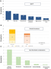
Measurements and main results: A total of 288 patients were implanted with venoarterial ECMO during the inclusion period. One hundred ninety-four patients were successfully weaned, and 109 underwent a CT examination to assess for vascular complications until 4 days after the weaning procedure. The median age of the cohort was 58 years (interquartile range [IQR], 46-64 yr), with a median duration of ECMO support of 7 days (IQR, 5-12 d). Vascular complications were observed in 88 patients (81%). The most frequent complication was thrombosis, either cannula-associated deep vein thrombosis (CaDVT) ( n = 63, 58%) or arterial thrombosis ( n = 36, 33%). Nonthrombotic arterial complications were observed in 48 patients (44%), with 35 (31%) presenting with bleeding. The most common site of CaDVT was the inferior vena cava, occurring in 33 (50%) of cases, with 20% of patients presenting with pulmonary embolism. There was no association between thrombotic complications and ECMO duration, anticoagulation level, or ECMO rotation flow. CT scans influenced management in 83% of patients. In-hospital mortality was 17% regardless of vascular complications.
Conclusions: Vascular complications related to venoarterial ECMO cannula are common after ECMO implantation. CT allows early detection of complications after weaning and impacts patient management. Patients should be routinely screened for vascular complications by CT after decannulation.
Copyright © 2024 The Author(s). Published by Wolters Kluwer Health, Inc. on behalf of the Society of Critical Care Medicine and Wolters Kluwer Health, Inc.
Conflict of interest statement
Dr. Luyt received funding from Advanz Pharma and Merck; he received funding from Merck. The remaining authors have disclosed that they do not have any potential conflicts of interest.
Figures


References
-
- Murphy D, Hockings L, Andrews R, et al. : Extracorporeal membrane oxygenation-hemostatic complications. Transfus Med Rev 2015; 29:90–101 - PubMed
-
- Riccabona M, Kuttnig-Haim M, Dacar D, et al. : Venous thrombosis in and after extracorporeal membrane oxygenation: Detection and follow-up by color Doppler sonography. Eur Radiol 1997; 7:1383–1386 - PubMed
-
- Riccabona M, Zobel G, Kuttnig-Haim M, et al. : The role of sonography in pediatric patients on extracorporeal treatment at the intensive care unit. First Alpe Adia Symp on Intensive Care of Children, April 20–22, 1995, Ljubljana, Slovenia, p 30
-
- Shafii A, Brown C, Murthy S, et al. : High incidence of upper-extremity deep vein thrombosis with dual-lumen venovenous extracorporeal membrane oxygenation. J Thorac Cardiovasc Surg 2012; 144:988–989 - PubMed
-
- Cooper E, Burns J, Retter A, et al. : Prevalence of venous thrombosis following venovenous extracorporeal membrane oxygenation in patients with severe respiratory failure. Crit Care Med 2015; 43:e581–e584 - PubMed

