. 2024 Nov 6:13:RP96755.
doi: 10.7554/eLife.96755.

DNAH3 deficiency causes flagellar inner dynein arm loss and male infertility in humans and mice

Affiliations

DNAH3 deficiency causes flagellar inner dynein arm loss and male infertility in humans and mice

Xiang Wang et al. Elife. .

Abstract

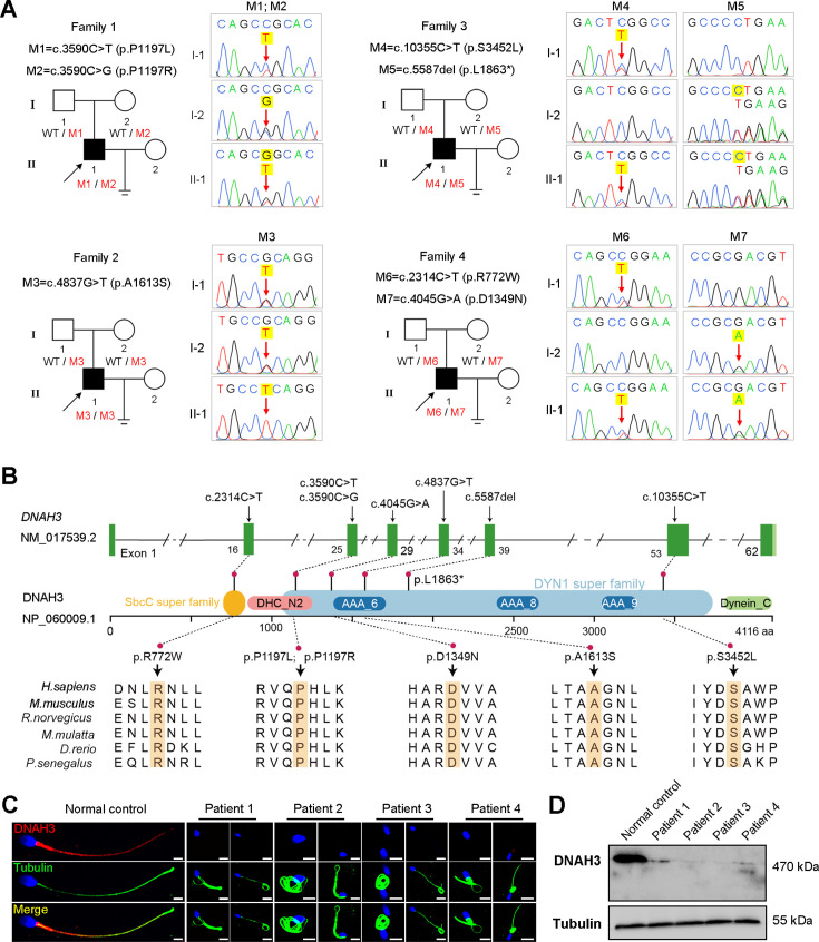
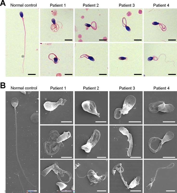
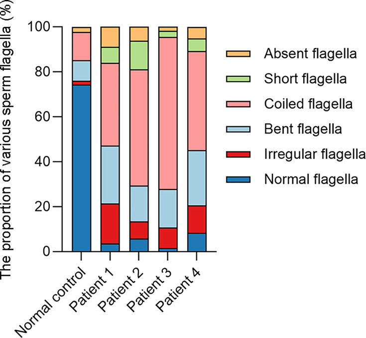
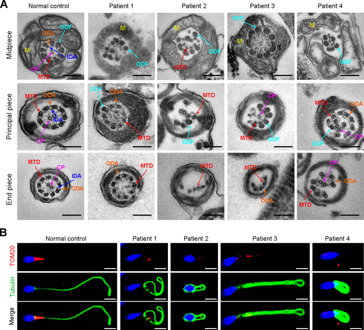
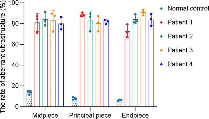
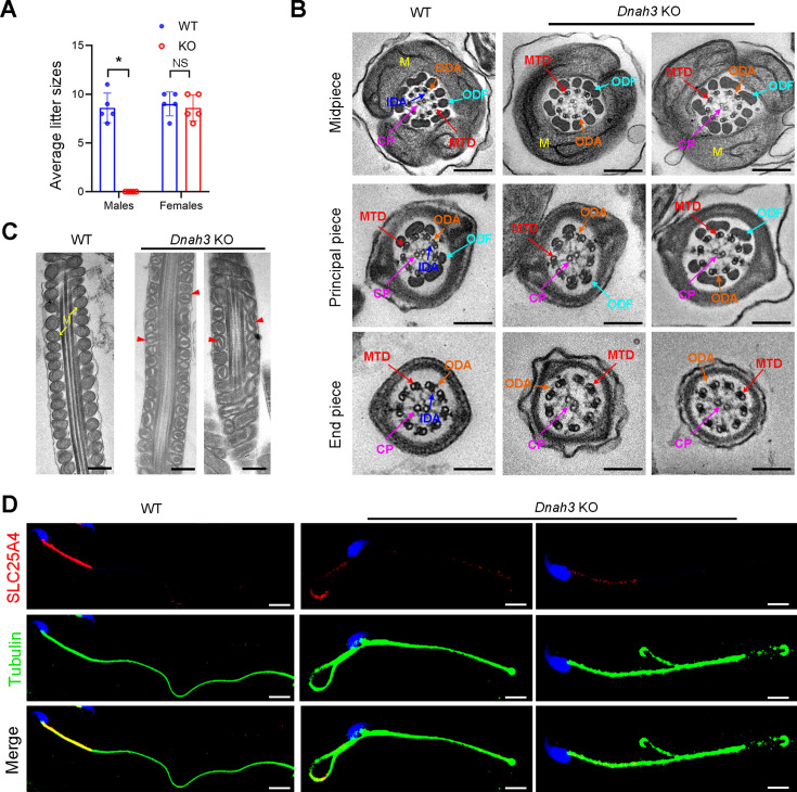
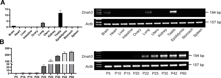
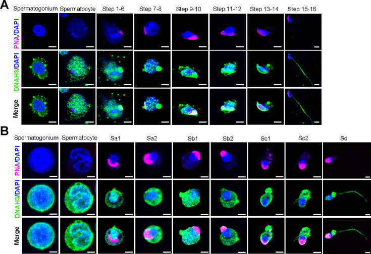
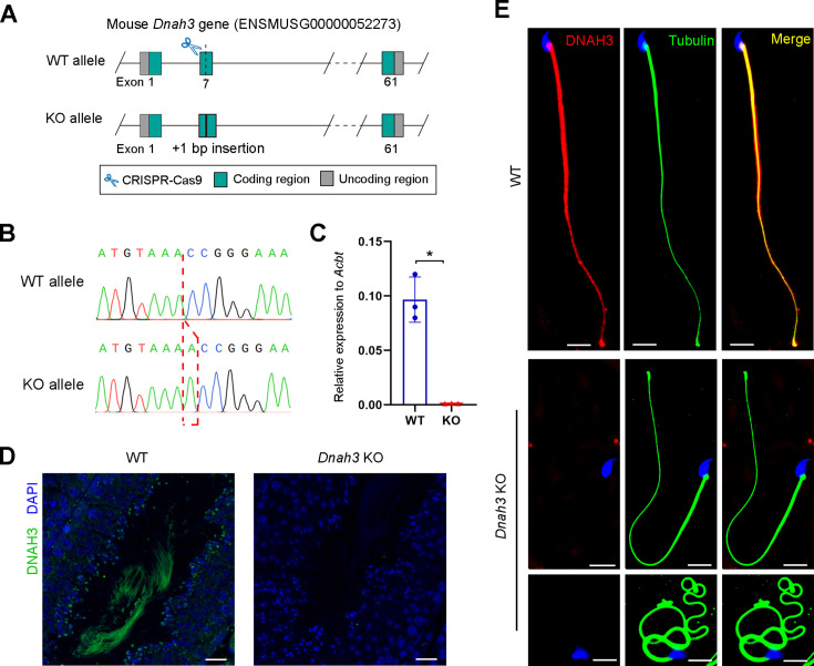
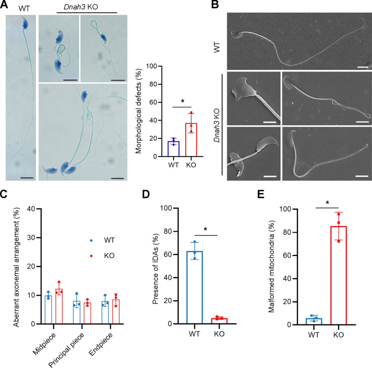
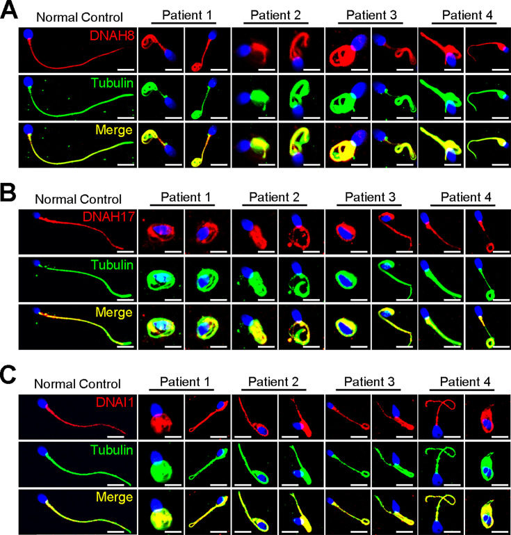
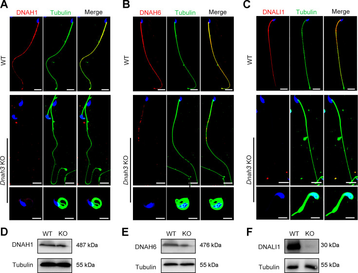
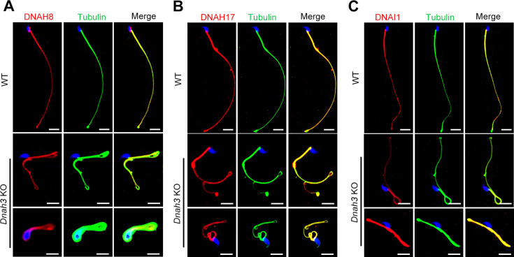
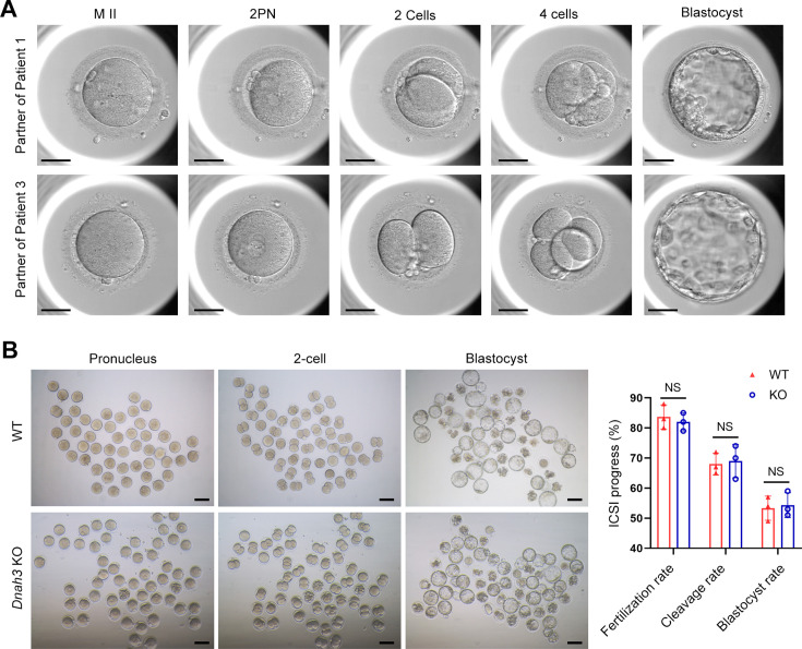
Axonemal protein complexes, including the outer and inner dynein arms (ODA/IDA), are highly ordered structures of the sperm flagella that drive sperm motility. Deficiencies in several axonemal proteins have been associated with male infertility, which is characterized by asthenozoospermia or asthenoteratozoospermia. Dynein axonemal heavy chain 3 (DNAH3) resides in the IDA and is highly expressed in the testis. However, the relationship between DNAH3 and male infertility is still unclear. Herein, we identified biallelic variants of DNAH3 in four unrelated Han Chinese infertile men with asthenoteratozoospermia through whole-exome sequencing (WES). These variants contributed to deficient DNAH3 expression in the patients' sperm flagella. Importantly, the patients represented the anomalous sperm flagellar morphology, and the flagellar ultrastructure was severely disrupted. Intriguingly, Dnah3 knockout (KO) male mice were also infertile, especially showing the severe reduction in sperm movement with the abnormal IDA and mitochondrion structure. Mechanically, nonfunctional DNAH3 expression resulted in decreased expression of IDA-associated proteins in the spermatozoa flagella of patients and KO mice, including DNAH1, DNAH6, and DNALI1, the deletion of which has been involved in disruption of sperm motility. Moreover, the infertility of patients with DNAH3 variants and Dnah3 KO mice could be rescued by intracytoplasmic sperm injection (ICSI) treatment. Our findings indicated that DNAH3 is a novel pathogenic gene for asthenoteratozoospermia and may further contribute to the diagnosis, genetic counseling, and prognosis of male infertility.

Keywords: DNAH3; asthenoteratozoospermia; genetics; genomics; human; inner dynein arm; male infertility; mouse.


Conflict of interest statement

XW, GS, YY, CJ, TR, XY, LZ, YZ, YO, XZ, SL, XT, TL, YS No competing interests declared

Figures

Figure 1.
Figure 1.. Identification of biallelic pathogenic variants in DNAH3 from four unrelated infertile families.
(A) Pedigrees of four families affected by DNAH3 variants (M1–M7). Black arrows indicate the probands in these families. (B) Location of the variants and conservation of affected amino acids in DNAH3. Black arrows indicate the position of the variants. (C) Immunofluorescence staining of DNAH3 in sperm from the patients and normal control. Red, DNAH3; green, α-Tubulin; blue, DAPI; scale bars, 5 μm. (D) Western blotting analysis of DNAH3 expressed in spermatozoa from the patients and normal control.
Figure 2.
Figure 2.. Defects in sperm morphology of the patients harboring DNAH3 variants.
(A, B) Abnormal sperm morphology was observed through Papanicolaou staining (A), and SEM analysis (B) compared to normal control. Scale bars, 5 μm.
Figure 2—figure supplement 1.
Figure 2—figure supplement 1.. The histogram of various flagellar morphology in the normal control and patients.
Figure 3.
Figure 3.. Ultrastructural and mitochondrial defects in sperm from infertile men with DNAH3 variants.
(A) TEM analysis of sperm obtained from a normal control and patients harboring DNAH3 variants. Cross-sections of the midpiece, principal piece and endpiece of sperm from normal control showed the typical ‘‘9+2’’ microtubule structure, and an IDA and an ODA were displayed on the A-tube of each microtubule doublet. Cross-sections of the midpiece, principal piece and endpiece of sperm from the patients displayed absent or disordered CPs, MTDs and ODFs, as well as an evident missing of the IDAs in different pieces of the flagella. M, mitochondria sheath; ODF, outer dense fiber; MTD, microtubule doublets; CP, central pair; IDA, inner dynein arms; ODA, outer dynein arms. Scale bars, 200 nm. (B) Immunofluorescence staining of TOM20 in sperm from the patients and normal control. Red, TOM20; green, α-Tubulin; blue, DAPI; scale bars, 5 μm.
Figure 3—figure supplement 1.
Figure 3—figure supplement 1.. The percentage of aberrant ultrastructure in different cross-sections of sperm from the normal control and patients.
Figure 4.
Figure 4.. Dnah3 KO male mice are infertile.
(A) Fertility of Dnah3 KO mice. The KO male mice were infertile (n=five biologically independent WT mice or KO mice; Student’s t test; *, p<0.05; NS, no significance; error bars, s.e.m.). (B) TEM analysis of the cross-sections of spermatozoa from Dnah3 KO mice revealed an obvious absence of IDAs in different pieces of the flagella compared to WT mice. M, mitochondrion sheath; ODF, outer dense fiber; MTD, microtubule doublet; CP, central pair; IDA, inner dynein arm; ODA, outer dynein arm. Scale bars, 200 nm. (C) Disrupted mitochondria were observed in spermatozoa tail from Dnah3 KO mice by TEM analysis. The yellow arrows indicate the normal mitochondria. The red arrowheads indicate the dilated intermembrane spaces and dissolved mitochondrial material. M, mitochondrion sheath. Scale bars, 200 nm. (D) Immunofluorescence staining of SLC25A4 indicated impaired mitochondrial formation in Dnah3 KO mice compared to WT mice. Red, SLC25A4; green, α-Tubulin; blue, DAPI; scale bars, 5 µm.
Figure 4—figure supplement 1.
Figure 4—figure supplement 1.. The expression of DNAH3 in mouse testis.
(A) qPCR analysis revealed that Dnah3 was highly expressed in the mouse testis. (B) qPCR analysis showed that Dnah3 expression was significantly elevated beginning on postnatal Day 12, peaked at postnatal Day 30, and maintained a stable expression level thereafter.
Figure 4—figure supplement 2.
Figure 4—figure supplement 2.. DNAH3 is expressed during spermatogenesis in mice and humans.
(A) Immunofluorescence staining of DNAH3 in isolated mouse germ cells. Pink, PNA; green, DNAH3; blue, DAPI; scale bars, 5 μm. (B) Immunofluorescence staining of DNAH3 in isolated human germ cells. Pink, PNA; green, DNAH3; blue, DAPI; scale bars, 5 μm.
Figure 4—figure supplement 3.
Figure 4—figure supplement 3.. Generation of Dnah3 KO mice.
(A) Schematic illustration of the strategy for the generation of Dnah3 KO mice. (B, C) PCR sequencing (B) and qPCR (C) were used to confirm the genotype and KO efficiency (n=three biologically independent WT mice or KO mice; Student’s t test; *, p<0.05; error bars, s.e.m.). (D) Immunofluorescence staining of DNAH3 in testis of Dnah3 KO mice and WT mice. Green, DNAH3; blue, DAPI; scale bars, 75 μm. (E) Immunofluorescence staining of DNAH3 in spermatozoa isolated from the cauda epididymis of Dnah3 KO mice and WT mice. Red, DNAH3; green, α-Tubulin; blue, DAPI; scale bars, 5 μm.
Figure 4—figure supplement 4.
Figure 4—figure supplement 4.. Ciliary development of Dnah3 KO mice.
(A) H&E staining of lung, brain, eye, and oviduct from Dnah3 KO mice and WT mice. Scale bars, 100 μm. (B) Analysis of ciliary development in the lung, brain, eye, and oviduct from Dnah3 KO mice and WT mice by using immunofluorescence staining. Green, Ac-Tubulin; blue, DAPI; scale bars, 20 μm.
Figure 4—figure supplement 5.
Figure 4—figure supplement 5.. Fertility of Dnah3 KO mice.
(A) H&E staining of ovary tissue sections from 8-week-old Dnah3 KO female mice and WT female mice. Scale bars, 75 μm (n=three biologically independent WT mice or KO mice). (B) Sizes of the testis and epididymis of the 8-week-old Dnah3 KO and WT mice (n=three biologically independent WT mice or KO mice; Student’s t test; NS, no significance; error bars, s.e.m.).
Figure 4—figure supplement 6.
Figure 4—figure supplement 6.. Morphology and ultrastructure of sperm isolated from Dnah3 KO mice.
(A, B) Papanicolaou staining (A), and SEM analysis (B) revealed morphological defects in partial spermatozoa from Dnah3 KO mice compared to WT mice. Scale bars in (A), 5 μm; scale bars in (B), 2.5 μm. (n=three biologically independent WT mice or KO mice; Student’s t test; error bars, s.e.m.). (C) The percentage of aberrant axonemal arrangement in different cross-sections of sperm from WT mice and Dnah3 KO mice. (n=three biologically independent WT mice or KO mice; error bars, s.e.m.). (D) The percentage of microtubule doublets that presented IDAs in WT mice and Dnah3 KO mice. (n=three biologically independent WT mice or KO mice; Student’s t test; error bars, s.e.m.). (E) Statistics of malformed mitochondria in the midpiece of sperm from WT mice and Dnah3 KO mice. (n=three biologically independent WT mice or KO mice; Student’s t test; error bars, s.e.m.).
Figure 5.
Figure 5.. Immunofluorescence staining and western blotting analysis of IDA-associated proteins in spermatozoa obtained from normal control and patients with DNAH3 variants.
(A – C) Immunofluorescence staining of DNAH1 (A), DNAH6 (B) and DNALI1 (C) in spermatozoa from patients and normal controls. Red, DNAH1 in (A), DNAH6 in (B), DNALI1 in (C); green, α-Tubulin; blue, DAPI; scale bars, 5 μm. (D – F) Western blotting analysis of DNAH1(D), DNAH6 (E), DNALI1 (F) in sperm lysates from the patients and normal control.
Figure 5—figure supplement 1.
Figure 5—figure supplement 1.. Immunofluorescence staining of ODA-associated proteins in spermatozoa obtained from variants within DNAH3 patients.
(A – C) The expression of DNAH8 (A), DNAH17 (B) and DNAI1 (C) in spermatozoa of the patients was comparable to that in normal controls. Red, DNAH8 in (A), DNAH17 in (B), DNAI1 in (C); green, α-Tubulin; blue, DAPI; scale bars, 5 μm.
Figure 6.
Figure 6.. Immunofluorescence staining and western blotting analysis of IDA-associated proteins in spermatozoa from WT and Dnah3 KO mice.
(A – C) Immunofluorescence staining of DNAH1 (A), DNAH6 (B) and DNALI1 (C) in spermatozoa from Dnah3 KO and WT mice. Red, DNAH1 in (A), DNAH6 in (B), DNALI1 in (C); green, α-Tubulin; blue, DAPI; scale bars, 5 μm. (D – F) Western blotting analysis of DNAH1(D), DNAH6 (E) and DNALI1 (F) in spermatozoa lysates from Dnah3 KO and WT mice.
Figure 6—figure supplement 1.
Figure 6—figure supplement 1.. Immunofluorescence staining of ODA-associated proteins in spermatozoa of Dnah3 KO and WT mice.
(A – C) The expression of DNAH8 (A), DNAH17 (B) and DNAI1 (C) in spermatozoa from Dnah3 KO mice was comparable to that in spermatozoa from WT mice. Red, DNAH8 in (A), DNAH17 in (B), DNAI1 in (C); green, α-Tubulin; blue, DAPI; scale bars, 5 μm.
Figure 7.
Figure 7.. ICSI outcomes of DNAH3-deficient patients and Dnah3 KO mice.
(A) The embryonic development of Patient 1 and Patient 3 after ICSI treatment. MII, metaphase II; PN, pronucleus; scale bars, 40 μm. (B) There was no difference in the fertilization rate or 2 cell and blastocyst embryo formation rates between the Dnah3 KO and WT groups (n=three biologically independent WT mice or KO mice; Student’s t test; NS, no significance; error bars, s.e.m.).

Update of

  • doi: 10.1101/2024.02.19.580977
  • doi: 10.7554/eLife.96755.1
  • doi: 10.7554/eLife.96755.2
  • doi: 10.7554/eLife.96755.3

Similar articles

Cited by

References

    1. Adzhubei IA, Schmidt S, Peshkin L, Ramensky VE, Gerasimova A, Bork P, Kondrashov AS, Sunyaev SR. A method and server for predicting damaging missense mutations. Nature Methods. 2010;7:248–249. doi: 10.1038/nmeth0410-248. - DOI - PMC - PubMed
    1. Agarwal A, Mulgund A, Hamada A, Chyatte MR. A unique view on male infertility around the globe. Reproductive Biology and Endocrinology. 2015;13:37. doi: 10.1186/s12958-015-0032-1. - DOI - PMC - PubMed
    1. Aprea I, Raidt J, Höben IM, Loges NT, Nöthe-Menchen T, Pennekamp P, Olbrich H, Kaiser T, Biebach L, Tüttelmann F, Horvath J, Schubert M, Krallmann C, Kliesch S, Omran H. Defects in the cytoplasmic assembly of axonemal dynein arms cause morphological abnormalities and dysmotility in sperm cells leading to male infertility. PLOS Genetics. 2021;17:e1009306. doi: 10.1371/journal.pgen.1009306. - DOI - PMC - PubMed
    1. Ben Khelifa M, Coutton C, Zouari R, Karaouzène T, Rendu J, Bidart M, Yassine S, Pierre V, Delaroche J, Hennebicq S, Grunwald D, Escalier D, Pernet-Gallay K, Jouk P-S, Thierry-Mieg N, Touré A, Arnoult C, Ray PF. Mutations in DNAH1, which encodes an inner arm heavy chain dynein, lead to male infertility from multiple morphological abnormalities of the sperm flagella. American Journal of Human Genetics. 2014;94:95–104. doi: 10.1016/j.ajhg.2013.11.017. - DOI - PMC - PubMed
    1. Burgess SA, Walker ML, Sakakibara H, Knight PJ, Oiwa K. Dynein structure and power stroke. Nature. 2003;421:715–718. doi: 10.1038/nature01377. - DOI - PubMed

MeSH terms