. 2024 Nov 6;148(1):60.
doi: 10.1007/s00401-024-02807-w.

TMEM106B amyloid filaments in the Biondi bodies of ependymal cells

Affiliations

TMEM106B amyloid filaments in the Biondi bodies of ependymal cells

Bernardino Ghetti et al. Acta Neuropathol. .

Abstract

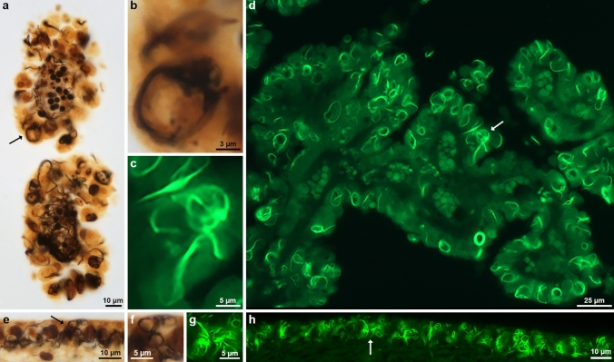
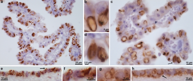
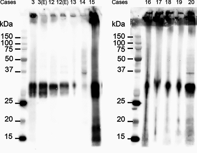
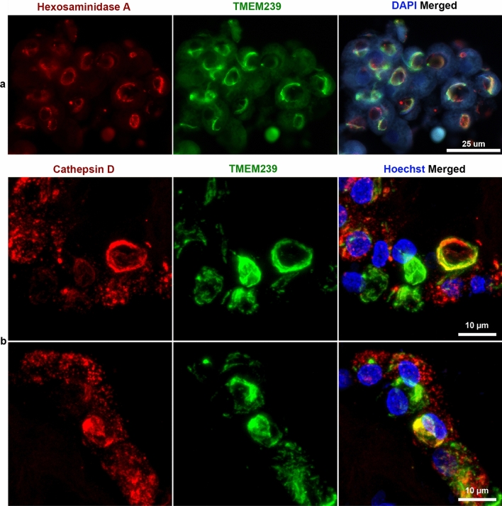
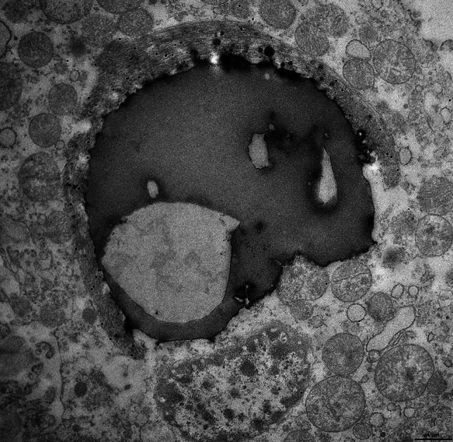
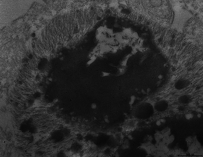
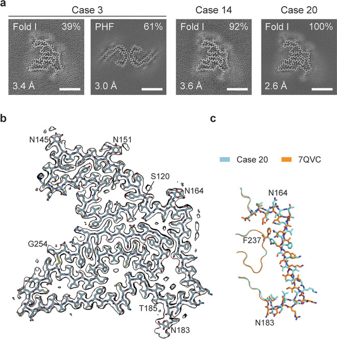
Biondi bodies are filamentous amyloid inclusions of unknown composition in ependymal cells of the choroid plexuses, ependymal cells lining cerebral ventricles and ependymal cells of the central canal of the spinal cord. Their formation is age-dependent and they are commonly associated with a variety of neurodegenerative conditions, including Alzheimer's disease and Lewy body disorders. Here, we show that Biondi bodies are strongly immunoreactive with TMEM239, an antibody specific for inclusions of transmembrane protein 106B (TMEM106B). Biondi bodies were labelled by both this antibody and the amyloid dye pFTAA. Many Biondi bodies were also labelled for TMEM106B and the lysosomal markers Hexosaminidase A and Cathepsin D. By transmission immuno-electron microscopy, Biondi bodies of choroid plexuses were decorated by TMEM239 and were associated with structures that resembled residual bodies or secondary lysosomes. By electron cryo-microscopy, TMEM106B filaments from Biondi bodies of choroid plexuses were similar (Biondi variant), but not identical, to the fold I that was previously identified in filaments from brain parenchyma.

Keywords: Amyloid; Biondi bodies; Electron cryo-microscopy; Ependymal cells; Lysosomes; Transmembrane protein 106B.


Figures

Fig. 1
Fig. 1
Histopathology of Biondi bodies using Bielschowsky silver and Thioflavin S. Choroid plexuses (a,d) and ependymal linings of the lateral ventricle (e,h) from cases 1 and 3 were stained with modified Bielschowsky silver (a,b,e,f) and Thioflavin S (c,d,g,h). Biondi bodies are seen in the cytoplasm of ependymal cells as argyrophilic (a,b,e,f) or fluorescent (c,d,g,h) inclusions. Arrows (a,d,e,h) point to Biondi bodies seen at high power in b, c, f and g. Scale bars: 10 µm (a,e,h), 3 µm (b), 5 µm (c,f,g), and 25 µm (d)
Fig. 2
Fig. 2
Immunohistochemistry of Biondi bodies using antibody TMEM239. Choroid plexuses (a,c) and ependymal linings of the lateral ventricle (e,h) from cases 3 and 5 were stained using antibody TMEM239, which labels TMEM106B inclusions specifically. Immunoreactive Biondi bodies are seen in (a-h). Biondi bodies are seen at high power (b,d,f,g) and arrows in c, e, and h point to Biondi bodies seen in d, f and g. Scale bars: 25 µm (a), 5 µm (b,f,g), 10 µm (c,e,h), and 2.5 µm (d)
Fig. 3
Fig. 3
Immunoblotting of Biondi bodies using antibody TMEM239 and genotyping. Sarkosyl-insoluble extracts from the choroid plexuses of cases 3 and 12–20 and extracts from the ependymal linings of the lateral ventricles from cases 3 and 12 were immunoblotted using antibody TMEM239, which labels TMEM106B inclusions specifically. All extracts showed an immunoreactive band of 29 kDa, diagnostic of the presence of TMEM106B filaments
Fig. 4
Fig. 4
Double-labelling of choroid plexuses with Biondi bodies using antibody TMEM239 and amyloid dye pFTAA. Choroid plexuses from cases 6–10 and 22. a-f, The panels on the left show single-labelling immunofluorescence by antibody TMEM239 (red); the middle panels show single-labelling fluorescence by the amyloid dye pFTAA (green); the panels on the right show double-labelling by TMEM239 and pFTAA, with the yellow colour appearing when TMEM239 fluorescence and pFTAA fluorescence merge. Nuclei were labelled using Hoechst dye (blue). Scale bar, 20 µm
Fig. 5
Fig. 5
Immunohistochemistry of Biondi bodies in choroid plexuses using double labelling with an anti-Hexosaminidase A antibody and antibody TMEM239, as well as with an anti-Cathepsin D antibody and antibody TMEM239. Choroid plexuses from cases 3 and 8. a, The panel on the left shows single-labelling immunofluorescence by anti-Hexosaminidase A antibody (red); the middle panel shows single-labelling immunofluorescence by antibody TMEM239 (green); the panel on the right shows double-labelling by anti-Hexosaminidase A antibody and antibody TMEM239, with the yellow colour indicating colocalisation. Nuclei were labelled using DAPI (blue). Scale bar, 25 µm. b, The panels on the left show single-labelling immunofluorescence by anti-Cathepsin D antibody (red); the middle panels show single-labelling immunofluorescence by antibody TMEM239 (green); the panels on the right show double-labelling, with the yellow colour indicating colocalisation. Nuclei were labelled using Hoechst dye (blue). Scale bar, 10 µm
Fig. 6
Fig. 6
Immunoelectron microscopy of a Biondi body. Choroid plexuses from case 8 and anti-TMEM106B antibody TMEM239. The cytoplasm of an ependymal cell contains a round body made of osmiophilic and electron-lucent areas that are surrounded by a crescent-shaped bundle of filaments (interpreted as a Biondi body) decorated by gold particles. The round body has the appearance of a secondary lysosome or a residual body. The nucleus of the cell and numerous mitochondria are seen around the inclusion. Scale bar, 1 µm
Fig. 7
Fig. 7
Immunoelectron microscopy of a Biondi body. Choroid plexuses from case 8 and anti-TMEM106B antibody TMEM239. A Biondi body is shown that consists of a ring of filaments decorated by gold particles surrounding a large osmiophilic inclusion. Note that the large osmiophilic inclusion, also decorated by gold particles, contains what we interpret to be filaments seen in cross-section. Smaller osmiophilic inclusions are intermixed with the filaments. Scale bar, 500 nm
Fig. 8
Fig. 8
Immunoelectron microscopy of Biondi bodies. Choroid plexuses from cases 8 and 23 and anti-TMEM106B antibody TMEM239. a, A membrane-bound, oval-shaped inclusion is decorated by gold particles and contains round osmiophilic bodies, an electron-lucent area and wisps of filaments. Two bundles of filaments decorated by gold particles are seen outside the membrane-bound inclusion. Scale bar, 1 µm b, Multiple osmiophilic bodies of different sizes are intermixed with a large bundle of filaments decorated by gold particles. Scale bar, 1 µm
Fig. 9
Fig. 9
Cryo-EM structure of TMEM106B filaments from Biondi bodies. Choroid plexuses from cases 3, 14 and 20. a, Cross-section through the cryo-EM reconstructions, perpendicular to the helical axis and with a projected thickness of approximately one rung, is shown for the filaments from the sarkosyl-insoluble fractions. The fractions of each filament type and the resolutions are indicated at the top right and bottom left corners, respectively. Scale bars, 5 nm. b, Cryo-EM density map (in transparent grey) and atomic model of the TMEM106B filament fold (cyan) from case 20. The amino- and carboxy-terminal residues S120 and G254, glycosylated residues N145, N151, N164 and N183, as well as residue T185 are labelled. c, Comparison of the TMEM106B fold from choroid plexuses of case 20 (cyan) with fold I of TMEM106B filaments from brain parenchyma (orange, PDB:7QVC). The divergent parts of the two folds are shown in sticks. In the rest, both folds are identical. The structures and orientations of their cropped-out parts are as in (b)

References

    1. Adams RD, Oksche A, and Haymaker W "Meninges, choroid plexuses, ependyma and their reactions. (1982) Part 2: Age-related changes and pathology." Histology and Histopathology of the Nervous System. Springfield, IL: Charles C. Thomas: 641–713.
    1. Åslund A, Sigurdson CJ, Klingstedt T, Gratwohl S, Bolmont T, Dickstein DL et al (2009) Novel pentameric thiophene derivatives for in vitro and in vivo imaging of a plethora of protein aggregates in cerebral amyloidoses. ACS Chem Biol 4:673–683 - PMC - PubMed
    1. Bacioglu M, Schweighauser M, Gray D, Lövestam S, Katsinelos A, Quaegebeur A et al (2024) Cleaved TMEM106B forms amyloid aggregates in central and peripheral nervous systems. Acta Neuropathol Commun 12:99 - PMC - PubMed
    1. Bargmann W, Katritsis E (1966) Über die sogenannten Filamente und das Pigment im Plexus chorioideus des Menschen. Z Zellforsch 75:366–370 - PubMed
    1. Bielschowsky M (1909) Eine Modifikation meines Silberimprägnationsverfahrens zur Darstellung der Neurofibrillen. J Psychol Neurol 12:135

Publication types

LinkOut - more resources