

A decision tree for predicting the causative pathogens of community-acquired pneumonia from thin-section computed tomography
- PMID: 39503821
- PMCID: PMC11868163
- DOI: 10.1007/s11604-024-01691-4
A decision tree for predicting the causative pathogens of community-acquired pneumonia from thin-section computed tomography
Abstract
Purpose: To determine whether decision trees are useful for predicting organisms that cause community-acquired pneumonia (CAP).
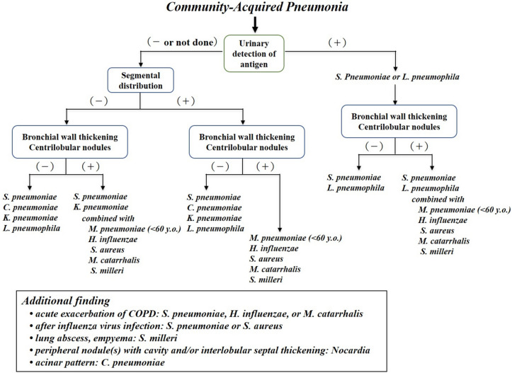
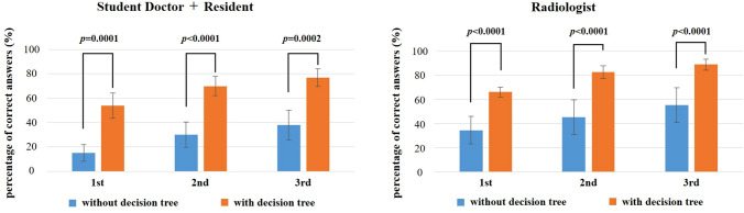
Materials and methods: We developed a decision tree for predicting the organisms that cause CAP based on previously reported characteristic computed tomography findings. Sixteen readers (two student doctors, six residents, and eight radiologists) separately diagnosed 68 randomly selected cases of CAP using chest computed tomography. The first, second, and third most likely causative organisms were estimated for each case, and the percentages of correct answers were evaluated for consistency with the isolated organisms. The same 68 cases were then read again using the decision tree, with the first three most likely organisms again being estimated, and the percentage of agreement was evaluated as the percentage of correct responses after using the decision tree.
Results: For student doctors, residents, and radiologists, the percentage of correct responses increased significantly (p < 0.0001) when the decision tree was used to predict the first, second, and third most probable causative organism. The radiologists all obtained an accuracy rate of 80% or higher when estimating up to three candidate organisms using the decision tree. The organism for which the decision tree was most useful was Mycoplasma pneumoniae, followed by Haemophilus influenzae and Chlamydophila pneumoniae (p < 0.001).
Conclusion: Use of the decision tree made it possible to estimate the organisms responsible for CAP with a high correct response rate.
Keywords: Chest; Community-acquired pneumonia; Computed tomography; Decision tree.
© 2024. The Author(s).
Conflict of interest statement
Declarations. Conflict of interest: None of the authors has a direct or indirect financial interest in the products under investigation or in the subject matter discussed in this article. Ethical approval: Our institutional review board approved this retrospective study (5–226). Informed consent: Our institutional review board waived the requirement for informed consent because of the study’s retrospective nature.
Figures


Similar articles
-
Predicting Mycoplasma pneumoniae and Chlamydophila pneumoniae in community-acquired pneumonia (CAP) pneumonia: epidemiological study of respiratory tract infection using multiplex PCR assays.Intern Emerg Med. 2021 Nov;16(8):2129-2137. doi: 10.1007/s11739-021-02744-6. Epub 2021 May 13. Intern Emerg Med. 2021. PMID: 33983474 Free PMC article.
-
Re-evaluation of the etiology and clinical and radiological features of community-acquired lobar pneumonia in adults.J Infect Chemother. 2018 Jun;24(6):463-469. doi: 10.1016/j.jiac.2018.02.001. Epub 2018 Mar 28. J Infect Chemother. 2018. PMID: 29605556
-
Efficacy and safety of ten day moxifloxacin 400 mg once daily in the treatment of patients with community-acquired pneumonia. Community Acquired Pneumonia Study Group.Respir Med. 2000 Feb;94(2):97-105. doi: 10.1053/rmed.1999.0710. Respir Med. 2000. PMID: 10714413 Clinical Trial.
-
Community-acquired and nosocomial pneumonia.Eur Radiol. 2004 Mar;14 Suppl 3(3):E2-20. doi: 10.1007/s00330-003-2162-7. Eur Radiol. 2004. PMID: 14749949 Free PMC article. Review.
-
[Community-acquired pneumonia].Radiologe. 2017 Jan;57(1):6-12. doi: 10.1007/s00117-016-0199-2. Radiologe. 2017. PMID: 28054135 Review. German.
References
-
- Beigelman-Aubry C, Godet C, Caumes E. Lung infections: the radiologist’s perspective. Diagn Interv Imaging. 2012;93:431–40. - PubMed
-
- Heitzman ER. Pneumonia and lung abscess. In: Heitzman ER, editor. The lung: radiologic–pathologic correlations. 2nd ed. St Louis, USA: Mosby; 1984. p. 194–234.
-
- Spencer H. The bacterial pneumonias. In: Spencer H, editor. Pathology of the lung. 4th ed. Oxford, UK: Pergamon Press; 1985. p. 167–212.
MeSH terms
LinkOut - more resources
Full Text Sources
Medical
Miscellaneous
