Review
. 2024 Nov 7;16(11):mfae049.
doi: 10.1093/mtomcs/mfae049.

Iron-sulfur cluster-dependent enzymes and molybdenum-dependent reductases in the anaerobic metabolism of human gut microbes

Affiliations
Review

Iron-sulfur cluster-dependent enzymes and molybdenum-dependent reductases in the anaerobic metabolism of human gut microbes

Leah E Zahn et al. Metallomics. .

Abstract

Metalloenzymes play central roles in the anaerobic metabolism of human gut microbes. They facilitate redox and radical-based chemistry that enables microbial degradation and modification of various endogenous, dietary, and xenobiotic nutrients in the anoxic gut environment. In this review, we highlight major families of iron-sulfur (Fe-S) cluster-dependent enzymes and molybdenum cofactor-containing enzymes used by human gut microbes. We describe the metabolic functions of 2-hydroxyacyl-CoA dehydratases, glycyl radical enzyme activating enzymes, Fe-S cluster-dependent flavoenzymes, U32 oxidases, and molybdenum-dependent reductases and catechol dehydroxylases in the human gut microbiota. We demonstrate the widespread distribution and prevalence of these metalloenzyme families across 5000 human gut microbial genomes. Lastly, we discuss opportunities for metalloenzyme discovery in the human gut microbiota to reveal new chemistry and biology in this important community.

Keywords: anaerobic metabolism; human gut microbiota; iron-sulfur cluster; metalloenzyme; molybdenum cofactor; redox chemistry.


Figures

Graphical Abstract
Graphical Abstract
Figure 1.
Figure 1.
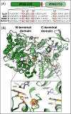
Fe–S cluster-dependent 2-hydroxyacyl-CoA dehydratases in reductive branch of amino acid Stickland fermentation. (A) Oxidative and reductive branches of Stickland fermentation of branched chain and aromatic amino acids (B) X-ray crystal structures of the (R)-2-hydroxyisocaproyl-CoA dehydratase component D (PDB 3O3N) and activase component A (PDB 4EHT). Fe–S clusters are shown as spheres, the 2-hydroxyisocaproyl-CoA substrate is shown as green sticks, and two ADP molecules are shown as yellow sticks. (C) Proposed mechanism for Fe–S cluster-mediated dehydration of 2-hydroxyacyl-CoA substrates, occurring in the active site of the component D α-subunit.
Figure 2.
Figure 2.
Radical SAM GRE activating enzymes involved in disease-associated chemistry. (A) Active site of pyruvate formate-lyase activating enzyme (PDB 3CB8) with SAM (pink sticks) bound to the Fe–S cluster and a peptide substrate mimic (grey sticks) bound in close proximity to the 5ʹ-carbon of SAM. (B) Current proposed mechanism of radical SAM enzymes, involving reductive cleavage of SAM to form the organometallic omega intermediate, followed by 5ʹ-deoxyadenosyl radical formation. GRE activation involves abstraction of the pro-S hydrogen atom from a glycine residue by the 5ʹ-deoxyadenosyl radical. (C) Key reactions in the human gut microbiota catalyzed by GREs, which require activation by radical SAM enzymes.
Figure 3.
Figure 3.
Two-domain architecture of Fe–S cluster-dependent flavoenzymes. (A) Cartoon depiction of the Fe–S flavoenzyme protein sequence, highlighting regions of domain homology and the location of the conserved cysteine motif. A segment of a multiple sequence alignment of gut microbial Fe–S flavoenzymes (UniProt IDs: P32370, A0A829NF98, M9NZ71, P19410) is shown with residue numbering corresponding to the BaiH protein sequence. (B) X-ray crystal structure of archetypal family member 2,4-dienoyl-CoA reductase from E. coli (PDB 1PS9). Organic cofactors and substrate analog are shown as sticks and the [Fe4S4] cluster is shown as spheres.
Figure 4.
Figure 4.
Reduction reactions catalyzed by Fe–S cluster-dependent flavoenzymes in the gut microbiota. (A) Biochemical pathway for 7-α-dehydroxylation of primary bile acids. (B) Reduction of heme-derived bilirubin to urobilinogen. (C) Metabolism of soy isoflavone daidzein to (S)-equol.
Figure 5.
Figure 5.
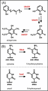
O2-independent hydroxylation reactions catalyzed by Fe–S cluster-dependent U32 oxidases. (A) Proposed hydroxylation reactions performed by the pair of U32 oxidases UbiU and UbiV in anaerobic ubiquinone biosynthesis. (B) rRNA and tRNA nucleotide modifications installed by U32 oxidases.
Figure 6.
Figure 6.
Molybdenum-dependent enzymes of the DMSO reductase family catalyzing oxygen atom transfer reactions using inorganic and organic substrates. (A) Structure of the bis-MGD molybdenum cofactor common to the DMSO reductase family. (B) Active site depiction of DMSO reductase (PDB 4DMR) showing the molybdenum bis-MGD cofactor with a terminal oxo ligand (red sphere), coordinating serine residue, and the DMSO substrate bound to the molybdenum ion (teal sphere). (C) Proposed reaction mechanism for DMSO reductase. (D) Known substrates of molybdenum-dependent catechol dehydroxylase (Cdh) enzymes. (E) Corticoid dehydroxylation catalyzed by a molybdenum-dependent enzyme from E. lenta.
Figure 7.
Figure 7.
Bioinformatic analysis of metalloenzyme family representation in human gut microbes. (A) Fraction of all genera or strains encoding at least one member of the metalloenzyme family. (B) Histogram plots for each metalloenzyme family of gene counts per genome. (C) Fraction of each genus encoding at least one member from the metalloenzyme family. Phyla from left to right: Actinomycetota (blue), Bacillota (orange), Bacteroidota (lime green), Campylobacterota (yellow), Euryarchaeota (cyan), Fusobacteriota (purple), Lentisphaerota (dark blue), Pseudomonadota (pink), Spirochaetota (light orange), Synergistota (magenta), Thermodesulfobacteriota (green), Verrucomicrobiota (brown), unclassified (grey).

Similar articles

Cited by

References

    1. Qin J, Li Y, Cai Z et al. A metagenome-wide association study of gut microbiota in type 2 diabetes. Nature 2012;490:55–60. 10.1038/nature11450 - DOI - PubMed
    1. Nicholson JK, Holmes E, Kinross J et al. Host-gut microbiota metabolic interactions. Science 2012;336:1262–7. 10.1126/science.1223813 - DOI - PubMed
    1. David LA, Maurice CF, Carmody RN et al. Diet rapidly and reproducibly alters the human gut microbiome. Nature 2014;505:559–63. 10.1038/nature12820 - DOI - PMC - PubMed
    1. Koppel N, Balskus EP. Exploring and understanding the biochemical diversity of the human microbiota. Cell Chem Biol 2016;23:18–30. 10.1016/j.chembiol.2015.12.008 - DOI - PubMed
    1. Chittim CL, Irwin SM, Balskus EP. Deciphering human gut microbiota—nutrient interactions: a role for biochemistry. Biochemistry 2018;57:2567–77. 10.1021/acs.biochem.7b01277 - DOI - PubMed

Publication types

LinkOut - more resources