. 2025 May;35(5):2378-2386.
doi: 10.1007/s00330-024-11176-7. Epub 2024 Nov 6.

Inter-reader agreement of the BI-RADS CEM lexicon

Affiliations

Inter-reader agreement of the BI-RADS CEM lexicon

Calogero Zarcaro et al. Eur Radiol. 2025 May.

Abstract

Purpose: The purpose of this study was to assess the inter-reader agreement of the breast imaging reporting and data system (BI-RADS) contrast-enhanced mammography (CEM) lexicon.

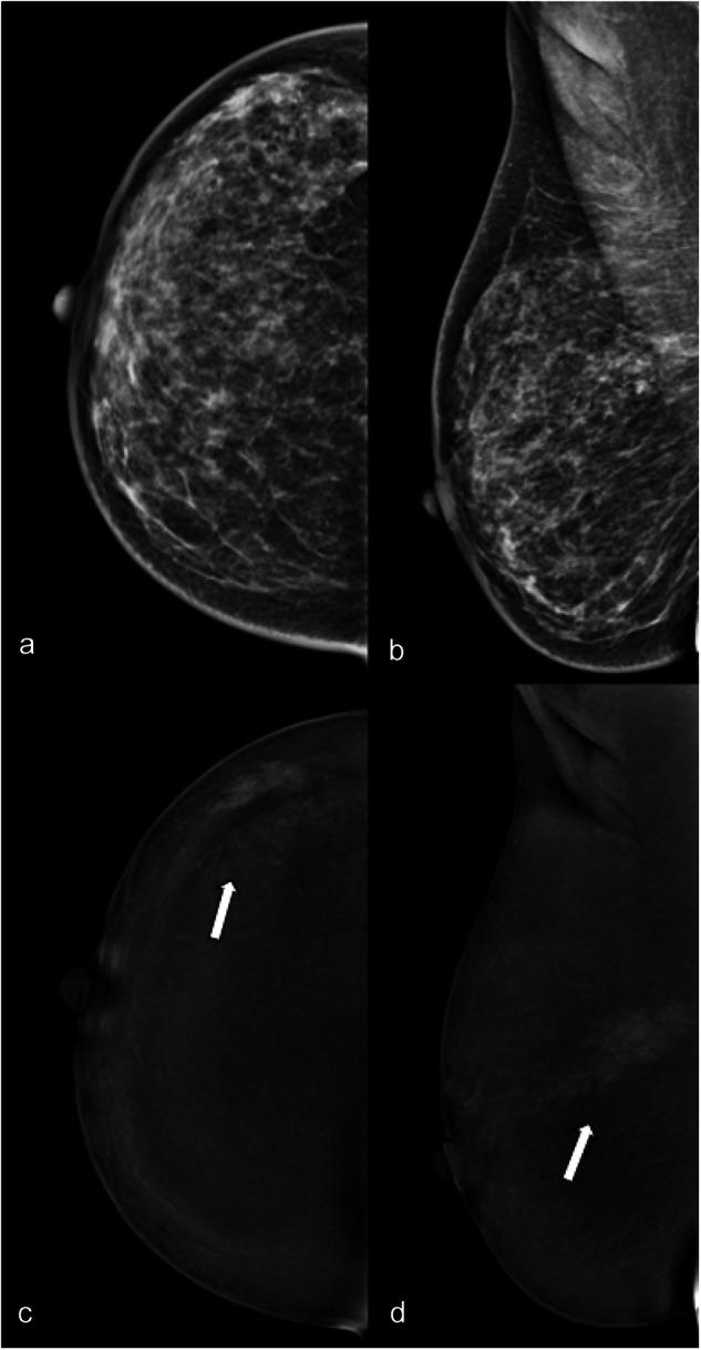
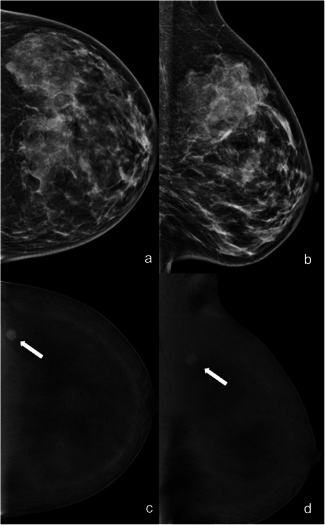
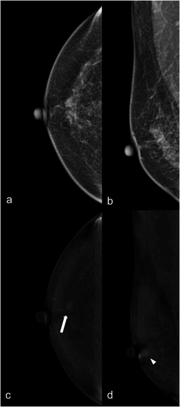
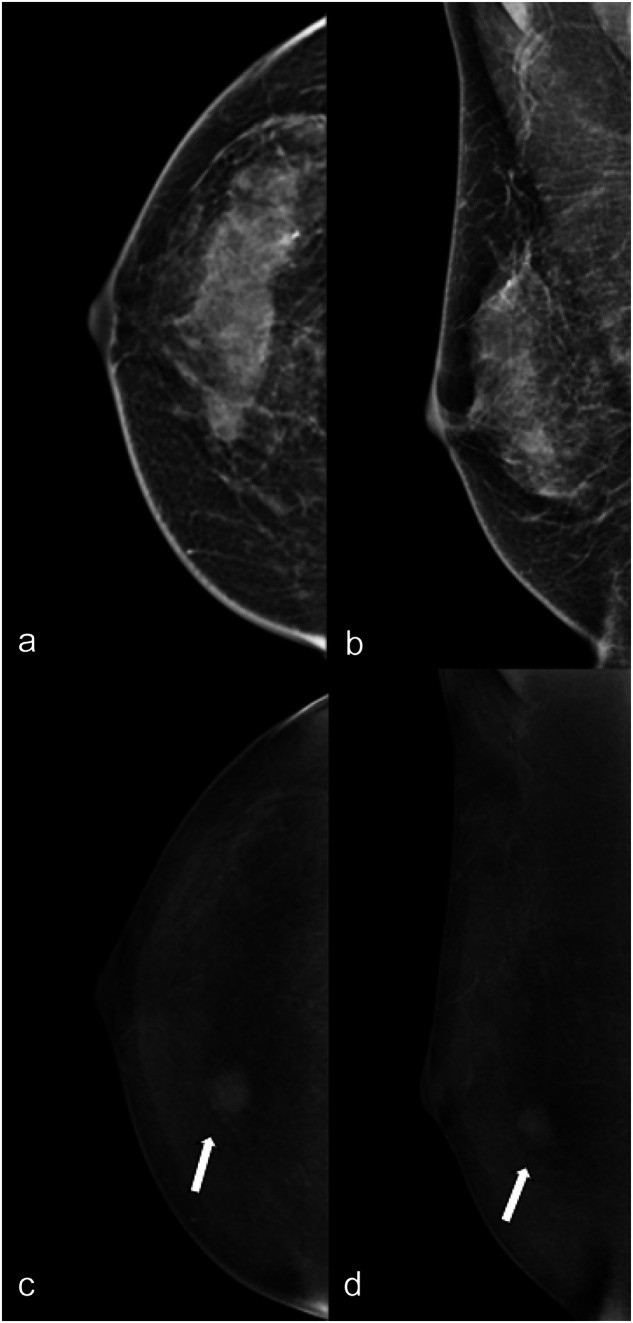
Materials and methods: In this IRB-approved, single-center, retrospective study, three breast radiologists, each with different levels of experience, reviewed 462 lesions in 421 routine clinical CEM according to the fifth edition of the BI-RADS lexicon for mammography and to the first version of the BI-RADS lexicon for CEM. Readers were blinded to patient outcomes and evaluated breast and lesion features on low-energy (LE) images (breast density, type of lesion, associated architectural distortion), lesion features on recombined (RC) images (type of enhancement, characteristic of mass enhancement, non-mass enhancement or enhancing asymmetry), and provided a final BI-RADS assessment. The inter-reader agreement was calculated for each evaluated feature using Fleiss' kappa coefficient. Sensitivity and specificity were calculated.

Results: The inter-reader agreement was moderate to substantial for breast density (ĸ = 0.569), type of lesion on LE images (ĸ = 0.654), and type of enhancement (ĸ = 0.664). There was a moderate to substantial agreement on CEM mass enhancement descriptors. The agreement was fair to moderate for non-mass enhancement and enhancing asymmetry descriptors. Inter-reader agreement for LE and LE with RC BI-RADS assessment was moderate (ĸ = 0.421) and fair (ĸ = 0.364). Diagnostic performance was good and comparable for all readers.

Conclusion: Inter-reader agreement of the CEM lexicon was moderate to substantial for most features. There was a low agreement for some RC descriptors, such as non-mass enhancement and enhancing asymmetry, and BI-RADS assessment, but this did not impact the diagnostic performance.

Key points: Question Data on the reproducibility and inter-reader agreement for the first version of the BI-RADS lexicon dedicated to CEM are missing. Finding The inter-reader agreement for the lexicon was overall substantial to moderate, but it was lower for the descriptors for non-mass enhancement and enhancing asymmetry. Clinical relevance A common lexicon simplifies communication between specialists in clinical practice. The good inter-reader agreement confirms the effectiveness of the CEM-BIRADS in ensuring consistent communication. Detailed definitions of some descriptors (non-mass, enhancing asymmetry) are needed to ensure higher agreements.

Keywords: Contrast media; Mammography; Observer variation, Breast neoplasms.


Conflict of interest statement

Compliance with ethical standards. Guarantor: The scientific guarantor of this publication is Paola Clauser. Conflict of interest: The authors of this manuscript declare relationships with the following companies: P.C.: speaker for Siemens Healthineers. P.C. is also a member of the Scientific Editorial Board for European Radiology (section: Breast) and as such did not participate in the selection nor review processes for this article. Statistics and biometry: No complex statistical methods were necessary for this paper. Informed consent: Written informed consent was waived by the Institutional Review Board due to the retrospective nature of the study. Ethical approval: Institutional Review Board approval was obtained (2112/2020). Study subjects or cohorts overlap: Not applicable. Methodology: Retrospective Observational Performed at one institution

Figures

Fig. 1
Fig. 1
LE (a, b) and RC (c, d) CEM images of the right breast of a 32-year-old patient with nipple discharge. The three readers evaluated in separate sessions ACR density (c, unanimously); type of lesion (microcalcifications for two of the three readers, mixed for the third); presence/absence of associated architectural distortion (absence, unanimously) on LE images; type of enhancement (white arrows: non-mass, unanimously); lesion conspicuity (high, unanimously) and non-mass enhancement descriptors (non-mass distribution segmental, unanimously; and the non-mass internal pattern of enhancement heterogeneous for two of the three readers, clumped for the third) on RC images. The level of suspicion on LE was BI-RADS 4, unanimously, and on RC BI-RADS 4 for two of the three readers and 5 for the third. Histology was ductal carcinoma in situ with micro invasion
Fig. 2
Fig. 2
LE (a, b) and RC (c, d) CEM images of the left breast of a 35-year-old patient. The three readers evaluated in separate sessions ACR density (d, unanimously); type of lesion (mass for two of the three readers, asymmetry for the third); presence/absence of associated architectural distortion (absence and unanimously) on LE images; type of enhancement (white arrows: mass, unanimously); and lesion conspicuity (high for two of the three readers, moderate for the third) and mass enhancement descriptors (mass shape round, mass margin irregular, mass internal pattern of enhancement homogeneous, unanimously) on RC images. Finally, they assessed LE BI-RADS (4 for two of the three readers, 3 for the third) and RC BI-RADS (5 for two of the three readers, 4 for the third). Histology was triple-negative breast cancer
Fig. 3
Fig. 3
LE (a, b) and RC (c, d) CEM images of the right breast of a 69-year-old patient. The three readers evaluated in separate sessions ACR density (b, unanimously); type of lesion (no lesion for two of the three readers, mixed for the third); presence/absence of associated architectural distortion (absence, unanimously) on LE images; type of enhancement (enhancing asymmetry for two of the three readers, evidenced by white arrows on CC view; non-mass for the third, evidenced by white arrowhead on MLO view); and lesion conspicuity (moderate for two of the three readers, low for the third) and mass enhancement descriptors (enhancing asymmetry internal pattern of enhancement homogeneous for two of the three readers, non-mass distribution focal and non-mass internal pattern of enhancement homogeneous for the third) on RC images. The level of suspicion was BI-RADS 1 for two of the three readers and for the third on LE and on RC BI-RADS 4, unanimously. Histology after US-guided biopsy was atypical ductal hyperplasia, final diagnosis at surgery was ductal carcinoma in situ
Fig. 4
Fig. 4
LE (a, b) and RC (c, d) CEM images of the right breast of a 49-year-old patient. The three readers evaluated in separate sessions ACR density (c, unanimously); type of lesion (mass for two of the three readers, no lesion for the third); presence/absence of associated architectural distortion (absence, unanimously) on LE images; type of enhancement (white arrows: mass, unanimously); lesion conspicuity (high for two of the three readers, moderate for the third) and mass enhancement descriptors (mass shape round for two of the three readers, oval for the third; mass margin circumscribed for two of the three readers, irregular for the third; the mass internal pattern of enhancement heterogeneous for two of the three readers, homogeneous for the third; and mass internal pattern of enhancement heterogeneous for two of the three readers, homogeneous for the third) on RC images. Finally, they assessed a LE BI-RADS (4 for two of the three readers, 1 for the third) and RC BI-RADS (4, unanimously). Histology was invasive ductal carcinoma

Comment in

References

    1. D’Orsi C, Sickles EA, Mendelson EB et al (2013) ACR BI-RADS® Atlas, breast imaging reporting and data system. American College of Radiology, Reston
    1. Jochelson MS, Lobbes MBI (2021) Contrast-enhanced mammography: state of the art. Radiology 299:36–48. 10.1148/radiol.2021201948 - DOI - PMC - PubMed
    1. Patel BK, Lobbes MBI, Lewin J (2018) Contrast enhanced spectral mammography: a review. Semin Ultrasound CT MR 39:70–79. 10.1053/j.sult.2017.08.005 - DOI - PubMed
    1. Dromain C, Thibault F, Diekmann F et al (2012) Dual-energy contrast-enhanced digital mammography: initial clinical results of a multireader, multicase study. Breast Cancer Res 14:R94. 10.1186/bcr3210 - DOI - PMC - PubMed
    1. Lalji UC, Houben IPL, Prevos R et al (2016) Contrast-enhanced spectral mammography in recalls from the Dutch breast cancer screening program: validation of results in a large multireader, multicase study. Eur Radiol 26:4371–4379. 10.1007/s00330-016-4336-0 - DOI - PMC - PubMed

LinkOut - more resources