. 2024 Nov 6;14(1):193.
doi: 10.1038/s41408-024-01175-8.

Mosaic chromosomal alterations (mCAs) in individuals with monoclonal B-cell lymphocytosis (MBL)

Affiliations

Mosaic chromosomal alterations (mCAs) in individuals with monoclonal B-cell lymphocytosis (MBL)

Aswin Sekar et al. Blood Cancer J. .

Abstract

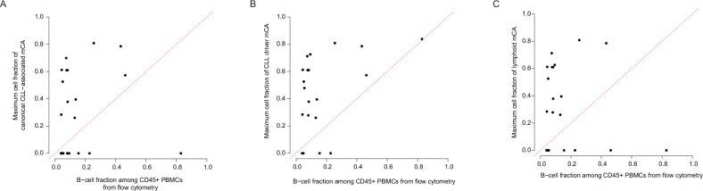
MBL is a precursor condition to chronic lymphocytic leukemia (CLL), characterized by monoclonal B-cells in blood. Mosaic chromosomal alterations (mCAs) are a form of clonal hematopoiesis that include gains, losses, and copy-neutral loss-of-heterozygosity of large DNA segments. Both MBL and mCAs have been found to increase the risk of CLL and lymphoid malignancies, and the aim of our study was to investigate how mCAs relate to MBL, which is currently unknown. We analyzed genetic, flow cytometric, and hematologic data from 4632 individuals from the Mayo Clinic Biobank and CLL Database. MBL was detected using flow cytometry and classified as high-count (HC) or low-count (LC) MBL based on clone size. mCAs were detected primarily from whole blood DNA using sensitive SNP-array-based analyses. mCAs commonly altered in CLL (deletion of 6q, 11q, 13q, 17p, and trisomy 12) were specific (>99%) to individuals with MBL and CLL. HC-MBL and LC-MBL individuals were 881-fold and 8-fold, respectively, more likely to harbor CLL-associated mCAs than those without MBL. The cell fraction bearing these mCAs typically exceeded the B-cell fraction, suggesting their origin prior to the B-cell lineage. Integrating genetic and blood count data enabled detecting HC-MBL with high specificity in a biobank sample. These results quantify the contribution of mCAs to MBL and could enable large studies of HC-MBL without the need for flow cytometric screening.


Conflict of interest statement

AS stock in Vertex Pharmaceuticals and consulting for Alight. SAP Research funding has been provided to the institution from Janssen, AstraZeneca, Merck, and Genentech for clinical studies in which Sameer A. Parikh is a principal investigator. Honoraria has been provided to the institution from Pharmacyclics, Merck, AstraZeneca, Janssen, Genentech, Amgen, MingSight Pharmaceuticals, TG Therapeutics, Eli Lilly, Novalgen Limited, Kite Pharma, and AbbVie for Sameer A. Parikh’s participation in consulting activities/advisory board meetings. PJH: None. NEK: Advisory Board for AbbVie, AstraZeneca, Beigene, Behring, Boehringer Ingelheim Pharmaceuticals, Inc., Dava Oncology, Janssen, Juno Therapeutics, Pharmacyclics. DSMC (Data Safety Monitoring Committee) for Agios Pharm, AstraZeneca, BMS –Celgene, Dren Bio Janssen. Research funding from: AbbVie, Acerta Pharma, Bristol Meyer Squib, Celgene, Genentech, Pharmacyclics, Sunesis, Vincerx. BLE: research funding from Celgene, Deerfield, Novartis, and Calico and consulting fees from Abbvie and GRAIL. He is a member of the scientific advisory board and shareholder for Neomorph Inc., TenSixteen Bio, Skyhawk Therapeutics, and Exo Therapeutics.

Figures

Fig. 1
Fig. 1. Relationship of mosaic chromosomal alterations (mCAs) with MBL.
Proportion of individuals with at least one canonical CLL-associated mCA (A) and autosomal mCA (B). Association of different categories of mCAs (defined below) with HC-MBL vs. controls in brown and LC-MBL vs. controls in dark gray (C) and HC-MBL vs. LC-MBL (D). Controls’ refers to individuals in whom flow cytometry screening did not identify a B-cell clone in peripheral blood mononuclear cells. Covariates in (C) and (D) included age, sex, European ancestry, SNP-array type, source of DNA (whole blood or PBMC), and batch effects. del 17p is not shown in (C) as it was not found in any of the controls, and association statistics for del 11q, trisomy 12, and del 13q are not displayed for LC-MBL given their rarity in both this category and controls. Canonical CLL-associated mCA: del 6q, del 11q, trisomy 12, del 13q, del 17p, and copy-number neutral loss-of heterozygosity at 13q/ MIR16-1 at 13q/ MIR16-1. CLL-driver mCA: includes canonical CLL-associated mCAs as well as those that were among 179 candidate drivers of CLL identified in two large genomic studies of CLL [27, 28]. Lymphoid mCA: mCAs whose frequency was specifically enriched in individuals with lymphoid malignancies in comparison to myeloid malignancies [17]. Autosomal mCA without CLL-driver mCA or lymphoid mCA: autosomal mCAs in individuals who have neither a CLL-driver mCA nor a lymphoid mCA.
Fig. 2
Fig. 2. Evaluation of sensitivity to detect mCAs in blood DNA as an explanation for lower frequency of mCAs within low-count MBL.
Clonal B-cell % from flow cytometry, which is clonal B-cells as a percentage of total B-cells, is shown for individuals with low-count MBL as a function of the type of mCAs present in each individual. Black horizontal bars and adjacent text indicate median values and p-values comparing clone size distribution are from a two-sided Mann–Whitney test.
Fig. 3
Fig. 3. Inference of lineage distribution of mCAs by comparison of mCA cell fraction against B-cell fraction.
Data are shown for canonical CLL-associated mCAs (A), CLL-driver mCAs (B), and lymphoid mCAs (C), the classification of which is detailed in the “Methods” section. Each point represents a single individual with HC-MBL from the Mayo Clinic MBL Biobank. For all individuals shown, flow cytometry was performed on frozen peripheral blood mononuclear cells (PBMCs) and DNA was extracted from PBMCs sampled on the same day as for flow cytometry. Data points with mCA cell fraction of 0 indicate individuals in whom the specified mCA type was not detected. Data points above the dashed red line indicate individuals in whom the fraction of cells containing a canonical CLL-associated mCA exceeds the B-cell fraction, suggesting the presence of the mCA beyond the B-cell lineage and origin prior to B-cell lineage commitment.
Fig. 4
Fig. 4. Test characteristics for distinguishing individuals with HC-MBL from controls and those with LC-MBL.
The mCA parameter modeled here is the presence of at least one CLL-driver mCA. Demographics refers to age and sex. ALC absolute lymphocyte count. PRS polygenic risk score associated with CLL. This analysis is based on individuals with available data across all the predictors among HC-MBL cases in the Mayo Clinic MBL Biobank (n = 60), controls (n = 2740), and LC-MBL (n = 669).

References

    1. Landgren O, Albitar M, Ma W, Abbasi F, Hayes RB, Ghia P, et al. B-cell clones as early markers for chronic lymphocytic leukemia. N Engl J Med. 2009;360:659–67. - PMC - PubMed
    1. Slager SL, Lanasa MC, Marti GE, Achenbach SJ, Camp NJ, Abbasi F, et al. Natural history of monoclonal B-cell lymphocytosis among relatives in CLL families. Blood. 2021;137:2046–56. - PMC - PubMed
    1. Vojdeman FJ, Helby J, Pedersen LB, Brieghel C, Andersen MA, Nordestgaard BG, et al. Chronic lymphocytic leukaemia clones are detectable decades before diagnosis. Br J Haematol. 2022;196:784–7. - PubMed
    1. Marti GE, Rawstron AC, Ghia P, Hillmen P, Houlston RS, Kay N, et al. Diagnostic criteria for monoclonal B-cell lymphocytosis. Br J Haematol. 2005;130:325–32. - PubMed
    1. Hallek M, Cheson BD, Catovsky D, Caligaris-Cappio F, Dighiero G, Dohner H, et al. iwCLL guidelines for diagnosis, indications for treatment, response assessment, and supportive management of CLL. Blood. 2018;131:2745–60. - PubMed

Publication types

LinkOut - more resources