. 2024 Nov 6;15(1):9602.
doi: 10.1038/s41467-024-54031-5.

AS160 is a lipid-responsive regulator of cardiac Ca2+ homeostasis by controlling lysophosphatidylinositol metabolism and signaling

Affiliations

AS160 is a lipid-responsive regulator of cardiac Ca2+ homeostasis by controlling lysophosphatidylinositol metabolism and signaling

Shu Su et al. Nat Commun. .

Abstract

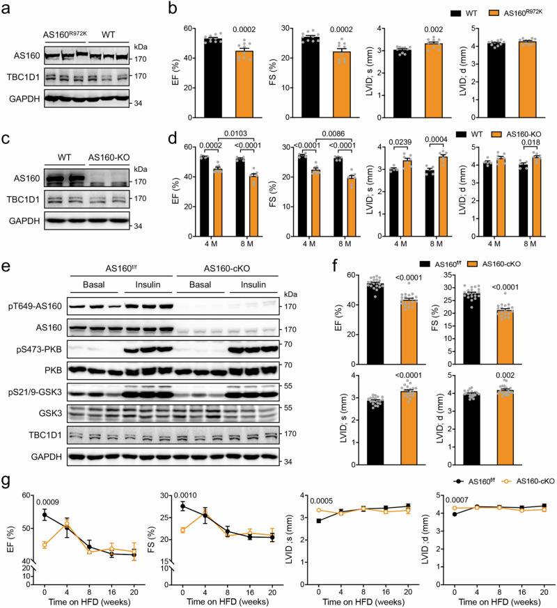
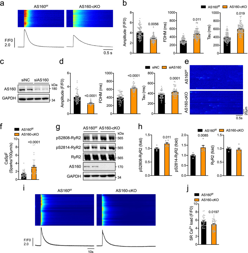
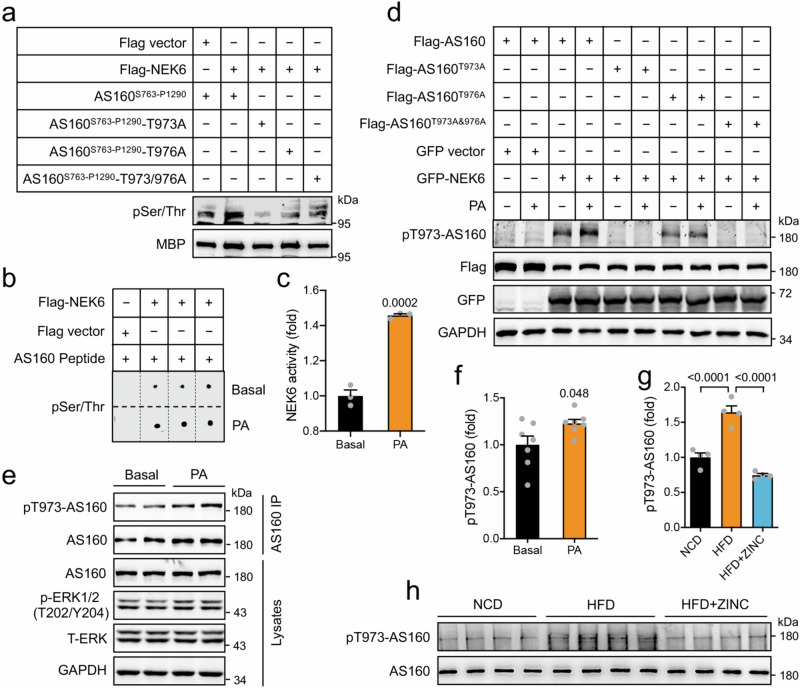
The obese heart undergoes metabolic remodeling and exhibits impaired calcium (Ca2+) homeostasis, which are two critical assaults leading to cardiac dysfunction. The molecular mechanisms underlying these alterations in obese heart are not well understood. Here, we show that the Rab-GTPase activating protein AS160 is a lipid-responsive regulator of Ca2+ homeostasis through governing lysophosphatidylinositol metabolism and signaling. Palmitic acid/high fat diet inhibits AS160 activity through phosphorylation by NEK6, which consequently activates its downstream target Rab8a. Inactivation of AS160 in cardiomyocytes elevates cytosolic Ca2+ that subsequently impairs cardiac contractility. Mechanistically, Rab8a downstream of AS160 interacts with DDHD1 to increase lysophosphatidylinositol metabolism and signaling that leads to Ca2+ release from sarcoplasmic reticulum. Inactivation of NEK6 prevents inhibition of AS160 by palmitic acid/high fat diet, and alleviates cardiac dysfunction in high fat diet-fed mice. Together, our findings reveal a regulatory mechanism governing metabolic remodeling and Ca2+ homeostasis in obese heart, and have therapeutic implications to combat obesity cardiomyopathy.


Conflict of interest statement

The authors declare no competing interests.

Figures

Fig. 1
Fig. 1. Effects of PA/HFD on AS160 Thr642-phosphorylation and GAP activity.
a Expression and phosphorylation of AS160 and PKB in PA- or OA-treated H9C2 cardiomyocytes. b GAP activity of endogenous AS160 in PA- or OA-treated H9C2 cardiomyocytes. n = 3 biological replicates. c Total and GTP-bound active Rab8a in PA- or OA-treated H9C2 cardiomyocytes. Expression (d) and GAP activity (e) of endogenous AS160 in the heart of mice administered with PA or OA for 3 h. n = 6. Ctrl, control. f Expression and phosphorylation of AS160 and PKB in the heart of mice fed normal chow diet (NCD) or HFD in response to insulin. g GAP activity of endogenous AS160 in the heart of mice fed NCD or HFD. n = 3. h Total and GTP-bound active Rab8a in the heart of mice fed NCD or HFD in response to insulin. Expression (i) and GAP activity (j) of endogenous AS160 in the heart of lean and obese monkeys (male, 5.6–26.9-year-old). n = 6 (Lean) and 7 (Obese). km Total and GTP-bound Rab8a in the heart of lean and obese monkeys (male, 5.6–26.9-year-old). n = 6 (Lean) and 7 (Obese). The data are given as the mean ± SEM. Numbers on the graphs represent p values. Statistical analyses were carried out using one-way ANOVA for (b), (e), two-sided t-test for (j), (l), (m), and two-way ANOVA for (g). Source data are provided as a Source Data file.
Fig. 2
Fig. 2. Cardiac function in AS160 deficient or inactive mutant mice.
a Expression of AS160 and TBC1D1 in the heart of AS160R972K mice. b Cardiac function of AS160R972K mice. EF, FS, LVID;s and LVID;d were measured in male AS160R972K mice (6-month-old). n = 10. c Expression of AS160 and TBC1D1 in the heart of AS160-KO mice. d Cardiac function of AS160-KO mice. EF, FS, LVID;s and LVID;d were measured in male AS160-KO mice (4- and 8-month-old). n = 5 (WT) and 7 (AS160-KO). e Expression and phosphorylation of AS160, PKB, GSK3 and TBC1D1 in the heart of AS160-cKO mice. f Cardiac function of AS160-cKO mice. EF, FS, LVID;s and LVID;d were measured in male AS160-cKO mice (4-month-old). n = 19 (AS160-cKO) and 20 (AS160f/f). g Cardiac function of AS160-cKO mice fed HFD. EF, FS, LVID;s and LVID;d were measured in male AS160-cKO mice that were fed HFD from the age of 5-month. n = 6. The data are given as the mean ± SEM. Numbers on the graphs represent p values. Statistical analyses were carried out using two-way ANOVA for (d), and two-sided t-test for (b), (f), (g). Source data are provided as a Source Data file.
Fig. 3
Fig. 3. Ca2+ homeostasis in AS160 deficient cardiomyocytes.
a, b Ca2+ transients in primary cardiomyocytes isolated from the AS160f/f and AS160-cKO mice (2-month-old, female) upon field stimulation. a Representative Ca2+ transient images and curves. b Amplitude, FDHM and Tau of Ca2+ transients. AS160f/f: 91 cells from 3 mice. AS160-cKO: 106 cells from 4 mice. c AS160 protein expression in AS160-knockdown neonatal rat cardiomyocytes. d Ca2+ transients in AS160-knockdown neonatal rat cardiomyocytes upon field stimulation. Amplitude, FDHM and Tau of Ca2+ transients were measured from 57 (siNC) and 59 (siAS160) cells. e, f Ca2+ sparks in primary cardiomyocytes isolated from the AS160f/f and AS160-cKO (2-month-old, male). e Representative images of Ca2+ sparks. f Quantification of Ca2+ spark frequency. 34 cells from 4 AS160f/f mice and 30 cells from 3 AS160-cKO mice were measured. g, h Expression and phosphorylation of RYR2 in the heart of female AS160-cKO and AS160f/f mice (3-month-old). g Representative blots. h Quantification of RYR2 protein and phosphorylation levels. n = 5 (AS160f/f) and 6 (AS160-cKO). i, j SR Ca2+ load in primary cardiomyocytes isolated from the AS160f/f and AS160-cKO mice (6-month-old, female). i Representative SR Ca2+ load images and curves. j Quantification of SR Ca2+ load. 30 cells from 5 AS160f/f mice and 38 cells from 5 AS160-cKO mice were measured. The data are given as the mean ± SEM. Numbers on the graphs represent p values. Statistical analyses were carried out using two-sided nested t-test for (b), (f), (j), and two-sided t-test for (d), (h). Source data are provided as a Source Data file.
Fig. 4
Fig. 4. LPI metabolism and signaling in AS160 deficient cardiomyocytes.
a Volcano plot of fold-change of lipid metabolites in the heart of the AS160f/f and AS160-cKO mice (male, 8-week-old). Fold-change was given as the ratio of AS160-cKO to AS160f/f. n = 6 (AS160-cKO) and 7 (AS160f/f). b Levels of total PI, LPI, LPA and LPS in the heart of the AS160f/f and AS160-cKO mice (male, 8-week-old). n = 6 (AS160-cKO) and 7 (AS160f/f). c Levels of LPI species in the heart of the AS160f/f and AS160-cKO mice (male, 8-week-old). n = 6 (AS160-cKO) and 7 (AS160f/f). d Ca2+ transients in primary cardiomyocytes isolated from the AS160f/f and AS160-cKO mice (6-month-old, female) upon treatment with the GPR55 inhibitor CID16020046 (CID). AS160f/f: 65 (Basal) and 70 (CID) cells from 4 mice. AS160-cKO: 79 (Basal) and 75 (CID) cells from 4 mice. e, f Ca2+ sparks in primary cardiomyocytes isolated from the AS160f/f and AS160-cKO mice (3-month-old, female) upon CID treatment. AS160f/f: 40 (Basal) and 40 (CID) cells from 3 mice. AS160-cKO: 34 (Basal) and 41 (CID) cells from 3 mice. e quantification of Ca2+ spark frequency. f Representative images of Ca2+ sparks. g Diastolic Ca2+ in cardiomyocytes from the AS160f/f and AS160-cKO mice (4-month-old, male). AS160f/f: 52 (Basal) and 57 (CID) cells from 5 mice. AS160-cKO: 54 (Basal) and 58 (CID) cells from 5 mice. Contractility in primary cardiomyocytes isolated from the AS160f/f and AS160-cKO mice (4-month-old, male) upon CID treatment. FS (h), and maximum contraction rates and maximum relaxation rates (i) were determined via the IonOptix system. For FS, AS160f/f: 59 (Basal) and 47 (CID) cells from 5 mice, AS160-cKO: 56 (Basal) and 54 (CID) cells from 5 mice. For maximum contraction rates, AS160f/f: 57 (Basal) and 51 (CID) cells from 5 mice, AS160-cKO: 49 (Basal) and 56 (CID) cells from 5 mice. For maximum relaxation rates, AS160f/f: 57 (Basal) and 45 (CID) cells from 5 mice, AS160-cKO: 55 (Basal) and 54 (CID) cells from 5 mice. j Cardiac function in AAV9-shNC or AAV9-shGPR55 administered male mice that were subjected to HFD for 6 weeks. n = 10. The data are given as the mean ± SEM. Numbers on the graphs represent p values. Statistical analyses were carried out using two-sided t-test for (a), (b), (c), nested two-way ANOVA for (d, e), (gi), and one-way ANOVA for (j). Source data are provided as a Source Data file.
Fig. 5
Fig. 5. Regulation of LPI metabolism by AS160 and Rab8a.
a Schematic illustration of LPI metabolic pathway. b Co-immunoprecipitation of GFP-AS160 with Flag-DDHD1. Flag-DDHD1 was co-expressed with GFP-AS160 in HEK293 cells, and immunoprecipitated from cell lysates using the Flag antibody. c Co-immunoprecipitation of GFP-DDHD1 with Flag-AS160. Flag-AS160 was co-expressed with GFP-DDHD1 in HEK293 cells, and immunoprecipitated from cell lysates using the Flag antibody. d Co-immunoprecipitation of GFP-AS160 with Flag-cPLA2. Flag-cPLA2 was co-expressed with GFP-AS160 in HEK293 cells, and immunoprecipitated from cell lysates using the Flag antibody. e Co-immunoprecipitation of GFP-cPLA2 with Flag-AS160. Flag-AS160 was co-expressed with GFP-cPLA2 in HEK293 cells, and immunoprecipitated from cell lysates using the Flag antibody. f Co-immunoprecipitation of endogenous DDHD1 and cPLA2 with Flag-AS160. Flag-AS160 was expressed in HEK293 cells, and immunoprecipitated from cell lysates using the Flag antibody. g Co-immunoprecipitation of endogenous AS160 and Rab8a with endogenous DDHD1 in H9C2 cardiomyocytes. h, i GTP-bound active Rab8a in AS160f/f and AS160-cKO cardiomyocytes in response to insulin. h Immunoblots. i Rab8a activation in which GTP-Rab8a was normalized with total Rab8a. n = 3 biological replicates. j Co-immunoprecipitation of GFP-Rab8a with Flag-DDHD1 or Flag-cPLA2. Flag-DDHD1 or Flag-cPLA2 was co-expressed with GFP-Rab8a in HEK293 cells, and immunoprecipitated from cell lysates using the Flag antibody. k, l Co-immunoprecipitation of GFP-DDHD1 or GFP-cPLA2 with Flag-tagged Rab8a WT or mutants. Flag-tagged Rab8a WT or mutants were co-expressed with GFP-DDHD1 (k) or GFP-cPLA2 (l) in HEK293 cells, and immunoprecipitated from cell lysates using the Flag antibody. m In vitro DDHD1 activity in the presence of GDP- or GTP-loaded Rab8a. n = 4 biological replicates. The data are given as the mean ± SEM. Numbers on the graphs represent p values. Statistical analyses were carried out using two-way ANOVA for (i), and one-way ANOVA for (m). Source data are provided as a Source Data file.
Fig. 6
Fig. 6. Phosphorylation of AS160 by NEK6 in response to PA.
a In vitro phosphorylation of recombinant MBP-AS160S763-P1290 WT and mutant proteins by a purified Flag-NEK6. Phosphorylation of MBP-AS160S763-P1290 WT and mutant proteins was detected using a pSer/Thr antibody. b, c NEK6 activity in response to PA. Flag-NEK6 was immunoprecipitated from HEK293 cells that were stimulated with or without PA, and assayed for its kinase activity using AS160-Nektide (LVDLGRtFPtHP, Thr973/976 shown in lower case) as a substrate. b Dot blots. c NEK6 activity. n = 3 biological replicates. d Thr973 phosphorylation of AS160 by NEK6 in response to PA. Flag-AS160 WT or mutants were co-expressed with GFP-NEK6 in HEK293 cells. Thr973 phosphorylation of AS160 was detected using the site-specific phospho-antibody. e, f Thr973 phosphorylation of endogenous AS160 in NRVCs in response to PA. e Representative blots. f Quantification data. n = 7 biological replicates. g, h Thr973 phosphorylation of endogenous AS160 in the heart of HFD-fed mice administered with or without ZINC05007751 for 8 weeks. g Quantification data. h Immunoblots. n = 4. The data are given as the mean ± SEM. Numbers on the graphs represent p values. Statistical analyses were carried out using two-sided t-test for (c) and (f), and one-way ANOVA for (g). Source data are provided as a Source Data file.
Fig. 7
Fig. 7. Regulation of AS160 GAP activity and cardiac function by NEK6.
a GAP activity of recombinant MBP-AS160S763-P1290 WT and mutant proteins upon in vitro phosphorylation by NEK6. n = 3 biological replicates. b GAP activity of Flag-AS160 WT and mutant proteins in response to PA. HEK293 cells expressing Flag-AS160, Flag-AS160T973A or Flag-AS160T976A were treated with or without PA. n = 3 biological replicates. c GAP activity of Flag-AS160 WT and mutant proteins in response to PA. HEK293 cells expressing Flag-AS160 or Flag-AS160T973A/T976A were treated with or without PA. n = 3 biological replicates. d GAP activity of Flag-AS160 WT and mutant proteins in response to PA. AS160-KO HEK293 cells expressing Flag-AS160, Flag-AS160T973D or Flag-AS160T976D were treated with or without PA. n = 3 biological replicates. e Effects of NEK6 on GAP activity of Flag-AS160 in response to PA. HEK293 cells co-expressing Flag-AS160 with GFP-NEK6 or GFP were treated with or without PA. n = 3 biological replicates. f Effects of NEK6 on GAP activity of Flag-AS160 WT and mutant proteins. Flag-AS160 WT or mutants was co-expressed with GFP-NEK6 or GFP in HEK293 cells. n = 3 biological replicates. g GAP activity of endogenous AS160 upon NEK6 knockdown in HEK293 cells in response to PA treatment. n = 3 biological replicates. h GAP activity of Flag-AS160 in HEK293 cells in response to PA treatment in the presence or absence of NEK6 inhibitor ZINC05007751. n = 3 biological replicates. i Cytosolic Ca2+ in NEK6-knockdown NRVCs in response to PA. n = 7 (NC) and 8 (NC + PA, siNEK6+PA) biological replicates. j GAP activity of endogenous AS160 in the heart of HFD-fed mice administered with or without ZINC05007751 for 8 weeks. n = 5 (NC + PA) and 6 (NC, siNEK6+PA). k Cardiac function of HFD-fed mice administered with or without ZINC05007751. EF and FS were measured in HFD-fed mice administered with or without ZINC05007751 for 6 weeks. n = 8 (NCD, HFD) and 10 (HFD + ZINC05007751). l A diagram (Created in BioRender. Shu S (2022) BioRender.com/y90d330) illustrates the proposed model in which AS160 is a lipid-responsive regulator governing metabolic flexibility and Ca2+ homeostasis in the heart. PA/HFD inhibits AS160-GAP activity through phosphorylation by NEK6 in the heart. Inactivation of AS160 causes metabolic remodeling, enhances LPI production and signaling, and impairs Ca2+ homeostasis in cardiomyocytes, which may consequently contribute to the development of obesity cardiomyopathy. The data are given as the mean ± SEM. Numbers on the graphs represent p values. Statistical analyses were carried out using one-way ANOVA for (a), (hk), and two-way ANOVA for (bg). Source data are provided as a Source Data file.

Similar articles

References

    1. Wong, C. & Marwick, T. H. Obesity cardiomyopathy: pathogenesis and pathophysiology. Nat. Clin. Pract. Cardiovasc. Med.4, 436–443 (2007). - PubMed
    1. Wong, C. & Marwick, T. H. Obesity cardiomyopathy: diagnosis and therapeutic implications. Nat. Clin. Pract. Cardiovasc. Med.4, 480–490 (2007). - PubMed
    1. Ren, J., Wu, N. N., Wang, S., Sowers, J. R. & Zhang, Y. Obesity cardiomyopathy: evidence, mechanisms, and therapeutic implications. Physiol. Rev.101, 1745–1807 (2021). - PMC - PubMed
    1. Nakamura, M. & Sadoshima, J. Cardiomyopathy in obesity, insulin resistance and diabetes. J. Physiol.598, 2977–2993 (2020). - PubMed
    1. Ouwens, D. M. et al. Cardiac contractile dysfunction in insulin-resistant rats fed a high-fat diet is associated with elevated CD36-mediated fatty acid uptake and esterification. Diabetologia50, 1938–1948 (2007). - PMC - PubMed

Publication types

MeSH terms

LinkOut - more resources