. 2024 Nov 6;14(1):26949.
doi: 10.1038/s41598-024-78528-7.

40 Hz light preserves synaptic plasticity and mitochondrial function in Alzheimer's disease model

Affiliations

40 Hz light preserves synaptic plasticity and mitochondrial function in Alzheimer's disease model

Amir Barzegar Behrooz et al. Sci Rep. .

Abstract

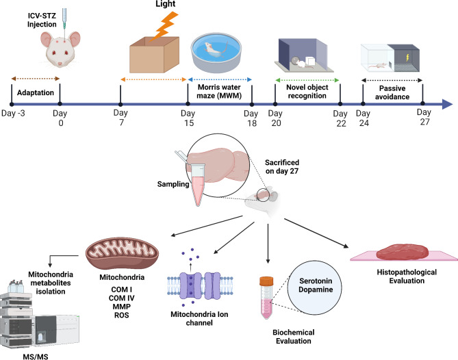
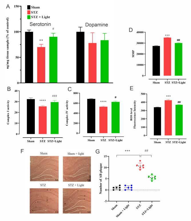
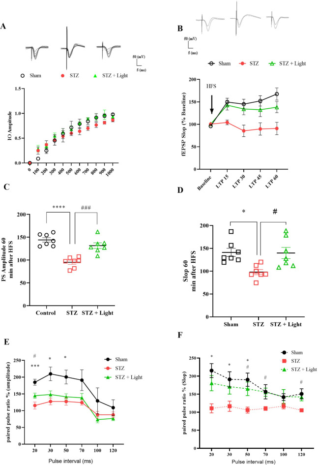
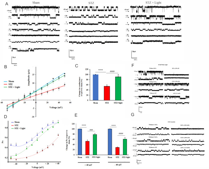
Alzheimer's disease (AD) is the most prevalent type of dementia. Its causes are not fully understood, but it is now known that factors like mitochondrial dysfunction, oxidative stress, and compromised ion channels contribute to its onset and progression. Flickering light therapy has shown promise in AD treatment, though its mechanisms remain unclear. In this study, we used a rat model of streptozotocin (STZ)-induced AD to evaluate the effects of 40 Hz flickering light therapy. Rats received intracerebroventricular (ICV) STZ injections, and 7 days after, they were exposed to 40 Hz flickering light for 15 min daily over seven days. Cognitive and memory functions were assessed using Morris water maze, novel object recognition, and passive avoidance tests. STZ-induced AD rats exhibited cognitive decline, elevated reactive oxygen species, amyloid beta accumulation, decreased serotonin and dopamine levels, and impaired mitochondrial function. However, light therapy prevented these effects, preserving cognitive function and synaptic plasticity. Additionally, flickering light restored mitochondrial metabolites and normalized ATP-insensitive mitochondrial calcium-sensitive potassium (mitoBKCa) channel activity, which was otherwise downregulated in AD rats. Our findings suggest that 40 Hz flickering light therapy could be a promising treatment for neurodegenerative disorders like AD by preserving synaptic and mitochondrial function.

Keywords: Alzheimer’s disease; Flickering 40 Hz white light; LTP; MitoBKCa+2; Mitochondrial function; Mitochondrial metabolites; Synaptic plasticity.


Conflict of interest statement

The authors declare no competing interests.

Figures

Fig. 1
Fig. 1
Schematic diagram of the experimental design.
Fig. 2
Fig. 2
Exposure to flickering 40 Hz white light improved cognitive function in a rat model of STZ-induced AD. MWM: Comparison of the time escape latency to reach the hidden platform in the acquisition phase over 3 days (A) and the time escape latency (B), total time spent in the target quadrant (C), and number of times crossing the platform (E) as assessed in the probe tests in sham, sham + light, STZ and STZ + light groups. Panel D shows a visual comparison of the average heatmap of MWM trials; the scale bar indicates the time spent in the target quadrant. Panels F and G depict the comparison of the exploring time of a novel object followed by a familiar object and the recognition index, respectively, in all 4 experimental groups. The effects of light treatment on passive avoidance learning acquisition in sham and STZ rats are indicated by differences in step through latency (STL; step-through latency) during the retrieval test performed 1 day after passive avoidance acquisition (H). One-way ANOVA analysis followed by Bonferroni’s post hoc test was performed to compare the mean between groups. Data are expressed as mean ± SEM (n = 7 rat/group). *P < 0.0.5, **P < 0.01, ***P < 0.001, **** P < 0.0001 compared to sham, and #P < 0.05, ##P < 0.01 compared to STZ.
Fig. 3
Fig. 3
Flickering 40 Hz white light prevented the deficits in serotonin levels and mitochondrial phenotype in the hippocampus of STZ-induced AD rats. Neurotransmitter (serotonin and dopamine) levels and mitochondrial phenotype were analyzed in hippocampal tissue homogenates obtained from sham, STZ, and STZ + light rats. Serotonin and dopamine levels were determined by ELISA assay (A; n = 6). Mitochondria were isolated for the measurement of complex I/IV activities, MMP, and ROS levels (B-E; n = 7). In addition, brain sections from sham, sham + light, STZ, and STZ + light rats were stained with Congo red, after which Aβ plaques were counted (F-G). One-way ANOVA analysis followed by the Bonferroni’s post hoc test was performed to compare the mean between groups. Data are expressed as mean ± SEM. *P < 0.0.5, **P < 0.01, ***P < 0.001, ****P < 0.0001 compared to sham, and #P < 0.05, ## P < 0.01 compared to STZ.
Fig. 4
Fig. 4
Flickering 40 Hz white light prevented the STZ-induced deficits in electrophysiological properties of hippocampal neurons in rats. Basal synaptic transmission and plasticity in the perforant pathway of the hippocampus was assessed in sham, STZ, and STZ + light animals. (A) The acquired input/output (I/O) curve from a 60 s recording and single traces of the DG neurons’ reaction to numerous stimulus intensities (100 to 1000 µA)(n = 6). (B) The effect of flickering 40 Hz white light on LTP induction and maintenance in the DG up to 60 min after high-frequency stimulation (HFS). Data were normalized against the baseline period (-30 to + 60 min), and single traces were recorded before and after HFS. fEPSP slope change (%) and time following HFS. (C-D) The PS amplitude and slop mean were recorded at 0 to 60 min after applying HFS (n = 7). (E-F) In short intervals between two strictly paired stimuli, the amplitude of PS and fEPSP slope were prompted by a paired pulse in all groups. One-way or Two-way ANOVA analyses followed by the Bonferroni’s post hoc test were performed to compare the mean between groups. Data are expressed as mean ± SEM (n = 6 rat/group for B-E). *P < 0.0.5, ** P < 0.01, ***P < 0.001, ****P < 0.0001 compared to sham, and #P < 0.05, ##P < 0.01, ###P < 0.001 compared to STZ.
Fig. 5
Fig. 5
Induction of AD reduced channel gating and conductance in mitochondrial mitoBKCa channels but was prevented by 40 Hz light therapy. Single channel recordings in 200/50 mM (cis/trans) KCl solution were performed using a bilayer lipid membrane setup. Single channel current-voltage relationships for mitoBKCa channels and percent of changes in the channel conductance and open probability (Po) as a function of voltage for mitoBKCa channels were evaluated in sham, STZ, and STZ + light animals (A-E). The closed state is marked by the black arrows. Each point represents the average Po as a function of voltages from five different experiments. The effects of IbTx, ChTx, and ATP on mitoBKCa channel activity were determined in STZ (F) and STZ + light animals (G). One-way ANOVA analyses followed by a Bonferroni’s post hoc test were performed to compare the mean between groups. Data are expressed as mean ± SEM (n = 5 (5 A-E) and 4 (5 F-G) rats per group). *P < 0.0.5, **P < 0.01, ***P < 0.001, ****P < 0.0001 compared to sham, and #P < 0.05, ##P < 0.01, ####P < 0.0001 compared to STZ.
Fig. 6
Fig. 6
Flickering 40 Hz white light counteracted the STZ-induced disruption in mitochondrial amino acid levels in rat brains. Differential levels of mitochondrial amino acids measured using MS-MS were compared in sham, sham + light, STZ, and STZ + light rats (A-B). Ten significant metabolites were downregulated in STZ rats vs. sham controls (C). A diagram of metabolite-protein joint pathways was generated (D), from which seven common pathways were extracted (E). The top 10 associated pathways affected by alanine, aspartate, and glutamine metabolism are illustrated (F). Data are expressed as mean ± SEM (n = 7 ± 1).

Similar articles

References

    1. Abubakar, M. B. et al. Alzheimer’s disease: An update and insights into pathophysiology. Front. Aging Neurosci.14, 742408. 10.3389/fnagi.2022.742408 (2022). From NLM. - PMC - PubMed
    1. Knopman, D. S. et al. Alzheimer disease. Nat. Rev. Dis. Primers 7 (1), 33. (2021). 10.1038/s41572-021-00269-y From NLM. - PMC - PubMed
    1. Jack, C. R. Jr. et al. NIA-AA research framework: Toward a biological definition of Alzheimer’s disease. Alzheimers Dement.14 (4), 535–562. 10.1016/j.jalz.2018.02.018 (2018). From NLM. - PMC - PubMed
    1. Sperling, R. A. et al. Toward defining the preclinical stages of Alzheimer’s disease: Recommendations from the National Institute on Aging-Alzheimer’s Association workgroups on diagnostic guidelines for Alzheimer’s disease. Alzheimers Dement.7 (3), 280–292. 10.1016/j.jalz.2011.03.003 (2011). From NLM. - PMC - PubMed
    1. Yiannopoulou, K. G. & Papageorgiou, S. G. Current and future treatments in Alzheimer disease: An update. J. Cent. Nerv. Syst. Dis.12, 1179573520907397. 10.1177/1179573520907397 (2020). From NLM. - PMC - PubMed

LinkOut - more resources