

CILP-2 expression in the intervertebral discs of patients with lumbar radiculopathy
- PMID: 39506696
- PMCID: PMC11539541
- DOI: 10.1186/s12891-024-07996-9
CILP-2 expression in the intervertebral discs of patients with lumbar radiculopathy
Abstract
Background: Intervertebral disc (IVD) degeneration (IVDD) is one of the main causes of low back pain. One of the most important features of IVDD is the loss of extracellular matrix (ECM) with its structural components. Cartilage intermediate layer proteins (CILPs), minor glycoproteins residing in ECM, have been found to be increased in IVD as degeneration and aging progresses. The aim of the present study was to evaluate the expression of CILP-2 in the IVD of patients with lumbar radiculopathy.
Methods: The IVD samples were collected from 25 patients during spinal surgery (interlaminectomy, herniated disc removal). The control IVD samples were obtained from nine patients who underwent lateral corpectomies in the thoracic region. CILP-2 expression was detected by immunohistochemistry. The patients were divided into two groups - aged under or over 50 years. A standardized clinical examination with assessment of radicular signs and deficits was performed. Subjective disability and pain were assessed using the visual analogue scale and Oswestry Disability Index (ODI). The pre-operative MRI was graded for the degree of IVD degeneration by Pfirrmann grading system. IVD samples obtained during operations were subjected to the standardized histopathological analysis applying modified Boos classification. The data were analysed by t-test, Mann-Whitney U-test, and Spearman correlation test.
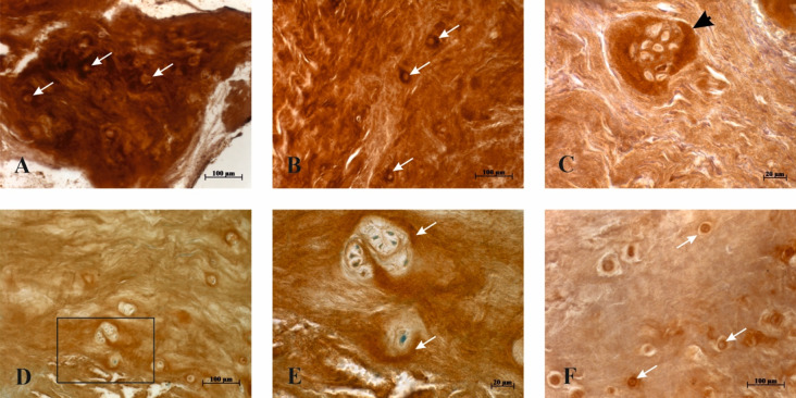
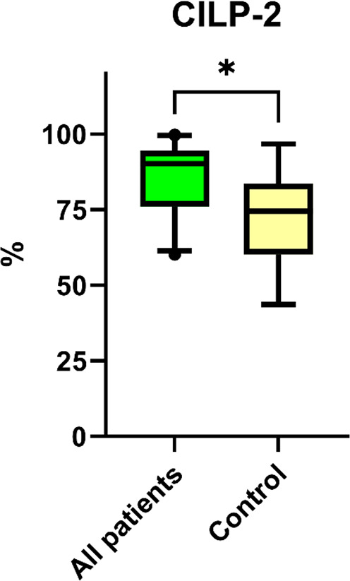
Results: Both histopathology scores and Pfirrmann grades did not differ between patients' groups. Also, no correlations were found between histopathology and Pfirrmann grades, neither were any differences seen when correlating both grades to ODI, back pain or leg pain scores. CILP-2 staining was noted in all studied samples, notably strong staining was seen around large cell clusters. However, no differences in CILP-2 staining were seen between the age groups of patients. No correlations were found between CILP-2 staining and Pfirrmann grades. Grading of CILP-2 immunostaining in nine control patient samples resulted in significantly lower values. The difference is statistically significant (P = 0.002) compared to CILP-2 staining scores of all 25 patients' samples.
Conclusion: In this study, we detected increased CILP-2 expression in the human IVD as compared to the control group patients. CILP-2 can be a possible IVDD marker; however, as knowledge about the role of CILP-2 is limited, further studies are required.
Keywords: Cartilage intermediate layer protein; Immunohistochemistry; Intervertebral disc degeneration; Radiculopathy.
© 2024. The Author(s).
Conflict of interest statement
The authors declare no competing interests.
Figures


References
-
- Hartvigsen J, Hancock MJ, Kongsted A, Louw Q, Ferreira ML, Genevay S, Hoy D, Karppinen J, Pransky G, Sieper J, Smeets RJ, Underwood M, Lancet Low Back Pain Series Working Group. What low back pain is and why we need to pay attention. Lancet. 2018;391(10137):2356–67. 10.1016/S0140-6736(18)30480-X. - DOI - PubMed
-
- Wang Z, Kim JH, Higashino K, Kim SS, Wang S, Seki S, Hutton WC, Yoon ST. Cartilage intermediate layer protein (CILP) regulation in intervertebral discs. The effect of age, degeneration, and bone morphogenetic protein-2. Spine (Phila Pa 1976). 2012;37(4):E203–8. 10.1097/BRS.0b013e31822dcf47. - DOI - PubMed
MeSH terms
Substances
LinkOut - more resources
Full Text Sources
