. 2024 Nov 7;40(1):38.
doi: 10.1186/s42826-024-00226-2.

Observation of neutrophil extracellular traps in the development of diabetic nephropathy using diabetic murine models

Affiliations

Observation of neutrophil extracellular traps in the development of diabetic nephropathy using diabetic murine models

You Hyun Jeon et al. Lab Anim Res. .

Abstract

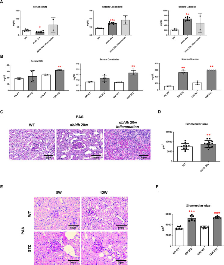
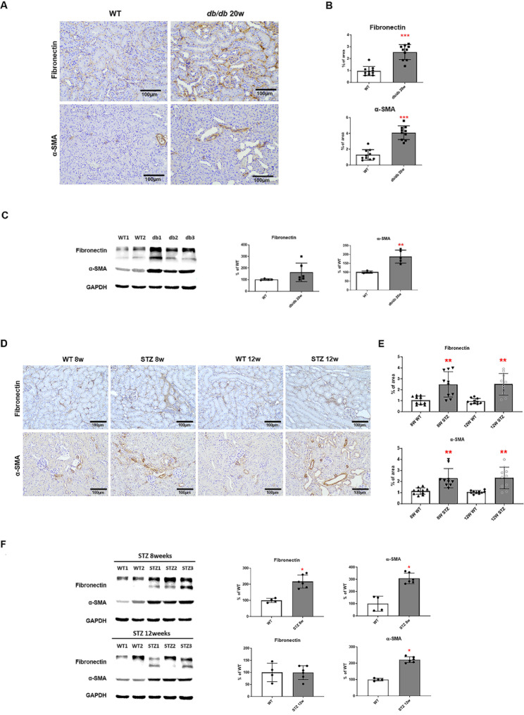
Background: Diabetic nephropathy (DN) is a progressive complication among patients with diabetes and the most common cause of end-stage kidney disease. Neutrophil extracellular traps (NETs) are known to play a role in kidney disease, thus this study aimed to determine their role in the development of diabetic kidney disease using diabetic murine models.

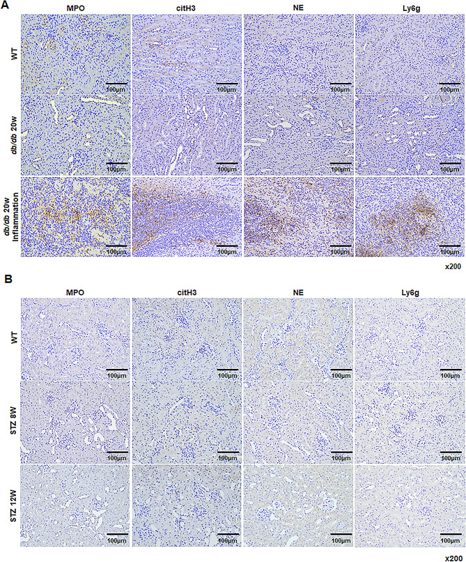
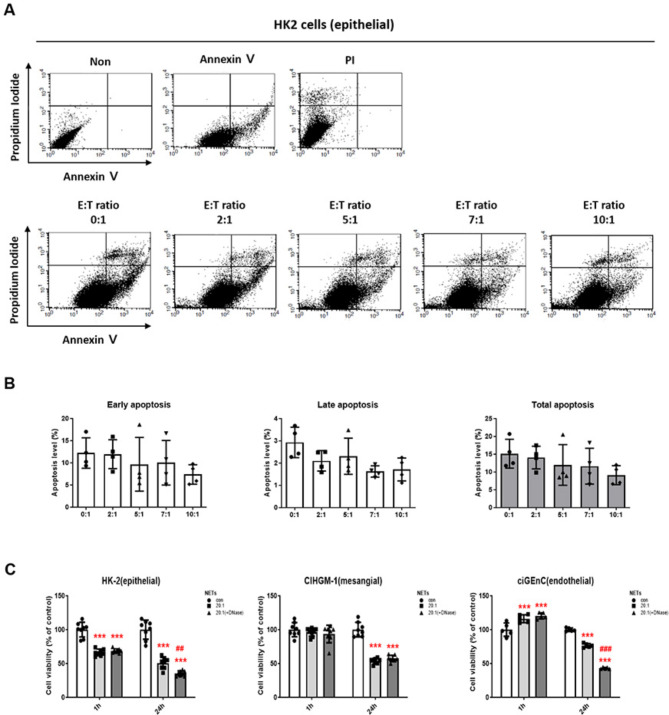
Results: Protein and histological analyses revealed that db/db mice and streptozotocin DN models expressed no significant NET-related proteins, myeloperoxidase, citrullinated histone H3 (citH3), neutrophil elastase, and lymphocyte antigen 6 complex locus G6D (Ly6G). However, the inflamed individuals in the DN model showed that citH3 and Ly6G were highly deposited in the renal system based on immunohistochemistry images. In vitro, NET treatment did not induce apoptosis in glomerular endothelial and renal tubular epithelial cells. NET inhibition by DNase administration demonstrated no significant changes in cell apoptosis.

Conclusions: NET-related proteins were only expressed in the DN model with tubulointerstitial inflammation. Our study revealed that NETs are only induced in mice with hyperglycemia-induced inflammation.

Keywords: Chronic kidney disease; Diabetic nephropathy; Hyperglycemia; Neutrophil; Neutrophil extracellular traps.


Conflict of interest statement

The authors declare no competing interests.

Figures

Fig. 1
Fig. 1
The biochemistry analysis of serum blood urea nitrogen (BUN), creatinine, and glucose levels and renal pathological structures in diabetic models. (A) Biochemistry analysis in db/db 20-week-old mice. n = 2–7. (B) Biochemistry analysis in streptozotocin (STZ)-induced diabetic models for 8-week- and 20-week-old mice. n = 5 each group. (C, E). Representative images of periodic acid-Schiff (PAS) staining (x400) and (D, F) glomerular size in both diabetic models. n ≥ 6. (Kruskal–Wallis test, *p < 0.05, **p < 0.01, ***p < 0.001 vs. WT)
Fig. 2
Fig. 2
High glucose serum levels induced the progression of fibrosis in diabetic kidney tissue. (A, B) Representative images of fibronectin and α-SMA staining of kidney tissue in db/db 20-week-old mice (x200). Bar graph summarizing the % of the positive staining area. n = 10 each group. (C) Fibronectin and α-SMA protein levels in db/db 20-week-old mice were determined by Western blot. n = 4–6 each group. (D, E) Representative images of fibronectin and α-SMA staining of kidney tissue in STZ-induced diabetic models (x200). Bar graph summarizing the % of the positive staining area. n = 10 each group. (F) Fibronectin and α-SMA protein levels in STZ-induced diabetic models were determined by Western blot. n = 4–6 each group (The Kruskal–Wallis test, *p < 0.05, **p < 0.01, ***p < 0.001 vs. WT)
Fig. 3
Fig. 3
The expression patterns of NETs-related proteins in a model with DN or inflammatory DN. (A) Representative images of NETs-related proteins staining of kidney tissues in db/db mice. (B) Representative images of NETs-related proteins staining of kidney tissues in STZ-induced diabetic models (x200)
Fig. 4
Fig. 4
Apoptosis and viability with NET or NET blocking in HK2 cells. (A) Annexin V-FITC and propidium iodide (PI) staining and (B) quantitative analyses for apoptosis after co-culture of neutrophils in HK2 cells. n = 4 each group. (C) MTT assay results according to NET blocking in HK2, CIHGM-1, and ciGEnC cells. n = 8 each group. (One-way ANOVA, ***p < 0.001 vs. con, ## p < 0.01, p < 0.001 vs. 20:1)

References

    1. Alicic RZ, Rooney MT, Tuttle KR. Diabetic kidney disease: challenges, Progress, and possibilities. Clin J Am Soc Nephrol. 2017;12(12):2032–45. - PMC - PubMed
    1. GBD Chronic Kidney Disease Collaboration. Global, regional, and national burden of chronic kidney disease, 1990–2017: a systematic analysis for the global burden of Disease Study 2017. Lancet. 2020;395(10225):709–33. - PMC - PubMed
    1. Chatterjee S, Khunti K, Davies MJ. Type 2 diabetes. Lancet. 2017;389(10085):2239–51. - PubMed
    1. Thomas MC, Cooper ME, Zimmet P. Changing epidemiology of type 2 diabetes mellitus and associated chronic kidney disease. Nat Rev Nephrol. 2016;12(2):73–81. - PubMed
    1. Saeedi P, Petersohn I, Salpea P, Malanda B, Karuranga S, Unwin N, et al. Global and regional diabetes prevalence estimates for 2019 and projections for 2030 and 2045: results from the International Diabetes Federation Diabetes Atlas, 9(th) edition. Diabetes Res Clin Pract. 2019;157:107843. - PubMed

LinkOut - more resources