. 2024 Nov 6;20(1):506.
doi: 10.1186/s12917-024-04350-y.

Iron deficiency in dogs suffering from atopic dermatitis

Affiliations

Iron deficiency in dogs suffering from atopic dermatitis

Carolina Frizzo Ramos et al. BMC Vet Res. .

Abstract

Background: Iron-deficiency is associated with increased morbidity and mortality in non-communicable diseases. However, iron parameters are rarely assessed in dogs. Here, we aimed to assess and correlate iron parameters in dogs suffering from Canine Atopic Dermatitis (CAD) compared to non-atopic, healthy dogs.

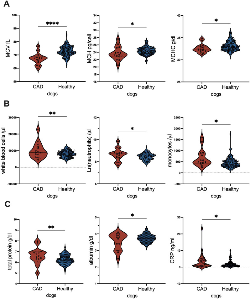
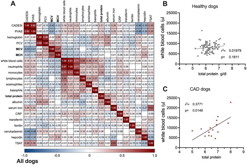
Results: For this retrospective study, blood values and sera of 34 dogs with confirmed CAD were compared with 94 healthy non-atopic dogs. In our cohort, dogs with CAD had significantly lower mean corpuscular volume (MCV, ) mean corpuscular hemoglobin (MCH) but higher white blood cell counts due to increased levels of circulating neutrophils and monocytes. CAD patients also had elevated total protein and c-reactive protein (CRP), but lower albumin levels compared to our healthy control dogs, indicated low-grade inflammation in the CAD cohort. Spearman correlations associated negatively clinical symptom (CADESI-4/PVAS) with MCV; ceruloplasmin and hepcidin, but positively with serum iron. Only in the CAD-cohort, MCV, CRP and albumin-levels negatively affected serum iron-levels and were positively associated with ceruloplasmin. Linear regression analysis revealed that serum iron-levels in CAD subjects, were positively dependent on hematocrit (packed cell volume, PCV) and albumin, and negatively dependent with white blood cells and neutrophils numbers. In contrast, in the healthy cohort, hepcidin was the sole factor associated with serum iron.

Conclusions: A decreased iron status was associated with a higher symptom burden. Iron homeostasis differed markedly in healthy and atopic dermatitis dogs. CAD patients had depleted iron-stores and presented themselves with subclinical inflammation.


Conflict of interest statement

FRW is lead- and EJJ co-inventor of patent EP2894478, owned by Biomedical International R+D GmbH, of which EJJ is shareholder. FRW is founder of ViaLym, has served as an investigator and received personal fees from Biomedical Int R&D, Allergy Therapeutics, Bencard Allergy and Lofarma. The other authors declare no competing interests.

Figures

Fig. 1
Fig. 1
Blood cells and inflammation marker differ in dogs with or without canine atopic dermatitis CAD. Boxplot comparing (A) Red blood cell parameters: mean corpuscular volume (MCV) and mean corpuscular hemoglobin (MCH) and mean corpuscular hemoglobin concentration (MCHC); (B) White blood cells: white blood cells, total neutrophils and monocyte numbers; and (C) Humoral parameters: total protein, albumin and CRP levels in treated dogs with CAD or healthy control dogs. For statistical analyses, groups were tested for normality with Anderson-Darling-test, before parameters were compared with unpaired t-test (MCV MCH, white blood cells, neutrophils, total protein and albumin) or Mann-Whitney U test (monocytes) and Kolmogorov-Smirnov test (CRP). p < 0.5, **** p < 0.0001
Fig. 2
Fig. 2
Increased serum iron and TSAT-levels, but lower hepcidin and ceruloplasmin-activity in CAD patients, Comparison of A. serum iron, B. transferrin saturation TSAT, C. ceruloplasmin activity and D. hepcidin in dogs with atopic dermatitis (CAD) or not (healthy). Data were assessed for normality, before comparing with t-test for TSAT-levels, or with non-parametric Kolmogorov-Smirnov test for other parameters. ** p < 0.01, **** p < 0.0001
Fig. 3
Fig. 3
Correlation and regression analysis of clinical symptoms (CADESI/PVAS) in dogs. (A). Spearman correlation of CADESI/PVAS with blood parameters with positive association in red hues and negative associations annotated in blue hues. Values below − 0.18 or above 0.18 are significant. Linear regression analyses of total protein (as an indicator of inflammation) with white blood cells in (B) healthy (C) dogs with CAD. P < 0.05 is significant
Fig. 4
Fig. 4
Correlation and regression analyses of serum iron in dogs with CAD or considered healthy. A. Spearman correlation of serum iron are in CAD-patients negatively associate with ferritin, MCV, PCV, albumin and CRP, and positive with ceruloplasmin, while in the healthy cohort, serum iron was negatively associated with hepcidin and positive with total protein. B. Linear regression analysis (with transformed data to reach parametric distribution) to inversed serum iron showed that serum iron was significantly negatively dependent with PCV and albumin, whereas a positive dependency was seen with white blood cell number and neutrophils in dogs with CAD. In contrast in the healthy cohort, only hepcidin showed a negatively dependency with serum iron-levels. P < 0.05 is considered significant

References

    1. Outerbridge CA, Jordan TJM. Current knowledge on canine atopic dermatitis: Pathogenesis and treatment. Adv Small Anim Care. 2021;2:101–15. - PMC - PubMed
    1. Yang L, Sato M, Saito-Abe M, Miyaji Y, Shimada M, Sato C et al. Allergic Disorders and Risk of Anemia in Japanese Children: Findings from the Japan Environment and Children’s Study. Nutrients. 2022;14(20). - PMC - PubMed
    1. Rhew K, Oh JM. Association between atopic disease and anemia in pediatrics: a cross-sectional study. BMC Pediatr. 2019;19(1):455. - PMC - PubMed
    1. Sato S. Iron deficiency: structural and microchemical changes in hair, nails, and skin. Semin Dermatol. 1991;10(4):313–9. - PubMed
    1. Stojkovic A, Radlovic N, Vuletic B, Nestorovic B, Lekovic Z, Obradovic S, et al. Presentation of an infant with nutritional deficiency dermatitis as the initial manifestation of cystic fibrosis. Srp Arh Celok Lek. 2013;141(11–12):810–3. - PubMed