

Clinical, pathological, and computed tomography morphological features of lung cancer with spread through air spaces
- PMID: 39507029
- PMCID: PMC11535835
- DOI: 10.21037/tlcr-24-715
Clinical, pathological, and computed tomography morphological features of lung cancer with spread through air spaces
Abstract
Background: Spread through air spaces (STAS) is significantly associated with decreased overall survival (OS) and reduced recurrence-free survival. However, there are no reliable methods to confirm the presence of STAS before surgery. The sensitivity and specificity of the intraoperative frozen section diagnosis of STAS are not satisfactory. This study sought to determine the clinical, pathological, and computed tomography (CT) features of lung cancer with STAS before surgery to guide treatment decisions.
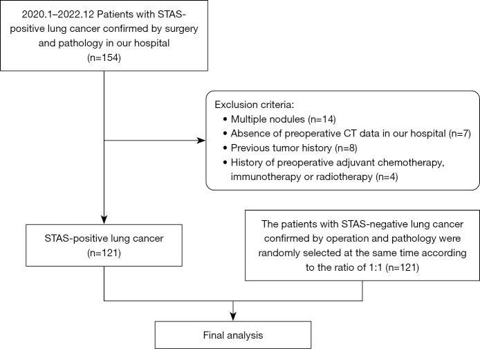
Methods: The data of 121 patients who were positive for STAS and 121 who were negative for STAS as confirmed by surgery and pathology were collected at Jiangsu Cancer Hospital from January 2020 to December 2022. The differences between the two groups in terms of the clinical, pathological, and CT characteristics were compared.
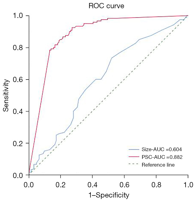
Results: STAS occurred not only in lung adenocarcinoma (LUAD) (106 of 121, 87.6%), but also in other pathological types of lung cancer (15 of 121, 12.4%). STAS was significantly correlated with pathological invasiveness [pathological differentiation, tumor, node, metastasis (TNM) staging, vascular invasion, and pleural invasion; all P<0.05]. STAS was most common in solid tumors (95 of 121, 78.51%). The receiver operating characteristic (ROC) curve showed that the optimal cut-off value for diagnosing STAS based on diameter is 1.55 cm with a sensitivity of 73.3% and a specificity of 47.9%. The percentage of solid components (PSC) is an independent influencing factor of lung cancer with STAS [odds ratio (OR) =111.27; P<0.05] with an optimal cut-off value of 63%, a sensitivity of 92.5%, and a specificity of 72.7%. In the part-solid nodules, the occurrence rate of STAS increased as the PSC increased. STAS was only observed in part-solid nodules with a PSC greater than 25%. Among the CT morphological features, lobulation was an independent influencing factor of lung cancer with STAS (OR =3.513; P<0.05), and persistent indistinct margin ground-glass opacity around the primary lesion of lung cancer (21 of 121, 17.36%) and satellite foci (9 of 121, 7.44%) strongly indicated the existence of STAS.
Conclusions: The clinical, pathological and CT features of STAS may guide clinicians to develop appropriate strategies and improve the survival rate of patients.
Keywords: Lung cancer; computed tomography (CT); ground-glass nodule; influencing factors; spread through air spaces (STAS).
2024 AME Publishing Company. All rights reserved.
Conflict of interest statement
Conflicts of Interest: All authors have completed the ICMJE uniform disclosure form (available at https://tlcr.amegroups.com/article/view/10.21037/tlcr-24-715/coif). The authors have no conflicts of interest to declare.
Figures
Similar articles
-
Predictive value of radiological features on spread through air space in stage cIA lung adenocarcinoma.J Thorac Dis. 2020 Nov;12(11):6494-6504. doi: 10.21037/jtd-20-1820. J Thorac Dis. 2020. PMID: 33282351 Free PMC article.
-
Predictive value of CT and 18F-FDG PET/CT features on spread through air space in lung adenocarcinoma.BMC Cancer. 2024 Apr 8;24(1):434. doi: 10.1186/s12885-024-12220-x. BMC Cancer. 2024. PMID: 38589832 Free PMC article.
-
Lung Adenocarcinoma: CT Features Associated with Spread through Air Spaces.Radiology. 2018 Dec;289(3):831-840. doi: 10.1148/radiol.2018180431. Epub 2018 Sep 4. Radiology. 2018. PMID: 30179108
-
Computed Tomography Features and Tumor Spread Through Air Spaces in Lung Adenocarcinoma: A Meta-analysis.J Thorac Imaging. 2023 Mar 1;38(2):W19-W29. doi: 10.1097/RTI.0000000000000693. Epub 2022 Dec 28. J Thorac Imaging. 2023. PMID: 36583661 Free PMC article.
-
[Research Progress on Spread Through Air Spaces of Lung Cancer].Zhongguo Fei Ai Za Zhi. 2022 Jan 20;25(1):54-60. doi: 10.3779/j.issn.1009-3419.2021.101.49. Epub 2021 Dec 23. Zhongguo Fei Ai Za Zhi. 2022. PMID: 34937151 Free PMC article. Review. Chinese.
Cited by
-
Which descriptor should spread through air spaces (STAS) be incorporated into? T descriptor versus residual tumor classification.J Pathol Clin Res. 2025 Jul;11(4):e70039. doi: 10.1002/2056-4538.70039. J Pathol Clin Res. 2025. PMID: 40689870 Free PMC article.
References
-
- Kadota K, Nitadori JI, Sima CS, et al. Tumor Spread through Air Spaces is an Important Pattern of Invasion and Impacts the Frequency and Location of Recurrences after Limited Resection for Small Stage I Lung Adenocarcinomas. J Thorac Oncol 2015;10:806-14. 10.1097/JTO.0000000000000486 - DOI - PMC - PubMed
-
- Travis WD, Eisele M, Nishimura KK, et al. The International Association for the Study of Lung Cancer (IASLC) Staging Project for Lung Cancer: Recommendation to Introduce Spread Through Air Spaces as a Histologic Descriptor in the Ninth Edition of the TNM Classification of Lung Cancer. Analysis of 4061 Pathologic Stage I NSCLC. J Thorac Oncol 2024;19:1028-51. - PubMed
LinkOut - more resources
Full Text Sources