. 2024 Nov 2:17:7993-8008.
doi: 10.2147/JIR.S481101. eCollection 2024.

Insulin Mediates Lipopolysaccharide-Induced Inflammatory Responses and Oxidative Stress in BV2 Microglia

Affiliations

Insulin Mediates Lipopolysaccharide-Induced Inflammatory Responses and Oxidative Stress in BV2 Microglia

Chi-Chen Huang et al. J Inflamm Res. .

Abstract

Introduction: Insulin, the key hormone for glucose regulation, has garnered attention for its role as an immune modulator. Impaired insulin signaling in the central nervous system is linked to neuroinflammation and neurodegenerative diseases. Microglia, the resident macrophage-like immune cells in the brain, are key regulators of neuroinflammation. However, the mechanisms by which insulin influences microglial immune responses remain relatively unknown.

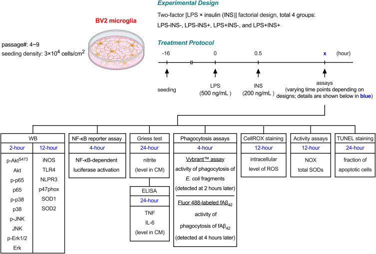
Methods: This study aimed to assess the effects of post-treatment with insulin [30 minutes after lipopolysaccharide (LPS) exposure] on LPS-induced inflammatory responses in BV2 microglial cells.

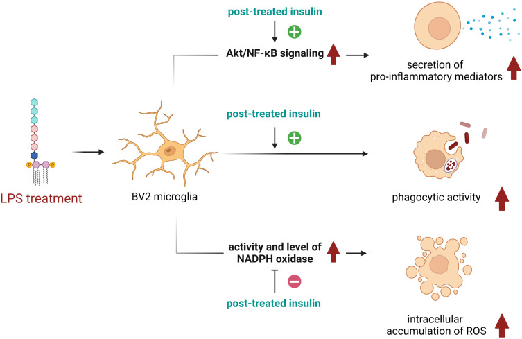
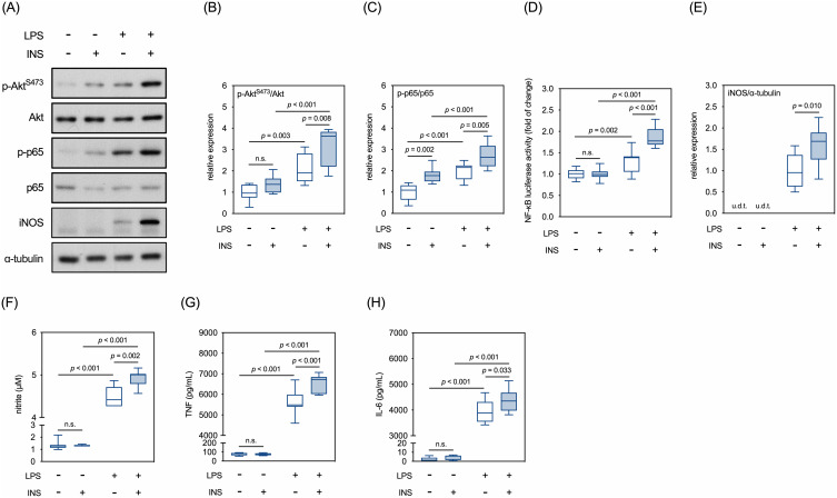
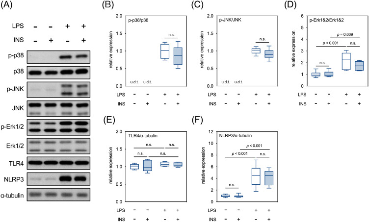
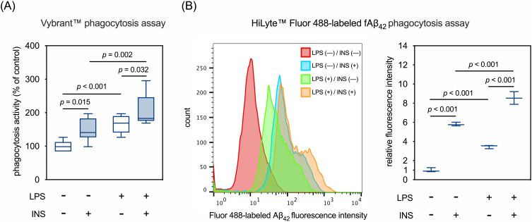
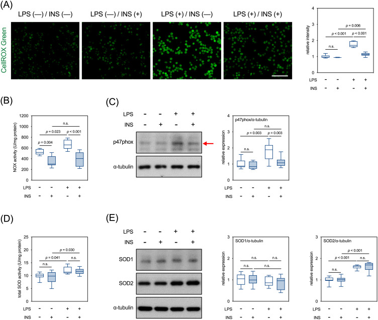
Results: Post-treatment with insulin potentiated LPS-induced production of nitric oxide and pro-inflammatory cytokines, such as TNF and IL-6, through activation of the Akt/NF-κB pathway. Insulin also enhanced the ability of BV2 cells to phagocytose bacteria particles and β-amyloid fibrils. Conversely, insulin inhibited activation of NADPH oxidase and reduced intracellular levels of reactive oxygen species in LPS-treated BV2 cells.

Conclusion: Insulin enhances microglial immune competence when challenged by endotoxins but mitigates oxidative stress in these cells.

Keywords: p47phox; phagocytosis; superoxide dismutase; β-amyloid.


Conflict of interest statement

The authors report no conflicts of interest in this work.

Figures

None
Graphical abstract
Figure 1
Figure 1
Experimental scheme of this study.
Figure 2
Figure 2
Effects of LPS and insulin on Akt/NF-κB signaling and secretions of pro-inflammatory mediators in BV2 microglial cells. (A) Representative Western blots for selected molecules involved in AKT/NF-κB signaling cascades. (B) Quantitative results of relative levels of p-Akt. (C) Quantitative results of relative levels of p-p65. (D) Quantitative results of NF-κB luciferase reporter assay. (E) Quantitative results of relative levels of iNOS. (F) Quantitative results of the Griess assay in the conditioned media. (G) Quantitative results of levels of TNF in the conditioned media. (H) Quantitative results of levels of IL-6 in the conditioned media. u.d.t., under the detective threshold. n.s., not significant. Sample size = 9 biological replicates in Western blots, Griess assay, and ELISA assays; 12 biological replicates in NF-κB luciferase reporter assay. See also Supplementary Table 1 for details of the statistical test results.
Figure 3
Figure 3
Effects of LPS and insulin on TLR4/MAPK signaling in BV2 microglial cells. (A) Representative Western blots for TLR4/MAPK signaling and NLRP3. (B-D) Quantitative results of relative levels of phosphorylated p38, JNK, and Erk1&2. (E and F) Quantitative results of relative levels of TLR4 and NLRP3. u.d.t., under detective threshold. n.s., not significant. Sample size = 9 biological replicates in each assay. See also Supplementary Table 1 for details of the statistical test results.
Figure 4
Figure 4
Effects of LPS and insulin on phagocytic activity of BV2 microglial cells. (A) Quantitative results of Vybrant™ phagocytosis assay. (B) Quantitative results of phagocytosis of HiLyte™ Fluor 488-labeled fAβ42. The left panel showed the overlayed histograms of flow cytometry. The right panel showed the quantitative results of flow cytometry. Sample size = 9 biological replicates in (A); 3 biological replicates in (B). See also Supplementary Table 1 for details of the statistical test results.
Figure 5
Figure 5
Effects of LPS and insulin on intracellular accumulation of ROS in BV2 microglial cells. (A) Representative micrographs and quantitative results of CellROX™ green staining. Scale bar: 100 µm. (B) Quantitative results of NOX activity. (C) Representative Western blots and quantitative results of p47phox. The red arrow indicates the accurate molecular size. (D) Quantitative results of total SOD activity. (E) Representative Western blots and quantitative results of SOD1 and SOD2. n.s., not significant. Sample size = 9 biological replicates in each assay. See also Supplementary Table 1 for details of the statistical test results.
Figure 6
Figure 6
Effects of LPS and insulin on apoptosis in BV2 microglial cells. (A) Representative micrographs of TUNEL staining. Scale bar: 100 µm. (B) Quantitative results of TUNEL staining. n.s., not significant. Sample size = 12 biological replicates. See also Supplementary Table 1 for details of the statistical test results.

Similar articles

Cited by

References

    1. Wang TF, Wu SY, Pan BS, Tsai SF, Kuo YM. Inhibition of nigral microglial activation reduces age-related loss of dopaminergic neurons and motor deficits. Cells. 2022;11(3). doi:10.3390/cells11030481 - DOI - PMC - PubMed
    1. Kiani Shabestari S, Morabito S, Danhash EP, et al. Absence of microglia promotes diverse pathologies and early lethality in Alzheimer’s disease mice. Cell Rep. 2022;39(11):110961. doi:10.1016/j.celrep.2022.110961 - DOI - PMC - PubMed
    1. Heppner FL, Ransohoff RM, Becher B. Immune attack: the role of inflammation in Alzheimer disease. Nat Rev Neurosci. 2015;16(6):358–372. doi:10.1038/nrn3880 - DOI - PubMed
    1. Nimmerjahn A, Kirchhoff F, Helmchen F. Resting microglial cells are highly dynamic surveillants of brain parenchyma in vivo. Science. 2005;308(5726):1314–1318. doi:10.1126/science.1110647 - DOI - PubMed
    1. Hanisch UK, Kettenmann H. Microglia: active sensor and versatile effector cells in the normal and pathologic brain. Nat Neurosci. 2007;10(11):1387–1394. doi:10.1038/nn1997 - DOI - PubMed

LinkOut - more resources