. 2024 Oct 23:15:1469686.
doi: 10.3389/fgene.2024.1469686. eCollection 2024.

Identification of novel 3D-genome altering and complex structural variants underlying retinitis pigmentosa type 17 through a multistep and high-throughput approach

Affiliations

Identification of novel 3D-genome altering and complex structural variants underlying retinitis pigmentosa type 17 through a multistep and high-throughput approach

Suzanne E de Bruijn et al. Front Genet. .

Abstract

Introduction: Autosomal dominant retinitis pigmentosa type 17 (adRP, type RP17) is caused by complex structural variants (SVs) affecting a locus on chromosome 17 (chr17q22). The SVs disrupt the 3D regulatory landscape by altering the topologically associating domain (TAD) structure of the locus, creating novel TAD structures (neo-TADs) and ectopic enhancer-gene contacts. Currently, screening for RP17-associated SVs is not included in routine diagnostics given the complexity of the variants and a lack of cost-effective detection methods. The aim of this study was to accurately detect novel RP17-SVs by establishing a systematic and efficient workflow.

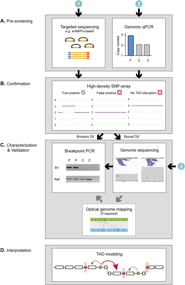
Methods: Genetically unexplained probands diagnosed with adRP (n = 509) from an international cohort were screened using a smMIPs or genomic qPCR-based approach tailored for the RP17 locus. Suspected copy number changes were validated using high-density SNP-array genotyping, and SV breakpoint characterization was performed by mutation-specific breakpoint PCR, genome sequencing and, if required, optical genome mapping. In silico modeling of novel SVs was performed to predict the formation of neo-TADs and whether ectopic contacts between the retinal enhancers and the GDPD1-promoter could be formed.

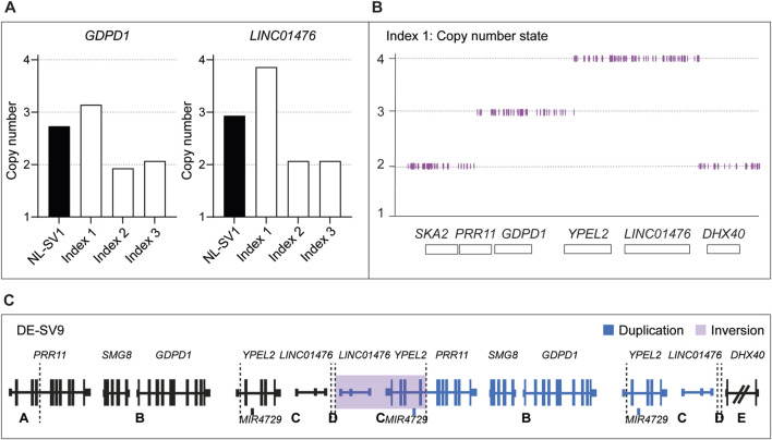
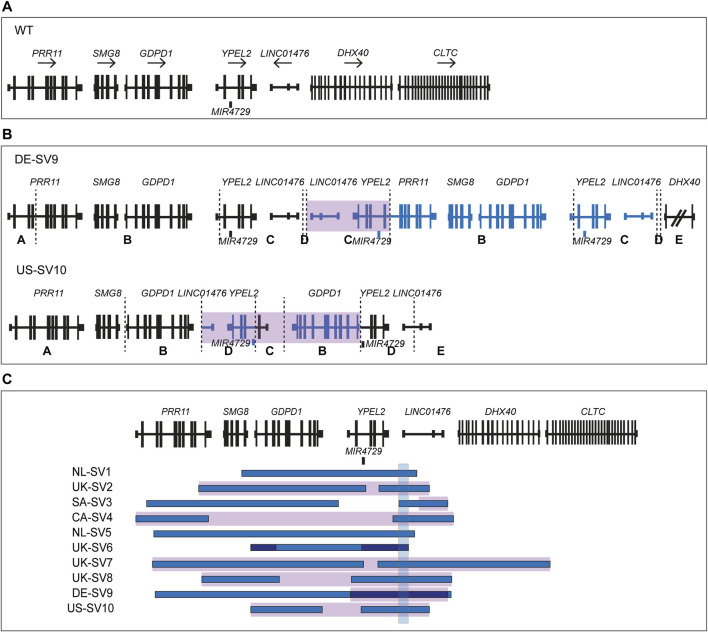
Results: Using this workflow, potential RP17-SVs were detected in eight probands of which seven were confirmed. Two novel SVs were identified that are predicted to cause TAD rearrangement and retinal enhancer-GDPD1 contact, one from Germany (DE-SV9) and three with the same SV from the United States (US-SV10). Previously reported RP17-SVs were also identified in three Australian probands, one with UK-SV2 and two with SA-SV3.

Discussion: In summary, we describe a validated multi-step pipeline for reliable and efficient RP17-SV discovery and expand the range of disease-associated SVs. Based on these data, RP17-SVs can be considered a frequent cause of adRP which warrants the inclusion of RP17-screening as a standard diagnostic test for this disease.

Keywords: gene diagnostics; gene regulation; inherited retinal dystrophies; retinitis pigmentosa; structural variants.


Conflict of interest statement

The authors declare that the research was conducted in the absence of any commercial or financial relationships that could be construed as a potential conflict of interest.

Figures

FIGURE 1
FIGURE 1
Schematic overview of the study design and RP17 screening targets. (A-I) The top panel provides a schematic overview of the RP17 locus including the GDPD1, YPEL2 and LINC01476 genes, a CTCF-enriched boundary element and active retinal enhancer elements that are located in LINC01476. (A-II) To allow efficient screening of copy gains overlapping with the RP17 locus, two sets of genomic qPCR probes were designed that span the exon 3 - intron 3 boundary of GDPD1 and located in intron 2 of LINC01476. (A-III) In addition, 417 smMIPs probes were designed that are distributed over the complete RP17 locus. LINC01476 is only partially displayed in the figure. (B) Overview of previously identified RP17-associated structural variants and duplicated and triplicated genomic regions involved.
FIGURE 2
FIGURE 2
Genomic qPCR detects a novel RP17-SV in German index patient. (A) In total 230 individuals affected with dominant retinitis pigmentosa were screened for copy number changes in the RP17 region by genomic qPCR. Genomic DNA from an individual carrying NL-SV1 was included as a positive control. In one index case of German origin (Index 1), genomic qPCR suggested a possible duplication of GDPD1, and a triplication of LINC01476 that is enriched for retinal enhancer elements. Index 2 and Index 3 were included in the figure as two representative negative samples that were screened by genomic qPCR. (B) qPCR results of index 1 were confirmed by SNP-array genotyping. (C) Since the identified structural variant (SV) did not resemble any of the previously reported RP17-SVs, genome sequencing was performed to determine the breakpoints of the variant. Genome sequencing confirmed the presence of a complex SV, which was termed DE-SV9. Breakpoints are indicated with dashed lines. Blue segments represent duplicated (B, D) or triplicated (C) regions. Inversions are highlighted in purple (segment C). The size of DHX40 is reduced for the purpose of the figure.
FIGURE 3
FIGURE 3
Overview of novel structural variants within the RP17 locus in adRP families. Breakpoints are indicated with dashed lines. Blue segments represent duplicated or triplicated regions, whereas inversions are highlighted in purple. (A) Wildtype (WT) chromosomal localization of the RP17 locus. (B) Novel RP17 structural variants identified in a German adRP family (DE-SV9) and a large US adRP family (US-SV10). (C) Overview of all SV breakpoints identified in the RP17 locus. An overlapping genomic region that is duplicated or triplicated in all reported pathogenic RP17-SVs is highlighted with a blue box (de Bruijn et al., 2020). This region was found to be duplicated or triplicated in all the newly identified SVs (chr17:59,421,853–59,433,404, 11.5 kb). Based on recent data acquired using this approach (Supplementary Results), the structure and nomenclature of UK-SV6 has been revised from the previous report (de Bruijn et al., 2020).
FIGURE 4
FIGURE 4
Schematic of the proposed workflow for the screening and characterization of RP17-SVs. The genetic investigation of RP17-SVs can be divided into three steps. (A) As a first step, prescreening to identify copy number changes can be performed using (Bardienb et al., 1995) genomic qPCR for specific targets in the RP17 locus or alternatively by (den Hollander et al., 1999) targeted sequencing such as smMIPs-based sequencing with probes designed to cover critical regions of the RP17 locus or exome sequencing. (B) Positive cases should be validated by high-density SNP array to discriminate between false- and true-positive variants. For diagnostic facilities that have already implemented (de Bruijn et al., 2020) genome-sequencing as a standard diagnostic test, prescreening steps I and II can be skipped. (C) If the SNP array data or variant calls from the genome sequencing data correspond to that of a known SV, a mutation-specific breakpoint (B1) PCR should be performed to confirm the SV identity. If the data suggest a novel SV, the breakpoints need to be determined and genome sequencing (if not performed yet) should be undertaken to characterize the SV in more detail. If the orientation of the SV cannot be resolved using short-read sequencing data only (e.g., as observed for UK-SV6, Supplementary Results) optical genome mapping or long-read sequencing should be performed to fully characterize the novel variant. (D) Finally, to assess pathogenicity of RP17-SVs the predicted effect the SV has on the TAD landscape of the RP17 locus should be assessed. For a RP17-SV to be pathogenic, the convergent feature is that there has to be a disruption of boundary elements to allow for ectopic contact between the retinal enhancers and the promoter of GDPD1 (Supplementary Figure S7). C, control; P, patient; Ref, Reference gene.

References

    1. Bardienb S., Ebenezer N., Greenberg J., Inglehearn C. F., Bartmann L., Goliath R., et al. (1995). An eighth locus for autosomal dominant retinitis pigmentosa is linked to chromosome 17q. Hum. Mol. Genet. 4 (8), 1459–1462. 10.1093/hmg/4.8.1459 - DOI - PubMed
    1. Chen S., Francioli L. C., Goodrich J. K., Collins R. L., Kanai M., Wang Q., et al. (2024). A genomic mutational constraint map using variation in 76,156 human genomes. Nature 625 (7993), 92–100. 10.1038/s41586-023-06045-0 - DOI - PMC - PubMed
    1. Chen X., Schulz-Trieglaff O., Shaw R., Barnes B., Schlesinger F., Källberg M., et al. (2016). Manta: rapid detection of structural variants and indels for germline and cancer sequencing applications. Bioinformatics 32 (8), 1220–1222. 10.1093/bioinformatics/btv710 - DOI - PubMed
    1. Cherry T. J., Yang M. G., Harmin D. A., Tao P., Timms A. E., Bauwens M., et al. (2020). Mapping the cis-regulatory architecture of the human retina reveals noncoding genetic variation in disease. Proc. Natl. Acad. Sci. U. S. A. 117 (16), 9001–9012. 10.1073/pnas.1922501117 - DOI - PMC - PubMed
    1. de Bruijn S. E., Fiorentino A., Ottaviani D., Fanucchi S., Melo U. S., Corral-Serrano J. C., et al. (2020). Structural variants create new topological-associated domains and ectopic retinal enhancer-gene contact in dominant retinitis pigmentosa. Am. J. Hum. Genet. 107 (5), 802–814. 10.1016/j.ajhg.2020.09.002 - DOI - PMC - PubMed

LinkOut - more resources