

Arthroscopic Management of Juxta-Articular Proximal Tibial Chondroblastoma: A Case Report and Literature Review
- PMID: 39508114
- PMCID: PMC11735375
- DOI: 10.1111/os.14287
Arthroscopic Management of Juxta-Articular Proximal Tibial Chondroblastoma: A Case Report and Literature Review
Abstract
Background: Chondroblastoma is a rare bone tumor that originates from the epiphysis, constitutes around 1% of all primary bone tumors and is recognized for its tendency to exhibit local invasiveness, as well as the possibility of metastasis and recurrence in nearby areas. Currently, the main surgical treatment for chondroblastoma is open surgery, involving excision of the lesion. There are relatively few reports on arthroscopic surgery for the treatment of chondroblastoma. However, open surgical curettage is associated with operation-related trauma and potential for damage to the osteoepiphysis resulting in growth disturbances.
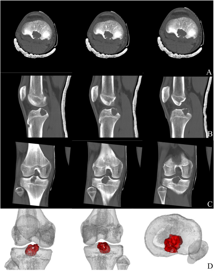
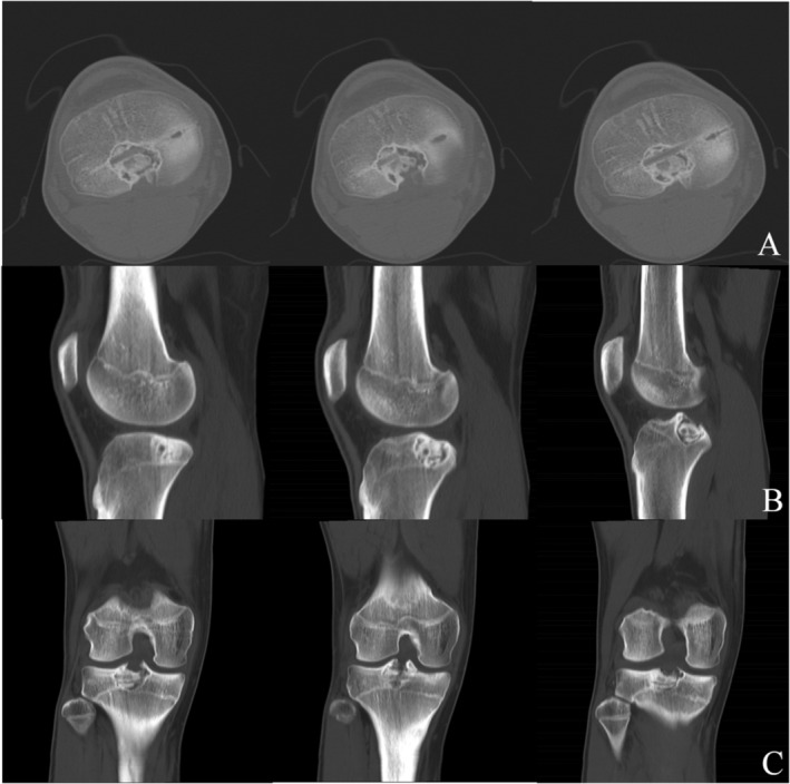
Case presentation: This case study presents the application of an arthroscopic technique in a 14-year-old male patient with chondroblastoma affecting the proximal tibia and tibial eminence. The procedure involved thorough removal of the lesion using direct visualization with the management of the cavity utilizing a substitute for autologous bone graft. After 1 year of follow-up, the patient remains free from symptoms, exhibits normal knee functionality, and radiographic analysis reveals a good autologous bone graft fusion without any signs of recurrence.
Conclusions: Based on the existing cases of arthroscopic treatment for chondroblastoma and the report of this case, arthroscopic treatment for chondroblastoma can be considered as a specific treatment option for certain patients. In some cases, this technique could be an effective alternative to open surgery.
Keywords: arthroscopy; bone substitute; chondroblastoma; proximal tibial.
© 2024 The Author(s). Orthopaedic Surgery published by Tianjin Hospital and John Wiley & Sons Australia, Ltd.
Conflict of interest statement
The authors declare no conflicts of interest.
Figures


References
-
- Chen W. and DiFrancesco L. M., “Chondroblastoma: An Update,” Archives of Pathology & Laboratory Medicine 141, no. 6 (2017): 867–871. - PubMed
-
- Xu H., Nugent D., Monforte H. L., et al., “Chondroblastoma of Bone in the Extremities: A Multicenter Retrospective Study,” Journal of Bone and Joint Surgery American Volume 97, no. 11 (2015): 925–931. - PubMed
-
- Wang J., Du Z., Yang R., Tang X., Yan T., and Guo W., “Analysis for Clinical Feature and Outcome of Chondroblastoma After Surgical Treatment: A Single Center Experience of 92 Cases,” Journal of Orthopaedic Science 27, no. 1 (2022): 235–241. - PubMed
-
- Choi J. H. and Ro J. Y., “The 2020 WHO Classification of Tumors of Bone: An Updated Review,” Advances in Anatomic Pathology 28, no. 3 (2021): 119–138. - PubMed
Publication types
MeSH terms
Grants and funding
LinkOut - more resources
Full Text Sources
Medical
