Review
. 2024 Nov;130(6):1203-1212.
doi: 10.1002/jso.27979. Epub 2024 Nov 7.

Imaging of Peritoneal Surface Malignancies

Affiliations
Review

Imaging of Peritoneal Surface Malignancies

Damiano Caruso et al. J Surg Oncol. 2024 Nov.

Abstract

Management of peritoneal surface malignancies is currently entrusted to a multimodality approach. Computed tomography (CT) scan remains the first imaging method despite the limitations in identifying small implants in critical regions. Magnetic resonance imaging is usually recommended for its performance in small implants, mesentery, and small bowel assessment. Positron emission tomography/CT plays an important role only in pseudomyxoma peritonei. Thus, becoming aware of the imaging strengths and drawbacks and having a multimodality imaging approach might be the best option for the patients.

Keywords: neoplasm metastasis; neoplasms; peritoneum; radiology.


Figures

Figure 1
Figure 1
Thickening and enhancement of peritoneal reflections (white arrowheads) with ascites (white star) (A and B). Thickening of right diaphragm (C) with the scalloping of liver surface (white arrows).
Figure 2
Figure 2
Female, 73‐year old, affected by gastric cancer with peritoneal involvement with micronodular (A, white arrowheads), nodular (B and C, white arrows), and plaque‐like (C, white stars) implants.
Figure 3
Figure 3
Cystic implants of pseudomyxoma peritonei (arrows) deriving from appendiceal tumor (arrowhead) on computed tomography images (A and B) with intraoperative and histological correlations (C and D).
Figure 4
Figure 4
Male, 68‐year old, affected by gastric cancer with stranding and thickening of the omentum (omental cake) (A, white arrowheads), confirmed at surgical inspection (B, white arrowheads).
Figure 5
Figure 5
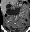
Diffuse thickening of mesentery with involvement of small bowel loop and occlusion (white arrows).
Figure 6
Figure 6
Female, 82‐year old, on computed tomography portal venous phase (A) and magnetic resonance imaging T2w (B), affected by colon cancer with peritoneal carcinomatosis and ascites (white and black stars in A and B, respectively).
Figure 7
Figure 7
Female, 56‐year old, affected by peritoneal mesothelioma with loculated ascites on computed tomography portal venous phase (A) and magnetic resonance imaging T2w (B) (white arrows).
Figure 8
Figure 8
Female, 68‐year old, affected by gastric cancer with calcifications (white arrowheads).
Figure 9
Figure 9
Schematic representation of Peritoneal Cancer Index and Completeness of Cytoreduction score.
Figure 10
Figure 10
Imaging modalities in preoperative setting: Current evidence. CT, computed tomography; 18F‐FDG; 18 F‐2‐deoxy‐2‐fluro‐d‐glucose; MRI, magnetic resonance imaging; PET, positron emission tomography; PMP, pseudomyxoma peritonei; PSMs, peritoneal surface malignancies.

References

    1. Flanagan M., Solon J., Chang K. H., et al., “Peritoneal Metastases from Extra‐Abdominal Cancer ‐ A Population‐Based Study,” European Journal of Surgical Oncology 44 (2018): 1811–1817. - PubMed
    1. Sugarbaker P. H., “Peritonectomy Procedures,” Annals of Surgery 221 (1995): 29–42. - PMC - PubMed
    1. Mogal H. and Shen P., “Top Peritoneal Surface Malignancy Articles from 2022 to Inform Your Cancer Practice,” Annals of Surgical Oncology 31 (2024): 5361–5369. - PubMed
    1. Cortés‐Guiral D., Hübner M., Alyami M., et al., “Primary and Metastatic Peritoneal Surface Malignancies,” Nature Reviews Disease Primers 7 (2021): 91. - PubMed
    1. Grange R., Si‐Mohamed S., Kepenekian V., Boccalini S., Glehen O., and Rousset P., “Spectral Photon‐Counting CT: Hype or Hope for Colorectal Peritoneal Metastases Imaging?” Diagnostic and Interventional Imaging 105 (2024): 118–120. - PubMed

MeSH terms