. 2025 Apr 26;25(6):1259-1279.
doi: 10.17305/bb.2024.11158.

Identification of potential hub genes associated with recurrent miscarriage through combined transcriptomic and proteomic analysis

Affiliations

Identification of potential hub genes associated with recurrent miscarriage through combined transcriptomic and proteomic analysis

Hao-Ran Xu et al. Biomol Biomed. .

Abstract

Recurrent miscarriage (RM) is currently difficult to prevent and treat due to a lack of comprehensive understanding of its molecular mechanisms. The aim of this study was to identify genes potentially involved in the pathogenesis of RM and to observe their expression in the decidual tissues of RM patients. A total of 1823 differentially expressed genes (DEGs) and 148 differentially expressed proteins (DEPs) in decidual tissues between RM and control groups were identified. Subsequently, DCN, DPT, LUM, MFAP4, and ISG15 were identified from the DEGs/DEPs as RM-related hub genes through systematic bioinformatics analysis. Bioinformatics analysis of the single-cell dataset GSE214607 revealed that the expression of these five hub genes in the decidual stromal cells (DSCs) of RM patients appeared to be upregulated, while the RT-qPCR assay showed that their decidual expression levels were significantly increased in RM patients. Uterine Isg15 expression was significantly increased, whereas the uterine expression of Dcn, Dpt, Lum, and Mfap4 was decreased in LPS-induced early pregnancy loss (EPL) mice. MiR-16-5p, -21-3p, -27a-3p, and -941 were identified as potentially involved in the regulation of these five hub genes, and their decidual expression levels were significantly decreased in RM patients. The abnormally increased ISG15 expression in the decidual tissues of RM patients and uterine tissues of LPS-induced mice was validated by WB analysis. ISG15 expression was significantly reduced during the in vitro decidualization of human endometrial stromal cells (hESCs). Collectively, DCN, DPT, LUM, MFAP4, and ISG15 were identified as RM-related hub genes, and their expression in the decidual tissues of RM patients was significantly increased. The decidualization of hESCs was accompanied by reduced ISG15 expression, suggesting that increased decidual ISG15 expression might lead to EPL by disrupting the decidualization process.


Conflict of interest statement

Conflicts of interest: Authors declare no conflicts of interest.

Figures
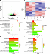
Figure 1.
Figure 1.
DEGs in decidual tissues of RM patients screened through RNA-seq analysis. (A) The volcano plot of DEGs between RM patients (RM, n ═ 3) and normal early pregnant women (Control, n ═ 3). Red dots represent upregulated genes in the RM group, green dots represent downregulated genes, and black dots represent genes with no significant differences between the RM and Control groups. (B) Heatmap clustering of DEGs between the RM and Control groups. Red indicates upregulation in the corresponding group, while blue indicates downregulation. RM: Decidual tissues of RM patients; Control: Decidual tissues of normal early pregnant women. (C) Top ten pathways associated with DEGs enriched by KEGG analysis. (D) Top ten BP associated with DEGs enriched by GO analysis. (E) Top ten CC associated with DEGs enriched by GO analysis. (F) Top ten MF associated with DEGs enriched by GO analysis. DEG: Differentially expressed gene; BP: Biological process; CC: Cellular component; MF: Molecular function; RM: Recurrent miscarriage; GO: Gene ontology; RNA-seq: RNA sequencing.
Figure 2.
Figure 2.
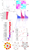
DEPs in decidual tissues of RM patients identified through iTRAQ analysis. (A) The volcano plot of DEPs between RM patients (RM group, n ═ 3) and normal early pregnant women (Control group, n ═ 3). Red dots represent upregulated proteins in the RM group, blue dots represent downregulated proteins, and gray dots represent proteins with no significant differences between the RM and Control groups. (B) Heatmap clustering of DEPs between the RM and Control groups. Red indicates upregulation in the corresponding group, while blue indicates downregulation. RM: Decidual tissues of RM patients (n ═ 3); Control: Decidual tissues of normal early pregnant women (n ═ 3). (C) Top ten pathways associated with DEPs enriched by KEGG analysis. (D) Top ten BP, CC, and MF associated with DEPs. (E) All pathways associated with DEPs enriched by GSEA analysis. (F) Top ten BP, CC, and MF items associated with DEPs enriched by GSEA analysis. (G) (a) represents the top ten hub proteins identified using the MCC algorithm; (b) and (c) represent 12 hub proteins identified using the two highest-scoring modules obtained with the MCODE plugin. BP: Biological process; CC: Cellular component; MF: Molecular function; RM: Recurrent miscarriage; GSEA: Gene set enrichment analysis; DEP: Differentially expressed protein; MCC: Maximum clique centrality.
Figure 3.
Figure 3.
Identification of RM-related hub genes through integrative analyses of DEGs and DEPs in decidual tissues of RM patients. (A) Nine-quadrant graph of correlation analysis from the transcriptomic and proteomic data of decidual tissues of RM patients and normal early pregnancies; (B) Top five pathways associated with both DEGs and DEPs in the decidual tissues of RM patients enriched by KEGG analysis; (C) Pathways associated with both DEGs and DEPs in the decidual tissues of RM patients enriched by GSEA analysis; (D) Venn diagram showing the identification of RM-related hub genes. MCC: Top 10 hub proteins identified from DEGs and DEPs using the MCC algorithm; MCODE: Top 12 hub proteins identified using the MCODE plugin; RNA_Protein: A total of 26 proteins common to both DEGs and DEPs; (E) TF network associated with the RM-related five hub genes using the JASPAR database; (F) Interaction network diagram of potential upstream microRNAs of the RM-related five hub genes using the miRNet database; (G) Top ten proposed drugs targeting RM-related hub genes identified using the DsigDB database. RM: Recurrent miscarriage; GSEA: Gene set enrichment analysis; DEP: Differentially expressed protein; DEG: Differentially expressed gene; TF: Transcription factor; MCC: Maximum clique centrality.
Figure 4.
Figure 4.
Expression patterns of RM-related hub genes in various cell populations between RM patients and normal early pregnant women using the single-cell dataset GSE214607. (A) UMAP visualization of 16 cell clusters contained in human decidual tissues during early pregnancy; (B) UMAP plot of nine cell types contained in human decidual tissues during early pregnancy; (C/E/G/I/K) Expression profiles of ISG15/DCN/LUM/MFAP4/DPT in different cell types of human decidua during early pregnancy displayed by UMAP plot; (D/F/H/J/L) Expression profiles of ISG15/DCN/LUM/MFAP4/DPT in different cell types displayed by violin plot. DSC: Decidual stromal cell; EVT: Extravillous trophoblast; CTB: Cytotrophoblast; RM: Recurrent miscarriage patients (n ═ 3); NC: Normal early pregnant women (n ═ 5).
Figure 5.
Figure 5.
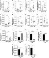
Expression levels of five hub genes and four upstream miRNAs in human decidual tissues and mouse uterine tissues detected by qPCR analysis. (A–E) The relative expression levels of ISG15, DCN, LUM, MFAP4, and DPT in human decidual tissues during early pregnancy; (F–I) The relative expression levels of miR-27a-3p, miR-16-5p, miR-21-3p, and miR-941 in human decidual tissues during early pregnancy; (J–N) The relative expression levels of Isg15, Dcn, Lum, Mfap4, and Dpt in mouse uterine tissues at day 7.5 of pregnancy (vaginal plug ═ day 0.5 of pregnancy). RM: Recurrent miscarriage patients (n ═ 9); Control: Normal early pregnant women (n ═ 9); 0 h: Day 7.5 pregnant mice without LPS treatment (n ═ 4); 24 h: Day 7.5 pregnant mice treated with LPS (0.25 mg/kg) at day 6.5 of pregnancy (n ═ 3); *P < 0.05, **P < 0.01, ***P < 0.001, ****P < 0.0001. qPCR: Quantitative PCR.
Figure 6.
Figure 6.
Expression levels of ISG15 in human decidual tissues, mouse uterine tissues, and T-HESC cells as analyzed by Western blot and RT-qPCR. (A) A representative Western blot image showing ISG15 protein levels in mouse uterine tissues. Control: Mice at day 7.5 of pregnancy without LPS treatment; LPS 1-3: Mice at day 7.5 of pregnancy treated with LPS (0.25 mg/kg) at day 6.5 of pregnancy. (B) A representative Western blot image showing ISG15 protein levels in human decidual tissues during early pregnancy. Control 1-4: Normal early pregnant women; RM 1-4: RM patients. (C) Relative uterine expression level of ISG15 protein in mice. Control: Mice at day 7.5 of pregnancy without LPS treatment (n ═ 4); LPS: Mice at day 7.5 of pregnancy treated with LPS (0.25 mg/kg) at day 6.5 of pregnancy (n ═ 3). (D) Relative expression level of ISG15 protein in human decidual tissues. Control: Normal early pregnant women (n ═ 9); RM: Recurrent miscarriage patients (n ═ 9). (E/G/F) Expression levels of PRL/ISG15/IGFBP1 mRNAs in T-HESCs. (H) A representative Western blot image showing ISG15 protein levels in T-HESCs. (I) Relative expression level of ISG15 protein during in vitro decidualization of T-HESCs. D0: Non-induced decidualization; D2: 48 h after induced decidualization with E2, MPA, and cAMP; D4: 96 h after induced decidualization with E2, MPA, and cAMP; *P < 0.05, **P < 0.01, ***P < 0.001, ****P < 0.0001; Cell experiments were independently repeated at least three times in triplicate (n ═ 3×2). RT-qPCR: Real-time quantitative PCR.
Figure S1.
Figure S1.
(A) Represents the top five upregulated pathways after GSEA analysis; (B) Represents the top five downregulated pathways after GSEA analysis; (C) Represents the top ten BP items after GSEA analysis; (D) Represents the top ten CC items after GSEA analysis; represents the top ten MF items after GSEA analysis. MF: Molecular function; GSEA: Gene set enrichment analysis; CC: Cellular component; BP: Biological process.
Figure S2.
Figure S2.
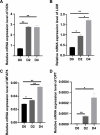
(A) Represents the relative expression level of DCN during in vitro decidualization of T-HESCs; (B) Represents the relative expression level of LUM during in vitro decidualization of T-HESCs; (C) Represents the relative expression level of MFAP4 during in vitro decidualization of T-HESCs; (D) Represents the relative expression level of DPT during in vitro decidualization of T-HESCs. *P < 0.05, **P < 0.01; Cell experiments were independently repeated at least three times in triplicate (n ═ 3×2). D0: Non-induced decidualization; D2: 48 h after induced decidualization with a combined treatment of E2, MPA, and cAMP; D4: 96 h after induced decidualization with a combined treatment of E2, MPA, and cAMP; DCN: Decorin; LUM: Lumican; MFAP4: Microfibrillar-associated protein 4; DPT: Dermatopontin.

Similar articles

References

    1. Quenby S, Gallos ID, Dhillon-Smith RK, Podesek M, Stephenson MD, Fisher J, et al. Miscarriage matters: the epidemiological, physical, psychological, and economic costs of early pregnancy loss. Lancet. 2021;397(10285):1658–67. https://doi.org/10.1016/S0140-6736(21)00682-6. - PubMed
    1. Pan HT, Ding HG, Fang M, Yu B, Cheng Y, Tan YJ, et al. Proteomics and bioinformatics analysis of altered protein expression in the placental villous tissue from early recurrent miscarriage patients. Placenta. 2018;61:1–10. https://doi.org/10.1016/j.placenta.2017.11.001. - PubMed
    1. Davalieva K, Terzikj M, Bozhinovski G, Kiprijanovska S, Kubelka-Sabit K, Plaseska-Karanfilska D. Comparative proteomics analysis of decidua reveals altered RNA processing and impaired ribosome function in recurrent pregnancy loss. Placenta. 2024;154:28–37. https://doi.org/10.1016/j.placenta.2024.06.005. - PubMed
    1. Sun F, Cui L, Qian J, Li M, Chen L, Chen C, et al. Decidual stromal cell Ferroptosis associated with abnormal iron metabolism is implicated in the pathogenesis of recurrent pregnancy loss. Int J Mol Sci. 2023;24(9):7836. https://doi.org/10.3390/ijms24097836. - PMC - PubMed
    1. Zhao X, Jiang Y, Luo S, Zhao Y, Zhao H. Intercellular communication involving macrophages at the maternal-fetal interface may be a pivotal mechanism of URSA: a novel discovery from transcriptomic data. Front Endocrinol (Lausanne) 2023;14:973930. https://doi.org/10.3389/fendo.2023.973930. - PMC - PubMed