. 2024 Nov 7;19(11):e0308974.
doi: 10.1371/journal.pone.0308974. eCollection 2024.

Effect of different running protocols on bone morphology and microarchitecture of the forelimbs in a male Wistar rat model

Affiliations

Effect of different running protocols on bone morphology and microarchitecture of the forelimbs in a male Wistar rat model

Andy Xavier et al. PLoS One. .

Abstract

Background: It is accepted that the metabolic response of bone tissue depends on the intensity of the mechanical loads, but also on the type and frequency of stress applied to it. Physical exercise such as running involves stresses which, under certain conditions, have been shown to have the best osteogenic effects. However, at high intensity, it can be deleterious for bone tissue. Consequently, there is no clear consensus as to which running modality would have the best osteogenic effects.

Aim: Our objective was to compare the effects of three running modalities on morphological and micro-architectural parameters on forelimb bones.

Methods: Forty male Wistar rats were randomly divided into four groups: high intensity interval training (HIIT), continuous running, combined running ((alternating HIIT and continuous modalities) and sedentary (control). The morphometry, trabecular microarchitecture and cortical porosity of the ulna, radius and humerus were analyzed using micro-tomography.

Results: All three running modalities resulted in bone adaptation, with an increase in the diaphyseal diameter of all three bones. The combined running protocol had positive effects on the trabecular thickness in the distal ulna. The HIIT protocol resulted in an increase in both medio-lateral diameter and cortical bone area over total area (Ct.Ar/Tt.Ar) at the ulnar shaft compared with sedentary condition. Moreover, the HIIT protocol decreased the mean surface area of the medulla (Ma.Ar) according to sedentary condition at the ulnar shaft.

Conclusion: This study has shown that HIIT resulted in a decrease in trabecular bone fraction in favor of cortical bone area at the ulna.


Conflict of interest statement

The authors have declared that no competing interests exist.

Figures
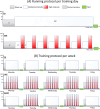
Fig 1
Fig 1. Exercise protocols.
(A) During a day, rats, according to their group, either practiced continuous running (CR), or high intensity interval training (HIIT). (B) Organization of the 5 training days per week according to running protocols. CR: Continuous Running group; HIIT: High Intensity Interval Training group; ComR: Combined Running group (i.e. alternating between the continuous and the HIIT running).
Fig 2
Fig 2. Example of the subchondral bone selection on a cross-section of the distal ulna using Dragonfly software.
The area colored in blue corresponds to the subchondral bone selected according to the grey level. The yellow line corresponds to the measurement of the thickness of the subchondral bone.
Fig 3
Fig 3. Illustration of the main differences in ulna morphology between running groups, based on morphometric and microarchitecture analyses.
Images acquired with Bruker SkyScan 1176 (Kontich, Belgium) and analyzed by DragonFly software (version 2022.2 Build 1399). The upper images represent the three-dimensional reconstruction of the ulna at mid-diaphysis. The upper line of table corresponds to the differences in the analysis of the cortical microarchitecture (Ct.Ar/Tt.Ar: Cortical Area/Total Area) of the ulna at the mid-diaphysis. The lower images represent the ulnar cortices at mid-diaphysis. The lower line of table corresponds to the morphometric measurements of the ulnar cortical at mid-diaphysis (the arrows correspond to the significant difference compared to the SED group: green arrow: medio-lateral diameter, blue arrow: medio-lateral diameter of the medullary canal). SED: Sedentary group; HIIT: High Intensity Interval Training group; CR: Continuous Running group; ComR: Combined Running group. The statistically significant differences (*p-value<0.05) are summarized in Tables 2 and 3.
Fig 4
Fig 4. Subchondral bone thickness in the distal radius and ulna.
Measurements were performed with Dragonfly software from μCT images. Values are expressed in mm. *: p-value < 0.05; ***: p-value < 0.001. SED: Sedentary group (green); HIIT: High Intensity Interval Training group (red); CR: Continuous Running group (blue); ComR: Combined Running group (yellow).
Fig 5
Fig 5. Summary schematic of the main statistically significant results of the effects of the three running modalities on the upper limbs compared with the sedentary group.
(humerus in red, radius in blue, ulna in green); HIIT: High Intensity Interval Training group. ⬊: Decrease; ⬈: Increase; Ct.Po: Cortical Porosity; Po.N: Pore Number; Po.Dm: Pore diameter; BV/TV: Bone Volume/Tissue Volume; SB.Th: Subchondral Bone Thickness.

References

    1. Barnsley J, Buckland G, Chan PE, et al. Pathophysiology and treatment of osteoporosis: challenges for clinical practice in older people. Aging Clinical and Experimental Research. 2021;33:759–73. doi: 10.1007/s40520-021-01817-y - DOI - PMC - PubMed
    1. Marques EA, Mota J, Carvalho J. Exercise effects on bone mineral density in older adults: a meta-analysis of randomized controlled trials. Age (Dordrecht, Netherlands). 2012;34:1493–515. doi: 10.1007/s11357-011-9311-8 - DOI - PMC - PubMed
    1. Bonewald LF. Osteocytes as dynamic multifunctional cells. Annals of the New York Academy of Sciences. 2007;1116:281–90. doi: 10.1196/annals.1402.018 - DOI - PubMed
    1. Weatherholt AM, Fuchs RK, Warden SJ. Cortical and trabecular bone adaptation to incremental load magnitudes using the mouse tibial axial compression loading model. Bone. 2013;52:372–9. doi: 10.1016/j.bone.2012.10.026 - DOI - PMC - PubMed
    1. Wolff J. The Law of Bone Remodelling. The Law of Bone Remodelling. Published Online First: 1986. doi: 10.1007/978-3-642-71031-5 - DOI

LinkOut - more resources