. 2024 Nov 7;20(11):e1012610.
doi: 10.1371/journal.ppat.1012610. eCollection 2024 Nov.

A major role of class III HD-ZIPs in promoting sugar beet cyst nematode parasitism in Arabidopsis

Affiliations

A major role of class III HD-ZIPs in promoting sugar beet cyst nematode parasitism in Arabidopsis

Xunliang Liu et al. PLoS Pathog. .

Abstract

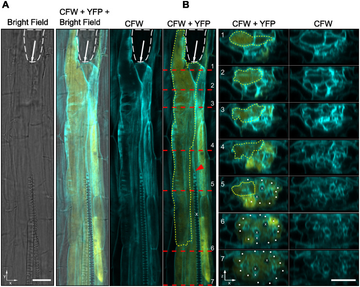
Cyst nematodes use a stylet to secrete CLE-like peptide effector mimics into selected root cells of their host plants to hijack endogenous plant CLE signaling pathways for feeding site (syncytium) formation. Here, we identified ATHB8, encoding a HD-ZIP III family transcription factor, as a downstream component of the CLE signaling pathway in syncytium formation. ATHB8 is expressed in the early stages of syncytium initiation, and then transitions to neighboring cells of the syncytium as it expands; an expression pattern coincident with auxin response at the infection site. Conversely, MIR165a, which expresses in endodermal cells and moves into the vasculature to suppress HD-ZIP III TFs, is down-regulated near the infection site. Knocking down HD-ZIP III TFs by inducible over-expression of MIR165a in Arabidopsis dramatically reduced female development of the sugar beet cyst nematode (Heterodera schachtii). HD-ZIP III TFs are known to function downstream of auxin to promote cellular quiescence and define stem cell organizer cells in vascular patterning. Taken together, our results suggest that HD-ZIP III TFs function together with a CLE and auxin signaling network to promote syncytium formation, possibly by inducing root cells into a quiescent status and priming them for initial syncytial cell establishment and/or subsequent cellular incorporation.


Conflict of interest statement

The authors have declared that no competing interests exist.

Figures

Fig 1
Fig 1. Identification of potential genes downstream of CLE signaling upon BCN infection.
A. Example of excised root segments used for RNA sequencing. Approximately 8 mm long root segments spanning BCN infection sites and corresponding uninfected or HsCLE2p-treated root tissues were cut under a stereoscope and immediately frozen in liquid nitrogen for RNA isolation. B. Criteria used for filtering CLE downstream genes. Genes (147) that fit these four criteria were considered as CLE downstream genes, 1). down-regulated in the clv triple mutant compared to wild-type in control sample; 2). Up-regulated by HsCLE2p treatment in wild-type seedlings, but not in the clv triple mutant (3); 4). Up-regulated by BCN infection in wild-type seedlings. C. Heatmap showing expression profiles of the 147 candidate genes positively regulated by CLE signaling upon BCN infection. Expressions of ATHB8 and WOX4 are denoted with red arrows. D. Expression of ATHB8 gene in the RNAseq dataset.E. qPCR validation of ATHB8 gene expression in wild-type and clv triple mutant upon HsCLE2p treatment and BCN infection. Letter above each bar represents statistical groups of Tukey’s HSD test following ANOVA analysis.
Fig 2
Fig 2. Expression of ProATHB8::GUS in the BCN infection site.
ProATHB8::GUS expression at different stages of syncytium development in wild-type. bar = 200 μm. J2, second-stage juvenile; J3, third-stage juvenile; S, syncytium.
Fig 3
Fig 3. Expression of ProATHB8::4xYFP and ProMIR165a::GFP at BCN infection sites in Arabidopsis roots.
A. Continuous monitoring of ProATHB8::4xYFP expression in early stages of BCN infection. Red brackets indicate increased YFP signal at early stages of BCN infection. Insets represent YFP signal of root segment about 850 μm away from the infection site. YFP signal intensity at 3 dpi was quantified at the syncytium (s, red line) or adjacent site (a, cyan line). B. Continuous monitoring of ProMIR165a::GFP in early stages of BCN-infected roots. Left panel of 2 dpi, and insets of 3, 4, and 5 dpi panels represent GFP signal of root segment adjacent to the infection site. GFP signal intensity at 3 dpi was quantified at the syncytium (s, red line) and adjacent site (a, cyan line). Red arrowhead, position of nematode head. bar = 200 μm.
Fig 4
Fig 4. Loss of function of ATHB8 and its closest homologue ATHB15/CNA do not affect BCN infection or syncytia development in Arabidopsis.
A. 7-day-old seedlings of athb8-11, cna-2, and athb8-11 can-2 grown on a vertical plate. bar = 1 cm. B. Measurement of root length shown in (A). C. 14-day-old seedlings grown in 12-well plates. Top panel, top view of representative seedlings. Bottom panel, root system of representative 14-day-old seedlings grown in 12-well plates and scooped out for photograph. bar = 1 cm. D. BCN penetration rate on the tested mutants, counted at 3–5 days post inoculation. E. Number of adult females developed on the tested mutants. F. Syncytium size developed on the tested mutants. All bar graphs represent mean ± SE. Dots represent each individual measurement. Samples sizes were shown in parentheses in each bar. Statistical tests were performed with Wald test following generalized linear mixed-effect model (for penetration and infection data) or linear mixed-effect model (for syncytium size). Data were repeated twice with similar results (S16 Fig).
Fig 5
Fig 5. Inducible expression of MIR165a suppresses BCN infection in Arabidopsis.
A. Induction of MIR165a suppressed expression of HD-ZIP III TFs in 12-well plates. Top panel, 48 hours post-induction. Bottom panel, 72 hours post-induction. Letter above each bar represents statistical group of Tukey’s HSD test following ANOVA analysis. B. Expression of HD-ZIP III TFs 3–14 days post induction of MIR165a with 5 μM estradiol (E2). Effect of MIR165a induction attenuates over time.C. Suppression of HD-ZIP III TFs did not affect nematode penetration into the root. Penetration rate counted at 5 dpi. D. Suppression of HD-ZIP III TFs suppressed BCN female development on Arabidopsis roots. E. Suppression of HD-ZIP III TFs resulted in smaller syncytia size. Bar graph represents mean ± SE. Dots represent each individual measurement. Sample sizes were shown in parentheses in each bar. Statistical tests were performed with the Wald test following a generalized linear mixed-effect model (for penetration and infection data) or linear mixed-effect model (for syncytium size). Infection assays were repeated four times with similar results (S17 Fig). Four reps of syncytium size data were unified using biological replications as a block effect to build the linear mixed-effect model. Only statistically significant results were labeled on the graph. E2, estradiol.
Fig 6
Fig 6. Effect of overexpressing ATHB8 on cyst nematode infection in Arabidopsis.
A. Expanded ATHB8d-YFP expression domain after introduction to Pro35SiGUS and ProG1090iGUS lines. 5-day-old seedlings were induced with 5 μM estradiol for 24 hours before imaging. bar = 100 μm. B. Expression of HD-ZIP III genes in 12-well plates after estradiol (5 μM) induction. C-E. Effect of ATHB8d overexpression on BCN penetration (C), female development (D), and syncytia size (E) in Arabidopsis root. Bar graph represents mean ± SE. Dots represent each individual measurement. Sample sizes were shown in parentheses in each bar. Statistical tests were performed with the Wald test following a generalized linear mixed-effect model (for penetration and infection data) or a linear mixed-effect model (for syncytium size). Data shown in C–E were repeated twice with similar results (S19 Fig).
Fig 7
Fig 7. Expression of ProATHB8::4xYFP in the BCN induced syncytium at 1 dpi.
A. A confocal optical section of developing syncytium with ProATHB8::4xYFP expression. B. Optical cross section of positions shown in (A). White dashed line, outline of nematode head. White arrow, position of the stylet. Red dashed line, positions of cross section shown in (B). Yellow dashed line, outline of the syncytium. x, xylem cells. Red arrowhead, disrupted xylem differentiation. White dot, pericycle cells. CFW, calcofluor white staining of cell wall. YFP, yellow fluorescent signal. bar = 20 μm.
Fig 8
Fig 8. Distribution of ProATHB8::4xYFP activity at BCN infection site at 3 dpi.
White dashed line, outline of nematode. Red box, zoomed in portion in (B). Red dashed lines in (B), position of optical cross sections shown in (C). Yellow dashed line, outline of the syncytium. x, xylem cells. x’, ectopic xylem cells. White dot, pericycle cells. CFW, calcofluor white staining of cell wall. YFP, yellow fluorescent signal. bar = 100 μm.

References

    1. Anjam MS, Shah SJ, Matera C, Rozanska E, Sobczak M, Siddique S, et al.. Host factors influence the sex of nematodes parasitizing roots of Arabidopsis thaliana. Plant Cell Environ. 2020;43(5):1160–74. - PubMed
    1. Golinowski W, Grundler FMW, Sobczak M. Changes in the structure of Arabidopsis thaliana during female development of the plant-parasitic nematode Heterodera schachtii. Protoplasma. 1996;194(1):103–16.
    1. Sobczak M, Golinowski W. Structure of cyst nematode feeding sites. In: Berg RH, Taylor CG, editors. Cell Biology Of Plant Nematode Parasitism. Berlin, Heidelberg: Springer Berlin Heidelberg; 2009. p. 153–87.
    1. Holtmann B, Kleine M, Grundler FMW. Ultrastructure and anatomy of nematode-induced syncytia in roots of susceptible and resistant sugar beet. Protoplasma. 2000;211(1):39–50.
    1. Hewezi T, Baum TJ. Manipulation of plant cells by cyst and root-knot nematode effectors. Mol Plant Microbe Interact. 2013;26(1):9–16. doi: 10.1094/MPMI-05-12-0106-FI - DOI - PubMed

MeSH terms

LinkOut - more resources