. 2024 Nov 7;19(11):e0307153.
doi: 10.1371/journal.pone.0307153. eCollection 2024.

3-nitropyridine analogues as novel microtubule-targeting agents

Affiliations

3-nitropyridine analogues as novel microtubule-targeting agents

Jean Herman et al. PLoS One. .

Abstract

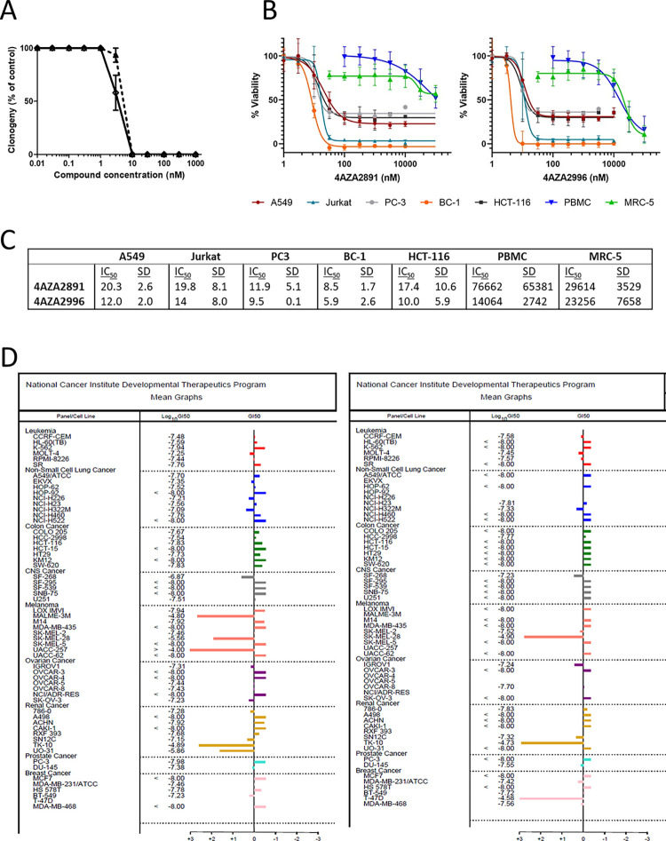
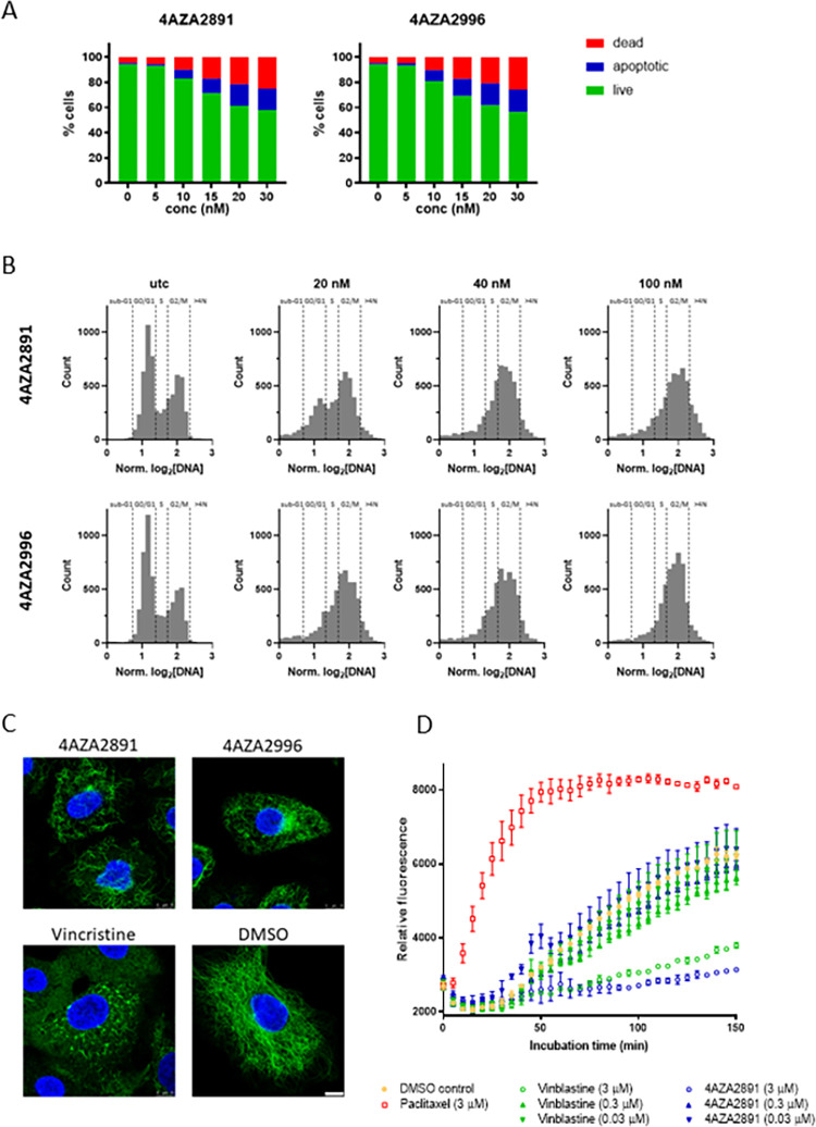
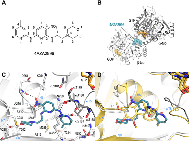
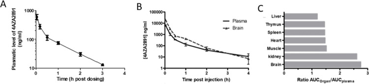
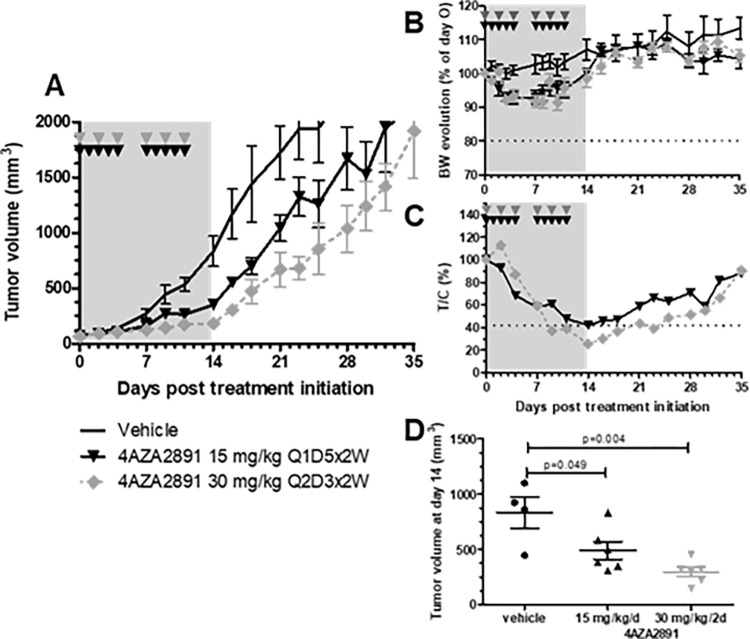
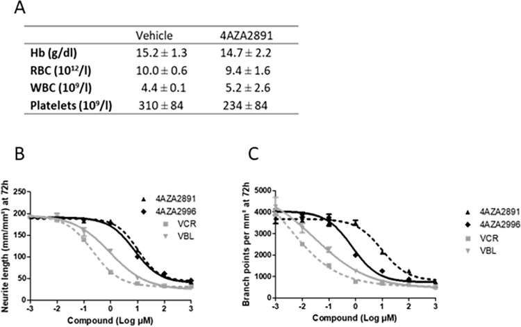
Microtubule-targeting agents are an important class of anti-cancer drugs; their full potential is however not realized because of significant myelotoxicity and neurotoxicity. We here report 3-nitropyridine analogues as a novel group of microtubule-targeting agents with potent anti-cancer effects against a broad range of cancer types. We show that these 3-nitropyridines induce cell cycle arrest in the G2-M phase and inhibit tubulin polymerization by interacting with tubulin. Determination of the tubulin-4AZA2996 structure by X-ray crystallography demonstrated that this class of compounds binds to the colchicine-site of tubulin. Furthermore, the anti-cancer effect was demonstrated both in vitro and in vivo in a murine heterotopic xenograft model of colon cancer. When administered intravenously, 4AZA2891 effectively inhibited cancer growth. Whereas 3-nitropyridine compounds do not induce myelotoxicity at pharmacological doses, the neurotoxicity associated with microtubule-targeting agents is still present.


Conflict of interest statement

The authors have declared that no competing interests exist.

Figures

Fig 1
Fig 1. Inhibition of the growth of cancer cell lines.
(A) Survival of HT-29 cells exposed to increasing concentrations of 4AZA2891 (dashed line) and 4AZA2996 (solid line) in a clonogenic assay. HT-29 cells were seeded and compounds were added 24 hours later. After 6 days of incubation, cells were fixed with methanol and stained with Trypan blue. Colonies were counted and numbers were normalized to the number of colonies in the non-treated wells. The results are expressed as mean ± SEM of three independent experiments. (B) 4AZA2891 and 4AZA2996 induce cytotoxicity in hematological and solid cancer cell lines while sparing normal cells. Cells were treated with different concentrations of compound, and 72 hours later viability was measured by MTS assay or in the case of PBMC by annexin V/PI flow cytometry. Datapoints are mean ± SD (n = 2–4). (C) IC50 values (nM) as calculated from the linear portions of the log dose response curves depicted in Fig 1B. Numbers represent average values of two to four experiments ± S.D. (D) 4AZA2891 (left) and 4AZA2996 (right) were assessed in the NIH panel of cancer cell lines at 5 concentrations and the GI50 was determined for each cell line from the dose-response curves. The mean GI50 for each compound across all cell lines was calculated and is represented as a vertical line in the graphs. This mean is assigned a value of zero and all the Gi50’s of cell lines are plotted relative to it. The bars representing cell lines that require concentrations higher than the mean for inhibition point to the left; those representing cell lines that are more sensitive to the compound point to the right. The mean GI50 (log10) for 4AZA2891 is -7.45 molar and for 4AZA2996–7.66 molar.
Fig 2
Fig 2. Mechanism of action of the 3-nitropyridine compounds.
(A) 4AZA2891 and 4AZA2996 induce apoptosis in Jurkat cells. Apoptosis was measured by annexin V/PI staining. Cells were treated with different concentrations of compound or carrier (DMSO) and 24 hours later apoptotic and dead cells were stained and subsequently analyzed by flow cytometry. (B) 4AZA2891 and 4AZA2996 induce G2 cell cycle arrest in Jurkat cells. Cells were treated with different concentrations of compound or carrier (DMSO) for 24 hours and then stained with DAPI cell cycle profiling by high content imaging. Both compounds cause a clear reduction of the G0/G1 while the G2 as well as the subG1 population increases. (C) 4AZA2891 and 4AZA2996 disintegrate the microtubule network. Immunofluorescence staining of alpha-tubulin in A549 cells treated for 4 hours with 250 nM of compound 4AZA2891, 4AZA2996 or reference compound vincristine. Green: α-tubulin, blue: DAPI. Scale bar: 10 μm. (D) 4AZA2891 dose-dependently inhibits tubulin polymerization in vitro. The polymerization of purified tubulin is measured by the increase in fluorescence due to the incorporation of a fluorescent reporter into microtubules as polymerization proceeds. 4AZA2891 dose-dependently causes, as vinblastine, a marked decrease in final polymer formation as compared to the DMSO control and in contrast to the effect observed for tubulin stabilizing reference compound paclitaxel.
Fig 3
Fig 3. Co-crystallization of 4AZA2996 with tubulin.
(A) Chemical structure of 4AZA2996 (N6-(4-methylpyridin-2-yl)-N2-(2-morpholinoethyl)-3-nitropyridine-2,6-diamine). (B) Ribbon representation of the tubulin-bound 4AZA2996 structure. The α- and β-tubulin chains are in dark and light gray, respectively. The ligand 4AZA2996 (teal) and the nucleotides (orange) are in sphere and stick representation, respectively. Oxygen and nitrogen atoms are colored in red and blue, respectively. (C) Close-up view of the atomic interaction network observed between 4AZA2996 (teal) and tubulin (gray). Interacting residues of tubulin within 4Å distance are shown in stick representation and are labeled. Oxygen and nitrogen atoms are colored in red and blue, respectively. Hydrogen bonds are depicted as black dashed lines. Secondary structural elements of tubulin are labeled in blue. For simplicity, only α-tubulin residues and secondary structural elements are indicated with an “α”. (D) Superposition of 4AZA2996 (teal) with colchicine (yellow-orange). The structures were superimposed onto the β-tubulin chains B of their respective T2R-TTL complexes. Only the ligands are shown in stick representation. Oxygen and nitrogen atoms are colored red and blue, respectively. Secondary structural elements of tubulin are labeled in blue.
Fig 4
Fig 4. Pharmakokinetics and tissue distribution of 4AZA2891.
(A) Sprague-Dawley rats (n = 3) were injected IV with 4AZA2891 at 2 mg/kg and blood was collected at the indicated time points. Plasma concentrations were measured using LC/MS/MS (lower limit of quantification of the bioanalysis: 10 ng/ml). (B, C) CD1 mice were injected IV with 4AZA2891 at 20 mg/kg and sacrificed at different time point post injection (3 mice per time point). (B) Concentration versus time curves of 4AZA2891 in plasma and brain. (C) Ratios of respective AUCs of organs over plasma.
Fig 5
Fig 5. Antitumor effect of 4AZA2891 in a HT-29 xenograft model.
(A) The efficacy of 4AZA2891 was evaluated in a xenograft model. 107 HT-29 cells were injected subcutaneously in the flank of male nude mice. After tumor development, the mice were assigned to different treatment groups: vehicle, 4AZA2891 at 15 mg/kg/d administered 5 days a week for 2 weeks or 30 mg/kg/d every other day for 2 weeks. Treatments were given by IV administration. (B) Body weight evolution of mice for each treatment group. (C) Ratio treated over control relative tumor volume (T/C). (D) Tumor volume at the end of the treatment period. (The grey zones represent the treatment period; the arrowheads indicate the schedule of treatment administration).
Fig 6
Fig 6. Myelotoxicity and neurotoxicity of 4AZA compounds.
(A) Balb/c mice were treated IV once daily for 6 days with 4AZA2891 at 15 mg/kg or with the vehicle (4 mice per treatment group). On day 7, blood was collected and subjected to an automated blood count (Sysmex XE 2100 Hematology Analyser). Hb: hemoglobine; RBC: red blood cell; WBC: white blood cell. Data are mean ± SD. (B, C) In vitro neurotoxicity of 4AZA compounds, Vincristine (VCR) and Vinblastine (VBL). Primary cortical neurons were cultivated in presence of increasing doses of compounds. (B) Neurite length and (C) branch points were quantified at 72 hours. The measurements were performed in triplicate in one experiment.

References

    1. Jordan MA, Wilson L. Microtubules as a target for anticancer drugs. Nat Rev Cancer Vol. 2004;4: 253–265. doi: 10.1038/nr1317 - DOI - PubMed
    1. Jordan MA, Kamath K. How do microtubule-targeted drugs work? An overview. Curr Cancer Drug Targets. 2007;7: 730–42. Available: https://pubmed.ncbi.nlm.nih.gov/18220533/ doi: 10.2174/156800907783220417 - DOI - PubMed
    1. Fanale D, Bronte G, Passiglia F, Calò V, Castiglia M, Di Piazza F, et al.. Stabilizing versus destabilizing the microtubules: A double-edge sword for an effective cancer treatment option? Anal Cell Pathol. 2015;2015. doi: 10.1155/2015/690916 - DOI - PMC - PubMed
    1. Steinmetz MO, Prota AE. Microtubule-Targeting Agents: Strategies To Hijack the Cytoskeleton. Trends Cell Biol. 2018;28: 776–792. doi: 10.1016/j.tcb.2018.05.001 - DOI - PubMed
    1. Musacchio A, Hardwick KG. The spindle checkpoint: Structural insights into dynamic signalling. Nat Rev Mol Cell Biol. 2002;3: 731–741. doi: 10.1038/nrm929 - DOI - PubMed

LinkOut - more resources