. 2025 Jan 9;31(2):e202403058.
doi: 10.1002/chem.202403058. Epub 2024 Dec 4.

Odorant Binding Proteins Facilitate the Gas-Phase Uptake of Odorants Through the Nasal Mucus

Affiliations

Odorant Binding Proteins Facilitate the Gas-Phase Uptake of Odorants Through the Nasal Mucus

Massimiliano Paesani et al. Chemistry. .

Abstract

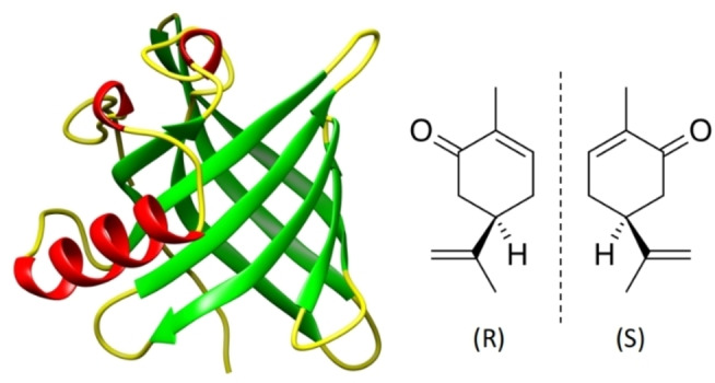
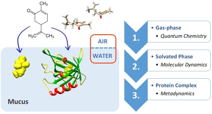
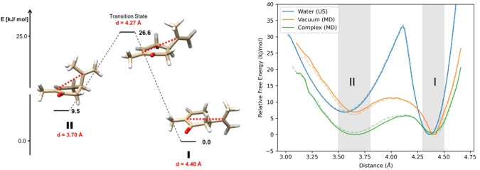
Mammalian odorant binding proteins (OBPs) have long been suggested to transport hydrophobic odorant molecules through the aqueous environment of the nasal mucus. While the function of OBPs as odorant transporters is supported by their hydrophobic beta-barrel structure, no rationale has been provided on why and how these proteins facilitate the uptake of odorants from the gas phase. Here, a multi-scale computational approach validated through available high-resolution spectroscopy experiments reveals that the conformational space explored by carvone inside the binding cavity of porcine OBP (pOBP) is much closer to the gas than the aqueous phase, and that pOBP effectively manages to transport odorants by lowering the free energy barrier of odorant uptake. Understanding such perireceptor events is crucial to fully unravel the molecular processes underlying the olfactory sense and move towards the development of protein-based biomimetic sensor units that can serve as artificial noses.

Keywords: Biosensors; Carvone; Enhanced Sampling; High-Resolution Spectroscopy; Odorant Binding.


Conflict of interest statement

The authors declare no conflict of interest.

Figures

Figure 1
Figure 1
Left: Structure of porcine odorant binding protein (pOBP, PDB‐ID: 1A3Y [5] ). The eight anti‐parallel beta‐sheets of pOBP are typical for members of the lipocalin family and form a strongly hydrophobic binding environment for small organic molecules. Secondary elements; beta‐sheets, alpha helices, and loops are colored in green, red, and yellow, respectively. Right: The two enantiomeric forms of carvone ((R)‐ and (S)‐enantiomer).
Figure 2
Figure 2
Overview of the multi‐scale approach implemented in this work to study the transfer of carvone from the gas‐ to the aqueous phase, and in its bound state with pOBP. This approach allows to compare the conformational space that is explored by carvone in the different environments using adapted computational techniques. The computational set up for each scale is provided in the SI.
Figure 3
Figure 3
Structural superposition of the optimized conformers of (R)‐carvone at the MP2/6‐311++G(d,p) level of theory using the UCSF Chimera program package. Note that the six energy minima can be classified into two distinct subsets of conformations with an (I) equatorial, and (II) axial orientation of the isopropenyl moiety. All optimizations were carried out using the GAUSSIAN16 program package The conformers clustered within subset I and subset II correspond to the conformers 1, 3, 5 and to the conformers 2, 4, 6, respectively (see Figure S2 for details). The full overview of the quantum chemical results is provided in section 1 of the SI.
Figure 4
Figure 4
Left: Reaction coordinate for the interconversion of (R)‐carvone from conformer 1 (global minimum and representative conformer for subset I, see Figure 3) through the joint transition state to conformer 6 (representative conformer for subset II, see Figure 3). The distance between the two carbon atoms used as order parameter d to distinguish the different conformational states of carvone in the gas phase is highlighted in red. The structures and relative energies in kJ/mol were obtained after optimization at the MP2/6‐311++g(d,p) level of theory; Right: Free energy surfaces of (R)‐carvone in 3 different phases: water (Water), gas (Vacuum), and protein environment (Complex), the free energy surfaces of (S)‐carvone are depicted as dotted lines. US and MD: umbrella sampling simulations and molecular dynamics simulations, respectively. See SI for further details.
Figure 5
Figure 5
Snapshots from simulation runs of (R)‐carvone in the two solvated environments. A: Inside the protein binding pocket. B: In explicit water. The center of the light‐grey spheres is superimposed on the center of mass of carvone. The radii of the spheres are fixed to 7 Å and the water molecules within a distance of 15 Å are shown. C: Zoom on the light‐grey sphere shown in B.
Figure 6
Figure 6
Free energy surface of the pOBP carvone complex generated using volume‐based metadynamics for a ρ of 28 Å (Upper trace: (R)‐carvone. Lower trace: (S)‐carvone)). CN: coordination number (measure for the number of contacts between the ligand and the protein binding pocket), A and B: bound and unbound states of the pOBP‐carvone complex, respectively. See SI for all technical details and the simulation setup.

References

    1. Pelosi P., Knoll W., Biol. Rev. 2022, 97, 20. - PubMed
    1. Zaremska V., Tan J., Lim S., Knoll W., Pelosi P., Chem. Eur. J. 2020, 26, 8720. - PubMed
    1. Heydel J. M., Coelho A., Thiebaud N., Legendre A., Bon A. M. L., Faure P., Neiers F., Artur Y., Golebiowski J., Briand L., Anat. Rec. 2013, 296, 1333. - PubMed
    1. Mulla M. Y., Tuccori E., Magliulo M., Lattanzi G., Palazzo G., Persaud K., Torsi L., Nat. Commun. 2015, 6, 6010. - PMC - PubMed
    1. Spinelli S., Ramoni R., Grolli S., Bonicel J., Cambillau C., Tegoni M., Biochemistry 1998, 37, 7913. - PubMed

LinkOut - more resources