. 2024 Dec;300(12):107962.
doi: 10.1016/j.jbc.2024.107962. Epub 2024 Nov 5.

Structure-function analysis of tRNA t6A-catalysis, assembly, and thermostability of Aquifex aeolicus TsaD2B2 tetramer in complex with TsaE

Affiliations

Structure-function analysis of tRNA t6A-catalysis, assembly, and thermostability of Aquifex aeolicus TsaD2B2 tetramer in complex with TsaE

Shuze Lu et al. J Biol Chem. 2024 Dec.

Abstract

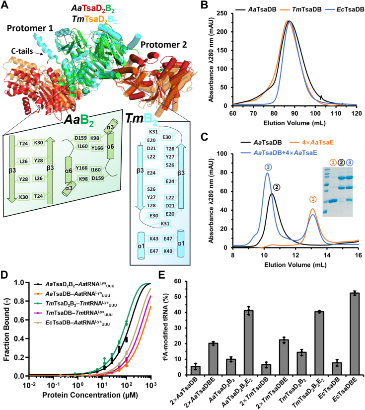
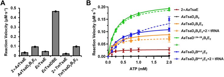
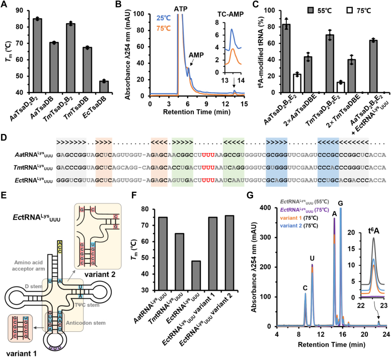
The universal N6-threonylcarbamoyladenosine (t6A) at position 37 of tRNAs is one of the core post-transcriptional modifications that are needed for promoting translational fidelity. In bacteria, TsaC uses L-threonine, bicarbonate, and ATP to generate an intermediate threonylcarbamoyladenylate (TC-AMP), of which the TC moiety is transferred to N6 atom of tRNA A37 to generate t6A by TsaD with the support of TsaB and TsaE. TsaD and TsaB form a TsaDB dimer to which tRNA and TsaE are competitively bound. The catalytic mechanism of TsaD and auxiliary roles of TsaB and TsaE remain to be fully elucidated. In this study, we reconstituted tRNA t6A biosynthesis using TsaC, TsaD, TsaB, and TsaE from Aquifex aeolicus and determined crystal structures of apo-form and ADP-bound form of TsaD2B2 tetramer. Our TsaD2B2-TsaE-tRNA model coupled with functional validations reveal that the binding of tRNA or TsaE to TsaDB is regulated by C-terminal tail of TsaB and a helical hairpin α1-α2 of TsaD. A. aeolicus TsaDB possesses a basal t6A catalytic activity that is stimulated by TsaE at the cost of ATP consumption. Our data suggest that the binding of TsaE to TsaDB induces conformational changes of α1, α2, α6, α7, and α8 of TsaD and C-terminal tail of TsaB, leading to the release of tRNA t6A and AMP. ATP-mediated binding of TsaE to TsaDB resets a t6A active conformation of TsaDB. Dimerization of TsaDB enhances thermostability and promotes t6A catalysis of TsaD2B2-tRNA, of which GC base pairs in anticodon stem are needed for the correct folding of thermophilic tRNA at higher temperatures.

Keywords: ATP hydrolysis; Aquifex aeolicus; GC base pairs content; TsaD(2)B(2) tetramer; TsaD–TsaB–TsaE–tRNA assembly; crystal structure; enzymatic reconstitution; oligomerization; tRNA t(6)A; thermostability.


Conflict of interest statement

Conflict of interest The authors declare that they have no conflicts of interest with the contents of this article.

Figures

Figure 1
Figure 1
Functional and structural characterization of TsaC, TsaD, TsaB, and TsaE fromAquifexaeolicus (Aa). A, reaction scheme of tRNA t6A biosynthesis by TsaC, TsaD, TsaB, and TsaE from bacteria. In the first step, TsaC protein uses L-threonine, HCO3-, and ATP to generate an unstable intermediate threonylcarbamoyladenylate (TC-AMP); in the second step, TsaDB complex transfers the TC-moiety from TC-AMP onto the N6 atom of tRNA A37, leading to tRNA t6A. The t6A-catalytic cycle of TsaDB is stimulated by TsaE at cost of ATP consumption. B, size-exclusion chromatography (SEC) profiles and SDS–PAGE analysis of A. aeolicus TsaD2B2, TsaC, TsaE, and Thermotoga maritima (Tm) TsaD2B2. C, crystal structure of A. aeolicus TsaD2B2 tetramer. TsaD and TsaB are shown in red and green, respectively. One TsaD–TsaB–Fe2+ protomer is cartooned as helices and the other TsaD–TsaB–ADP–Fe2+ protomer is cartooned as cylindrical helices. 2Fo–Fc electron densities are contoured at 1.0 σ for ADP and Fe2+ in TsaD and for H111–H115–D296 motif and CSO10 of TsaD. D, structural juxtaposition of A. aeolicus TsaD–ADP–Fe2+ (colored in red) and E. coli (Ec) TsaD–BK951–Zn2+ (PDB: 6Z81, colored in cyan). The close-up view shows the binding orientations of ADP or TC-AMP (BK951) in the catalytic sites of TsaD. The dashed line denotes the distance from CSO10 to the threonyl-moiety of TC-AMP. E, t6A-modification efficiencies of 5 μM A.aeolicus TsaD2B2E2 and A. aeolicus TsaDC10A2B2E2 in combination with 5 μM TsaC toward 60 μM in vitro transcribed (IVT) A. aeolicus tRNALysUUU, and t6A-modification efficiencies of 5 μM EDTA-dialyzed TsaD2B2 toward 60 μM metal-deprived IVT A. aeolicus tRNALysUUU in assays supplemented with 30 μM isolated TC-AMP and 100 μM MgCl2, FeCl2 or ZnCl2. Error bars represent standard deviations from triplicate measurements. F, structural juxtaposition of A. aeolicus TsaB and E. coli TsaB (PDB: 4YDU) manifests conserved N-terminal subdomains but varied C-terminal subdomains. The specific insertion segment (residues 143–162) of E. coli TsaB is highlighted in red. G, topological diagrams of A. aeolicus TsaB (green) and E. coli TsaB (gray). The secondary structures (β9α6β10) of the inserted segment of E. coli TsaB are shown in red. IVT, in vitro transcribed; PDB, Protein Data Bank; t6A, N6-threonylcarbamoyladenosine.
Figure 2
Figure 2
Structural and functional characterization of TsaD2B2tetramer and TsaDB dimer. A, structural juxtaposition of AaTsaD2B2 and TmTsaD2B2 (PDB: 6N9A) is labeled and shown in different colors. One TsaD–TsaB protomer is cartooned as helices and the other TsaD–TsaB protomer is cartooned as cylindrical helices. The close-up views show the interacting residues of AaTsaB–TsaB and TmTsaB–TsaB. B, SEC profiles of TsaDB dimers from Aquifex aeolicus, Thermotoga maritima, and Escherichia coli. C, SEC analysis of the interaction between A. aeolicus TsaDB and TsaE. Briefly, 400 μg TsaDB and 450 μg TsaE (1:4 in molar ratio) was applied to Superdex 75 10/300 GL column (GE HealthCare) and the inset shows SDS–PAGE analysis of proteins in fractions corresponding to the major peaks. D, microscale thermophoresis (MST) measurements of the interaction between 10 nM 5′ 6-FAM-tRNALysUUU and 195 nM–100 μM TsaD2B2 tetramer or TsaDB dimer from A. aeolicus, T. maritima or E. coli. E, comparison of tRNA t6A modification efficiencies in complete assays using 5 μM TsaD2B2 or TsaD2B2E2 from A. aeolicus and T. maritima, 10 μM TsaDB or TsaDBE from A. aeolicus and T. maritima, and 5 μM TsaDB or TsaDBE from E. coli. Error bars represent standard deviations from triplicate measurements. t6A, N6-threonylcarbamoyladenosine; SEC, size-exclusion chromatography.
Figure 3
Figure 3
Structural model of Aquifexaeolicus TsaD2B2in complex with TsaE or tRNA and functional validations. A, a structural model for A. aeolicus TsaD2B2–TsaE–tRNA complex. tRNA is bound to one TsaDB protomer and TsaE is bound to the other TsaDB protomer. B, a close view of the assemble of A. aeolicus TsaE onto TsaDB. C, a close view of the binding of anticodon stem loop of tRNA in the catalytic center of TsaD. The helical hairpin α1–α2 of A. aeolicus TsaD are shown in yellow; the equivalent α1 of Thermotoga maritima TsaD and C-terminal α7 of T. maritima TsaB are shown in gray and indicated with stars. The other parts of T. maritima TsaDB are omitted for clarity. D, local sequence alignment of residues 32 to 42 of TsaDs from A. aeolicus, T. maritima, and E. coli, and the schematic representation of A. aeolicus TsaDΔ34–38 mutant. E, local sequence alignment of the C-terminal tail of TsaBs from A. aeolicus, T. maritima, and E. coli, and the schematic representation of A. aeolicus TsaBmut mutant. FH, ITC analysis of the interaction between A. aeolicus TsaD2B2 (F), TsaDΔ34–382B2 (G) or TsaD2Bmut2 (H) and TsaE. Representative plots from an ITC experiment are shown with raw data in the upper panel and curve fit in the lower panel. I, MST measurements of the interaction between 10 nM 5′ 6-FAM-tRNALysUUU and 195 nM–100 μM TsaD2B2, TsaDΔ34–382B2 or TsaD2Bmut2. J, quantification of tRNA t6A modification efficiencies of 5 μM A.aeolicus TsaD2B2E2, TsaDΔ34–382B2E2 or TsaD2Bmut2E2 toward 60 μM IVT tRNALysUUU. Error bars represent standard deviations from triplicate measurements. K, EMSA analysis of the competition binding of TsaE and tRNA to TsaD2Bmut2. TsaE at indicated concentration was added to 5 μM AaTsaD2Bmut2 that was preincubated with 1 μM 5′ 6-FAM-tRNALysUUU. The upper panel shows the presence of 5′ 6-FAM-tRNALysUUU and bottom panel shows the presence of AaTsaD2Bmut2E2 and unbound TsaE. IVT, in vitro transcribed; 6-FAM, 6-carboxyfluorescein; ITC, isothermal titration calorimetry; MST, microscale thermophoresis; t6A, N6-threonylcarbamoyladenosine; EMSA, electrophoresis mobility shift assay.
Figure 4
Figure 4
Analysis of ATPase activity of TsaE in the presence of TsaDB and tRNA. A, initial velocities of 4 mM ATP to ADP hydrolysis by 10 μM A. aeolicus TsaE, 5 μM A.aeolicus TsaD2B2E2, 5 μM E.coli TsaE, 5 μM E.coli TsaDBE, 10 μM T. maritima TsaE, or 5 μM T.maritima TsaD2B2E2. Error bars represent standard deviations from triplicate measurements. B, steady-state kinetics analysis of the ATPase activity of 10 μM A.aeolicus TsaE in the presence of 5 μM A.aeolicus TsaD2B2 or mutants and 10 μM IVT tRNALysUUU. Kinetic parameters are summarized in Table 2. Error bars represent standard deviations from triplicate measurements. IVT, in vitro transcribed
Figure 5
Figure 5
Thermostability analysis of tRNA t6A biosynthetic systems from Aquifex aeolicus, Thermotoga maritima and Escherichia coli.A, melting temperature (Tm) values of TsaD2B2 or TsaDB from A. aeolicus, T. maritima, or E. coli determined by thermal shift assay. Error bars represent standard deviations from triplicate measurements. B, LC–MS chromatograms of TC-AMP formation in assays that contained 5 μM AaTsaC, 1 mM ATP, 4 mM L-threonine, and 10 mM NaHCO3 at 25 °C or 75 °C. C, quantification of tRNA t6A modification efficiency by 5 μM TsaD2B2E2 or 10 μM TsaDBE from A. aeolicus or T. maritima toward 60 μM cognate IVT tRNALysUUU at 55 °C or 75 °C. For the assay with 60 μM E.coli tRNALysUUU, 5 μM A.aeolicus TsaD2B2E2 was used. Error bars represent standard deviations from triplicate measurements. D, sequence alignment of tRNALysUUU from A. aeolicus, T. maritima, and E. coli. Amino acid acceptor arms, D–stems, anticodon stems, and TψC stems are shown in gray, pink, green, and blue, respectively. GC pairs and anticodons are highlighted in black and red, respectively. E, schematic representation of the GC base pairs mutations of E. coli tRNALysUUU variant 1 and variant 2. F, Tm values of A. aeolicus tRNALysUUU, T. maritima tRNALysUUU, E. coli tRNALysUUU, EctRNALysUUU variant 1 and variant 2, which were determined by circular dichroism spectra analysis. G, LC–MS chromatograms of tRNA t6A formation by AaTsaC and AaTsaD2B2E2 in assay using E. coli tRNALysUUU, variant 1 and variant 2 at 55 °C or 75 °C. IVT, in vitro transcribed; t6A, N6-threonylcarbamoyladenosine

References

    1. Grosjean H., Sprinzl M., Steinberg S. Posttranscriptionally modified nucleosides in transfer RNA: their locations and frequencies. Biochimie. 1995;77:139–141. - PubMed
    1. Giege R., Juhling F., Putz J., Stadler P., Sauter C., Florentz C. Structure of transfer RNAs: similarity and variability. Wiley Interdiscip. Rev. RNA. 2012;3:37–61. - PubMed
    1. Bjork G.R., Ericson J.U., Gustafsson C.E., Hagervall T.G., Jonsson Y.H., Wikstrom P.M. Transfer RNA modification. Annu. Rev. Biochem. 1987;56:263–287. - PubMed
    1. Yarian C., Townsend H., Czestkowski W., Sochacka E., Malkiewicz A.J., Guenther R., et al. Accurate translation of the genetic code depends on tRNA modified nucleosides. J. Biol. Chem. 2002;277:16391–16395. - PubMed
    1. Schultz S.K., Kothe U. RNA modifying enzymes shape tRNA biogenesis and function. J. Biol. Chem. 2024;300 - PMC - PubMed

Publication types

Supplementary concepts

LinkOut - more resources