

Comparison of ante- and postmortem ventricular wall thickness using echocardiography and autopsy findings
- PMID: 39511013
- PMCID: PMC12018510
- DOI: 10.1007/s00428-024-03960-z
Comparison of ante- and postmortem ventricular wall thickness using echocardiography and autopsy findings
Abstract
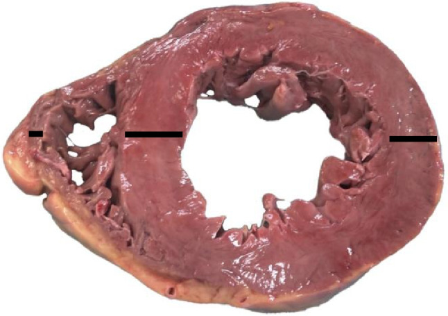
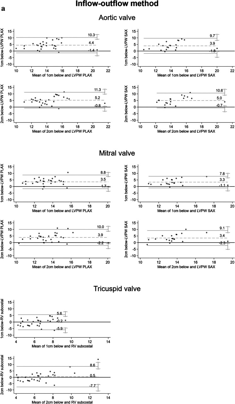
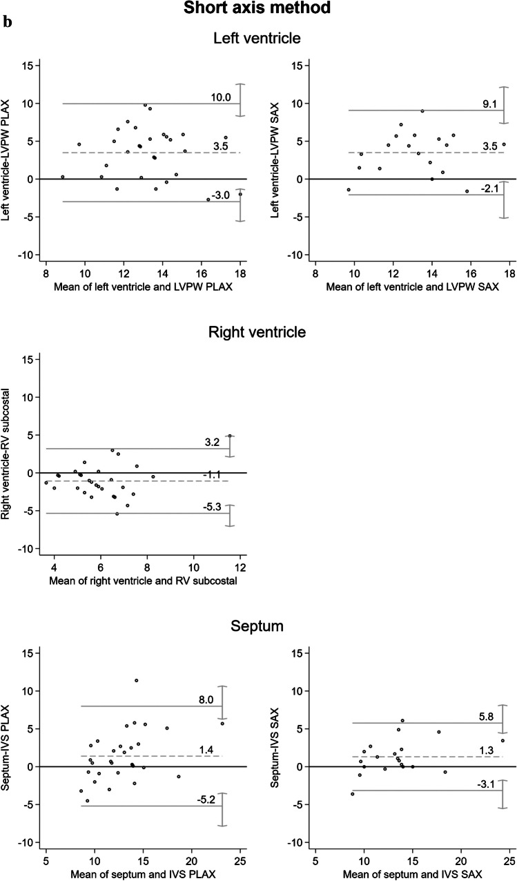
In autopsy practice, the thickness of ventricular walls is one of the parameters used to identify cardiac hypertrophy. The presented study aimed to compare ante- and postmortem measurements of ventricular wall thickness, (i) to determine a postmortem standardized localization and dissection method for ventricular wall measurements, and (ii) to determine the ability of postmortem measurements in recognition of antemortem hypertrophy. A single-center prospective study was conducted at the Institute of Legal Medicine in Hamburg, Germany. Sixty hearts were dissected alternating by the inflow-outflow or short-axis method, and the ventricular walls were measured at different locations and compared with the echocardiographic values of the end-diastolic phase during life of these individuals. The results showed measurement differences between the autoptic and echocardiographic values-for the left ventricle between 3.3 and 5.2 mm, for the right ventricle between 0.2 and 1.1 mm, and for the septum between 1.3 and 1.4 mm. Diagnostic performance of recognizing antemortem hypertrophy with postmortem measurement was poor, except for measuring the right ventricle and septum with the short-axis method (area under the ROC curve of 0.72 and 0.82, respectively). According to the results, cardiac changes may occur postmortem and need to be considered when used for diagnosing cardiac pathology. The postmortem diagnosis of left or right ventricular hypertrophy should always be made in conjunction with other, particularly cardiac, autopsy findings. An autoptic diagnosis of hypertrophy solely by a ventricular wall thickness > 15 mm or > 5 mm alone is not sufficient.
Keywords: Cardiac dissection; Cardiac hypertrophy; Cardiac pathologies; Forensic autopsy; Ventricular wall thickness.
© 2024. The Author(s).
Conflict of interest statement
Declarations. Ethics approval: This study was performed in line with the principles of the Declaration of Helsinki. The study protocol was approved by the Ethics Committee of the Hamburg Medical Association (Application Number 2020–10311-BO-ff). Consent to participate: We sincerely thank the relatives of the deceased for providing numerous consents for this study. Competing interests: The authors declare no competing interests.
Figures

Similar articles
-
Intravital and postmortem diagnostics of myocardial hypertrophy of the left ventricle:identity or convention?Ter Arkh. 2018 Sep 20;90(9):73-80. doi: 10.26442/terarkh201890973-80. Ter Arkh. 2018. PMID: 30701739
-
Usefulness of two-dimensional and speckle tracking echocardiography in "Gray Zone" left ventricular hypertrophy to differentiate professional football player's heart from hypertrophic cardiomyopathy.Am J Cardiol. 2011 Nov 1;108(9):1322-6. doi: 10.1016/j.amjcard.2011.06.053. Epub 2011 Aug 18. Am J Cardiol. 2011. PMID: 21855830
-
[The value of RV(6) > RV(5) of ECG in diagnosis of pneumocardiac disease complicated by left ventricular hypertrophy in coal-workers with pneumoconiosis].Zhonghua Lao Dong Wei Sheng Zhi Ye Bing Za Zhi. 2012 Sep;30(9):688-90. Zhonghua Lao Dong Wei Sheng Zhi Ye Bing Za Zhi. 2012. PMID: 23257097 Chinese.
-
Left myocardial wall measurements on postmortem imaging compared to autopsy.Cardiovasc Pathol. 2019 Nov-Dec;43:107149. doi: 10.1016/j.carpath.2019.107149. Epub 2019 Aug 22. Cardiovasc Pathol. 2019. PMID: 31639653
-
Pathological and physiological left ventricular hypertrophy: echocardiography for differentiation.Future Cardiol. 2009 Sep;5(5):495-502. doi: 10.2217/fca.09.34. Future Cardiol. 2009. PMID: 19715413 Review.
Cited by
-
Histopathological Analysis of Myocardial Remodeling Following Heart Failure: A Cadaveric Case Report.Cureus. 2025 Jul 14;17(7):e87889. doi: 10.7759/cureus.87889. eCollection 2025 Jul. Cureus. 2025. PMID: 40809625 Free PMC article.
References
-
- Grasner JT, Herlitz J, Tjelmeland IBM, Wnent J, Masterson S, Lilja G et al (2021) European Resuscitation Council Guidelines 2021: epidemiology of cardiac arrest in Europe. Resuscitation 161:61–79 - PubMed
-
- Kiguchi T, Okubo M, Nishiyama C, Maconochie I, Ong MEH, Kern KB et al (2020) Out-of-hospital cardiac arrest across the world: first report from the International Liaison Committee on Resuscitation (ILCOR). Resuscitation 152:39–49 - PubMed
-
- Collins KA, Hutchins GM (2005) An introduction to autopsy technique: step-by-step diagrams. Second edition. College of American Pathologists, USA
Publication types
MeSH terms
LinkOut - more resources
Full Text Sources
