. 2024 Nov 7;15(1):9294.
doi: 10.1038/s41467-024-53473-1.

Structural insights into the agonist selectivity of the adenosine A3 receptor

Affiliations

Structural insights into the agonist selectivity of the adenosine A3 receptor

Hidetaka S Oshima et al. Nat Commun. .

Abstract

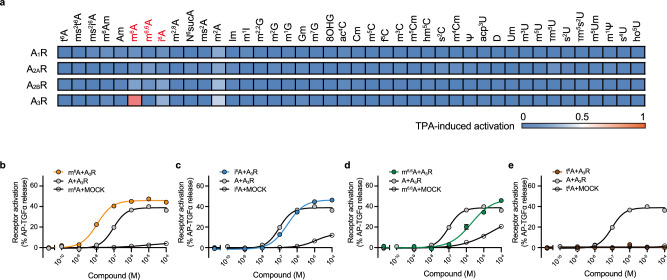
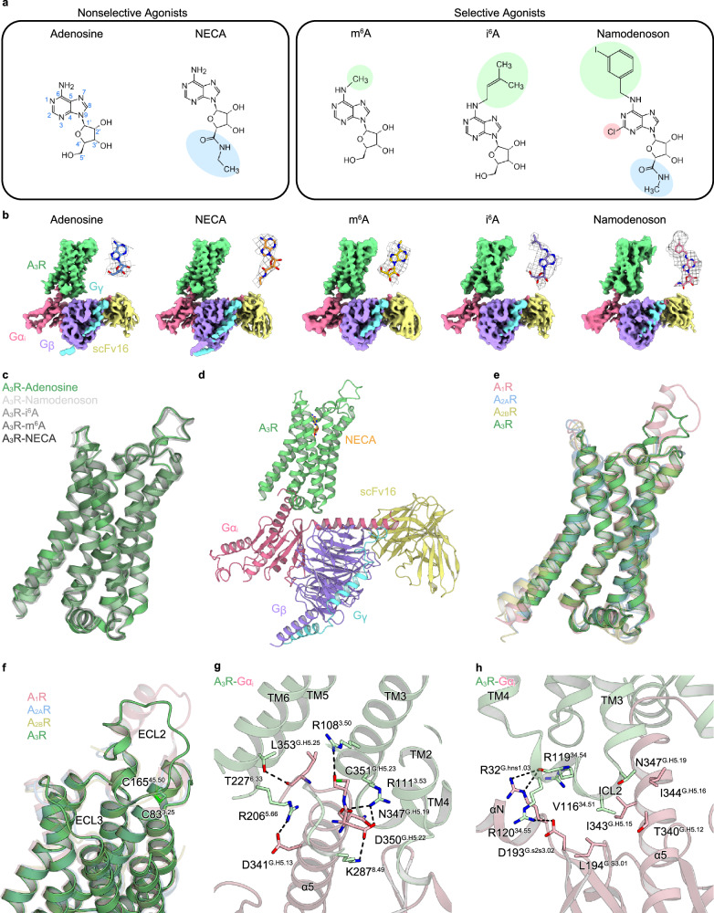
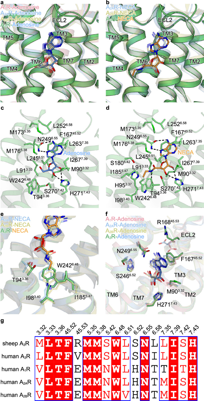
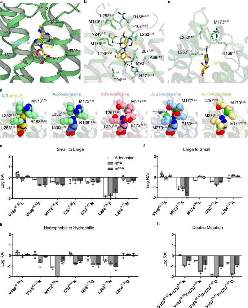
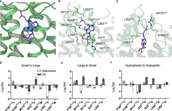
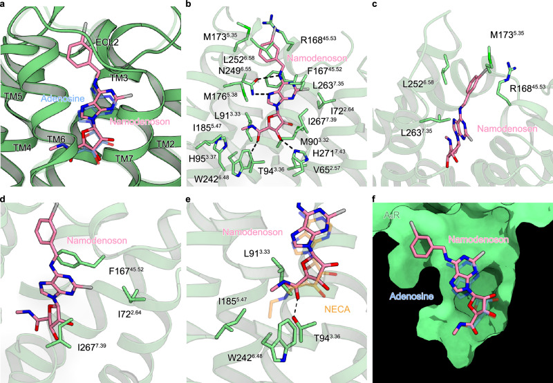
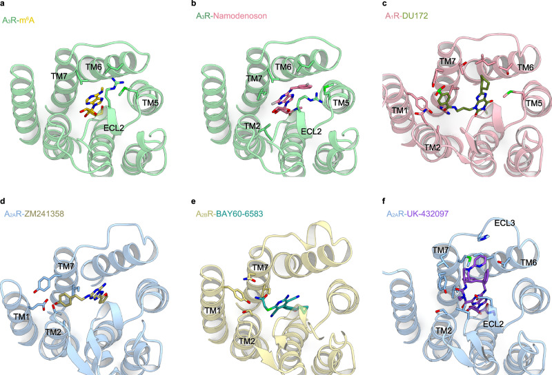
Adenosine receptors play pivotal roles in physiological processes. Adenosine A3 receptor (A3R), the most recently identified adenosine receptor, is expressed in various tissues, exhibiting important roles in neuron, heart, and immune cells, and is often overexpressed in tumors, highlighting the therapeutic potential of A3R-selective agents. Recently, we identified RNA-derived N6-methyladenosine (m6A) as an endogenous agonist for A3R, suggesting the relationship between RNA-derived modified adenosine and A3R. Despite extensive studies on the other adenosine receptors, the selectivity mechanism of A3R, especially for A3R-selective agonists such as m6A and namodenoson, remained elusive. Here, we identify tRNA-derived N6-isopentenyl adenosine (i6A) as an A3R-selective ligand via screening of modified nucleosides against the adenosine receptors. Like m6A, i6A is found in the human body and may be an endogenous A3R ligand. Our cryo-EM analyses elucidate the A3R-Gi complexes bound to adenosine, 5'-N-ethylcarboxamidoadenosine (NECA), m6A, i6A, and namodenoson at overall resolutions of 3.27 Å (adenosine), 2.86 Å (NECA), 3.19 Å (m6A), 3.28 Å (i6A), and 3.20 Å (namodenoson), suggesting the selectivity and activation mechanism of A3R. We further conduct structure-guided engineering of m6A-insensitive A3R, which may aid future research targeting m6A and A3R, providing a molecular basis for future drug discovery.


Conflict of interest statement

O.N. is a co-founder and an external director of Curreio, Inc. The remaining authors declare no competing interests.

Figures

Fig. 1
Fig. 1. Screening of modified nucleosides against human A3R.
a Heatmaps showing the activation of adenosine receptor subtypes by modified nucleosides, as measured by the TGF-α shedding assay. The color scale represents % receptor activation compared to TPA (12-O-tetradecanoylphorbol 13-acetate)-mediated receptor activation, which induces the maximum alkaline phosphatase(AP)-fused TGFα-shedding response independently of the receptor, and the adenosine receptor-dependence of signals were calculated by subtracting the response in mock-transfected cells from the response in adenosine receptor-expressing cells. The tested compounds’ concentrations were 100 nM for hA1R and hA3R, 1 µM for hA2AR, and 5 µM for hA2BR. Values are shown as an average of three independent experiments. Comparison of AP-TGFα-shedding response curves for hA3R between modified nucleosides (b: m6A, c: i6A, d: m6,6A, and e: t6A) and adenosine. Response curves for mock-transfected cells are shown in the same graph. Ligand-induced AP-TGF-α release ratio into conditioned media is quantified. Symbols and error bars represent mean ± SEM, respectively, of 3–6 independent experiments with each performed in triplicate. Source data are provided as a Source Data file.
Fig. 2
Fig. 2. Overall structure of the A3R-Gi complex.
a Structures of the agonists used for structural analyses. Atom numbers for the adenosine moiety are colored blue. Modifications at N6, C2, and 5′ positions are highlighted in green, red, and blue, respectively. b Cryo-EM maps of the A3R-Gi complex bound to adenosine, NECA, m6A, i6A, and namodenoson. Densities of each agonist are shown in the top-right corner of each map. c Superimposition of each agonist-bound A3R. d Overall structure of the A3R-Gi complex bound to NECA. e Superimposition of A1R (PDB 6D9H), A2AR (PDB 6GDG), A2BR (PDB 8HDP), and A3R. f Close-up view of ECLs of adenosine receptors. ECL2 and ECL3 show remarkable differences among the adenosine receptors. The disulfide bond between ECL2 and TM3 of A3R is labeled. Receptor-G-protein interactions around α5 (g) and ICL2 (h). The residues involved in the interactions are represented by stick models. Black dashed lines indicate hydrogen bonds.
Fig. 3
Fig. 3. Binding modes of adenosine and NECA.
a Superimposition of adenosine-bound A1R (PDB 6D9H), A2AR (PDB 2YDO), A2BR (PDB 8HDP), and A3R. b Superimposition of NECA-bound A2AR (PDB 6GDG), A2BR (PDB 7XY7), A3R. Binding modes of adenosine (c) and NECA (d). Black dashed lines indicate hydrogen bonds. e Comparison of the interactions around the 5′ tail of NECA. f Comparison of the binding modes of adenosine to each adenosine receptor. Representative residues involved in the ligand-receptor interaction are represented by stick models. Residues of A3R are labeled. Key interactions are conserved among adenosine receptors. g Alignment of residues comprising the orthosteric pockets of adenosine receptors.
Fig. 4
Fig. 4. Binding mode of m6A.
a Superimposition of adenosine-bound and m6A-bound A3R. b Binding mode of m6A. Black dashed lines indicate hydrogen bonds. c Close-up view of the hydrophobic pocket of the m6A-bound model. d CPK models of the hydrophobic pocket of A3R and those of A1R, A2AR, A2BR, and adenosine-bound A3R (PDB 6D9H, 2YDO, 8HDP, and this study, respectively). The methyl group of m6A is especially tightly packed. eh Relative RAi values of adenosine, m6A, and m6,6A for human A3R mutants, as determined by the TGFα-shedding assay. RAi values are expressed as fold change of the values for the WT. The LogRAi cutoff value was set to −2. Mutations that reduce (e) and enlarge (f) the size of the hydrophobic pocket. g Mutations that replace the original residue to a hydrophilic residue. h Designed mutants for selectively reducing the potency of m6A. Data are presented as mean values ± SEM from at least three independent experiments performed in triplicate (the n values are represented by circles). Source data are provided as a Source Data file.
Fig. 5
Fig. 5. Binding mode of i6A.
a Superimposition of adenosine- and i6A-bound A3R. b Binding mode of i6A. Black dashed lines indicate hydrogen bonds. c Close-up view of the hydrophobic pocket of the i6A-bound structure. df Relative RAi values of adenosine and i6A for human A3R mutants, as determined by the TGFα-shedding assay. The LogRAi cutoff value was set to −2. Mutations that reduce (d) and enlarge (e) the size of the hydrophobic pocket. f Mutations that replace the original residue to a hydrophilic residue. Data are presented as mean values ± SEM from at least three independent experiments performed in triplicate (the n values are represented by circles). The values for adenosine used references in (df) are the same as those for adenosine in Fig. 4e–g. Source data are provided as a Source Data file.
Fig. 6
Fig. 6. Binding mode of namodenoson.
a Superimposition of adenosine- and namodenoson-bound A3R. b Binding mode of namodenoson. Black dashed lines indicate hydrogen bonds. c Close-up view of the hydrophobic pocket of the namodenoson-bound structure. The 3-iodobenzyl group of namodenoson wedges into the hydrophobic pocket. d Close-up view of the interactions around the chloro group of namodenoson. e Close-up view of the interactions around the N-methylcarboxamide group of namodenoson. f Cross section of the orthosteric pocket of A3R. Namodenoson fills the orthosteric pocket comprehensively.
Fig. 7
Fig. 7. Structural insight into the selective recognition of adenosine receptors.
af Binding modes of the adenosine receptors and their selective drugs. a m6A-bound A3R. b Namodenoson-bound A3R. c DU172-bound A1R (PDB 5UEN). d ZM241358-bound A2AR (PDB 3PWH). e BAY60-6583-bound A2BR (PDB 7XY6). f UK-432097-bound A2AR (PDB 3QAK). While occupying the orthosteric pocket, each drug exhibits interactions with the extracellular portion of the receptor. These extracellular interactions would be essential for the selectivity among the adenosine receptors.

Similar articles

Cited by

References

    1. IJzerman, A. P., Jacobson, K. A., Müller, C. E., Cronstein, B. N. & Cunha, R. A. International union of basic and clinical pharmacology. CXII: adenosine receptors: a further update. Pharmacol. Rev.74, 340–372 (2022). - PMC - PubMed
    1. Effendi, W. I., Nagano, T., Kobayashi, K. & Nishimura, Y. Focusing on adenosine receptors as a potential targeted therapy in human diseases. Cells9, 785 (2020). - PMC - PubMed
    1. Borea, P. A., Gessi, S., Merighi, S., Vincenzi, F. & Varani, K. Pharmacology of adenosine receptors: the state of the art. Physiol. Rev.98, 1591–1625 (2018). - PubMed
    1. Zhou, Q. Y. et al. Molecular cloning and characterization of an adenosine receptor: the A3 adenosine receptor. Proc. Natl. Acad. Sci. USA89, 7432–7436 (1992). - PMC - PubMed
    1. Gao, Z.-G., Auchampach, J. A. & Jacobson, K. A. Species dependence of A3 adenosine receptor pharmacology and function. Purinergic Signal.19, 523–550 (2023). - PMC - PubMed

Publication types

MeSH terms

LinkOut - more resources