. 2024 Nov 7;15(1):9614.
doi: 10.1038/s41467-024-53981-0.

The BAF complex enhances transcription through interaction with H3K56ac in the histone globular domain

Affiliations

The BAF complex enhances transcription through interaction with H3K56ac in the histone globular domain

Kwangbeom Hyun et al. Nat Commun. .

Abstract

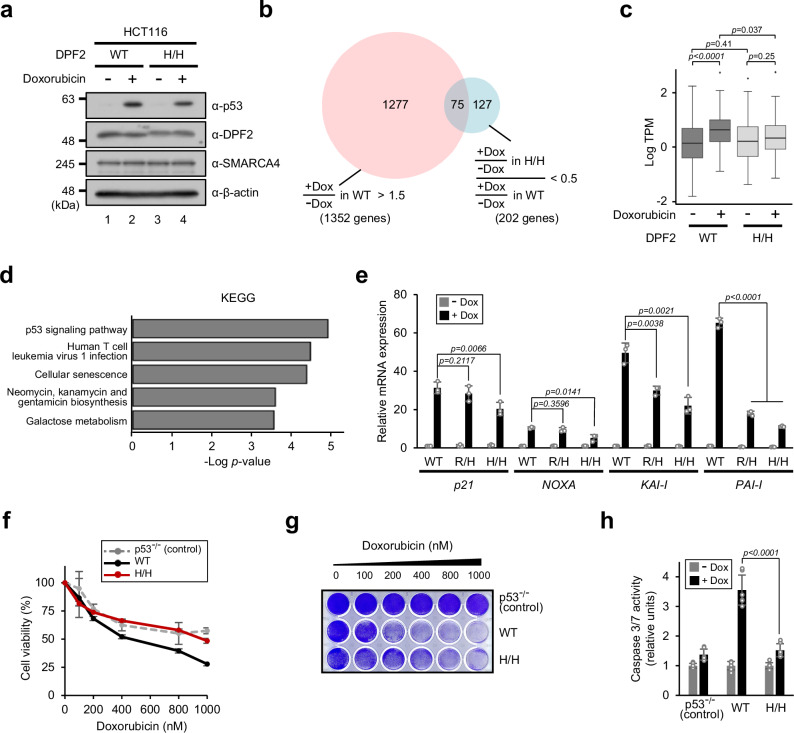
Histone post-translational modifications play pivotal roles in eukaryotic gene expression. To date, most studies have focused on modifications in unstructured histone N-terminal tail domains and their binding proteins. However, transcriptional regulation by chromatin-effector proteins that directly recognize modifications in histone globular domains has yet to be clearly demonstrated, despite the richness of their multiple modifications. Here, we show that the ATP-dependent chromatin-remodeling BAF complex stimulates p53-dependent transcription through direct interaction with H3K56ac located on the lateral surface of the histone globular domain. Mechanistically, the BAF complex recognizes nucleosomal H3K56ac via the DPF domain in the DPF2 subunit and exhibits enhanced nucleosome-remodeling activity in the presence of H3K56ac. We further demonstrate that a defect in H3K56ac-BAF complex interaction leads to impaired p53-dependent gene expression and DNA damage responses. Our study provides direct evidence that histone globular domain modifications participate in the regulation of gene expression.


Conflict of interest statement

The authors declare no competing interests.

Figures

Fig. 1
Fig. 1. Enhanced transcription from H3K56ac-containing chromatin in vitro.
p53- and p300-dependent transcription from recombinant chromatins containing full-length histone octamers harboring an individually acetylated lysine residue on H3 (a); full-length unmodified, H3K56ac and H3K56R histone octamers (b); and N-terminal tailless unmodified, H3K56ac, and H3K56R histone octamers (c), assembled using the ACF/NAP1 chromatin assembly system. d p53- and p300-dependent transcription from recombinant chromatins containing full-length unmodified, H3K56ac, and H3K56R histone octamers assembled using salt dialysis. Relative transcription levels were measured by autoradiography and normalized to those observed with the addition of p53 and p300 to chromatin containing unmodified histone octamers. Data are representative of at least two independent experiments. Source data are provided as a Source Data file.
Fig. 2
Fig. 2. Identification of nucleosomal H3K56ac-interacting proteins.
a Schematic flow for identification of the H3K56ac-mononucleosome interactome. b Volcano plot for the nucleosomal H3K56ac interactome created using label-free quantitation (LFQ) intensity values and p-values measured from biotin pull-down and MS analysis. Two independent biological replicates were analyzed, and each biological replicate was measured in triplicate by liquid chromatography-mass spectrometry (LC-MS) analysis. Human canonical BAF complex subunits are depicted as red squares. c Summary of the BAF complex subunits enriched by biotin pull-down. P-values for the differences in protein abundance were determined by two-tailed unpaired Student’s t-test (b, c). d Biotin pull-down assay using streptavidin-coupled mononucleosomes assembled with biotinylated 35N35 DNA and HeLa S3 nuclear extracts. Bound proteins were monitored by immunoblotting with the indicated antibodies. Data are representative of two independent experiments. Source data are provided as a Source Data file.
Fig. 3
Fig. 3. Direct interaction of the BAF complex with nucleosomal H3K56ac via the DPF2 subunit.
a Biotin pull-down assay using streptavidin-coupled mononucleosomes and purified BAF complex. Data are representative of two independent experiments. b Gel shift assay using mononucleosomes assembled with rhodamine-labeled 35N35 DNA and different concentrations of purified BAF complex (0, 0.25, 0.5, 1.0, 1.5, and 2.0 nM). Left: Binding of the BAF complex to nucleosomes measured by fluorography. Right: Levels of BAF complex-bound nucleosomes were quantitated using ImageJ. Data are presented as mean values ± SD of three independent experiments. c Schematic diagram of human DPF2 with Requiem, C2H2 zinc finger, and DPF domains. Coffin-Siris syndrome (CSS)-associated missense mutations found in the DPF domain are indicated. Numbers indicate amino acid residues. d Biotin pull-down assay using streptavidin-coupled mononucleosomes and purified BAF complexes containing a mutation in the DPF domain of DPF2. Top: Binding of the BAF complexes to nucleosomes monitored by immunoblotting with anti-DPF2 and anti-SMARCA4 antibodies. Bottom: Protein levels of nucleosome-bound DPF2 of each complex were determined using ImageJ and normalized to those of DPF2 in input. The level of DPF2 binding to H3K56ac-nucleosomes in WT DPF2-containing complex was defined as 1. Data are presented as mean values ± SD of three independent experiments. Source data are provided as a Source Data file.
Fig. 4
Fig. 4. Enhanced nucleosome remodeling and transcriptional activity by the BAF complex in the presence of H3K56ac.
a REA assay using mononucleosomes assembled with rhodamine-labeled 100N100 DNA and purified BAF complex. Left: Nucleosome-remodeling activity measured by fluorography. Note that the restriction enzyme, HhaI, was added to all reactions except lane 1. Right: Levels of remodeled nucleosomes were quantitated using ImageJ. Data are presented as mean values ± SD of three independent experiments. b REA assay with purified BAF complexes harboring a mutation in the DPF domain of DPF2. Data are presented as mean values ± SD of three independent experiments. P-values for the reactions with WT and R350H DPF2-containing complexes were determined by two-tailed unpaired Student’s t-test. c In vitro chromatin-transcription assay with purified BAF complexes containing WT or R350H DPF2. Note that p300 was added to all reactions. Relative transcription levels were measured by autoradiography and normalized to those observed with the addition of p53 and p300 to chromatin containing unmodified histone octamers (lane 2). Data are representative of two independent experiments. Source data are provided as a Source Data file.
Fig. 5
Fig. 5. Impaired p53-dependent gene expression and DNA damage responses in DPF2 R350H mutant cells.
a Doxorubicin-induced expression of p53. DPF2 WT and R350H (H/H) HCT116 cells were treated with 0.5 μM doxorubicin for 24 h, and protein levels were analyzed by immunoblotting. Data are representative of two independent experiments. b Venn diagram showing the overlap of genes upregulated ( > 1.5-fold, FDR < 0.05) by doxorubicin treatment in WT cells and genes whose doxorubicin-induced upregulation in H/H cells was reduced to less than 0.5-fold compared with that in WT cells (FDR < 0.05). c Box plot displaying expression levels of the 75 overlapping genes in b. The box plots indicate the median (central line), the third and first quartiles (box edges), and 1.5 × interquartile range (IQR) above and below the box (whiskers) (n = 1 as biological replicates were combined). P-values were determined by two-tailed unpaired Student’s t-test. TPM, transcripts per million. d KEGG (Kyoto Encyclopedia of Genes and Genomes) analysis of the 75 overlapping genes in b. The Fisher’s exact test p-values were obtained using the GeneSCF functional enrichment tool. e mRNA levels of p53-dependent p21, NOXA, KAI-I, and PAI-I genes, with or without 0.5 μM doxorubicin treatment for 48 h, were measured by RT-qPCR and normalized to that of TBP mRNA. The levels of transcription of each gene in WT cells without doxorubicin treatment were defined as 1. Data are presented as mean values ± SD of three independent experiments. Cell-viability (f), colony-formation (g), and caspase-activity (h) assays using p53-/- (control) and DPF2 WT and H/H HCT116 cells. Cells were treated with different concentrations (0, 100, 200, 400, 800, and 1000 nM) (f, g) and 1000 nM (h) of doxorubicin. Data are presented as mean values ± SD of six independent experiments (f, h). Data are representative of three independent experiments (g). P-values were determined by two-tailed unpaired Student’s t-test (e, h). Source data are provided as a Source Data file.
Fig. 6
Fig. 6. Schematic model of enhanced DNA damage-responsive gene expression mediated by H3K56ac-BAF complex interaction.
Upon DNA damage (depicted by red-colored DNA), the tumor suppressor p53 is induced and binds to p53-responsive elements in the promoters of DNA damage-responsive genes. p53 recruits transcriptional coactivators, including histone acetyltransferases (HATs) such as p300 and CBP, to these promoter regions through direct interactions. HATs acetylate H3K56 located on the lateral surface of the histone globular domain. The chromatin remodeler BAF complex recognizes H3K56ac via the DPF domain in the DPF2 subunit and facilitates nucleosome remodeling around the promoter regions, thereby leading to enhanced gene expression. HATs also acetylate multiple lysine residues on histones and transcription factors (not depicted), further promoting efficient transcription.

References

    1. Patel, D. J. & Wang, Z. Readout of epigenetic modifications. Annu. Rev. Biochem.82, 81–118 (2013). - PMC - PubMed
    1. Millán-Zambrano, G., Burton, A., Bannister, A. J. & Schneider, R. Histone post-translational modifications - cause and consequence of genome function. Nat. Rev. Genet.23, 563–580 (2022). - PubMed
    1. Mohammad, H. P., Barbash, O. & Creasy, C. L. Targeting epigenetic modifications in cancer therapy: erasing the roadmap to cancer. Nat. Med.25, 403–418 (2019). - PubMed
    1. Zhao, S., Allis, C. D. & Wang, G. G. The language of chromatin modification in human cancers. Nat. Rev. Cancer21, 413–430 (2021). - PMC - PubMed
    1. Janssen, S. M. & Lorincz, M. C. Interplay between chromatin marks in development and disease. Nat. Rev. Genet.23, 137–153 (2022). - PubMed

Publication types

MeSH terms

Associated data