. 2024 Nov 7;7(1):1455.
doi: 10.1038/s42003-024-07157-7.

SNX9 family mediates βarrestin-independent GPCR endocytosis

Affiliations

SNX9 family mediates βarrestin-independent GPCR endocytosis

Valeria L Robleto et al. Commun Biol. .

Abstract

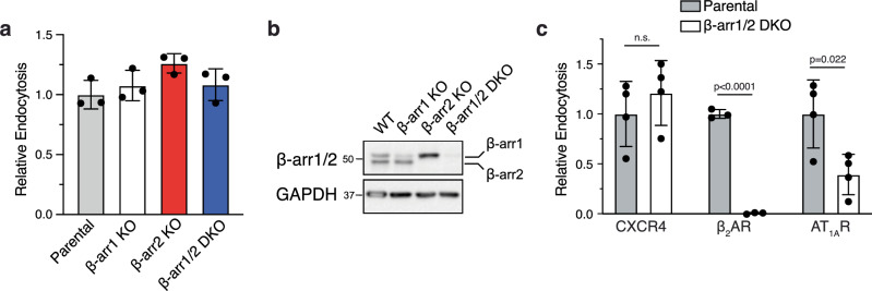
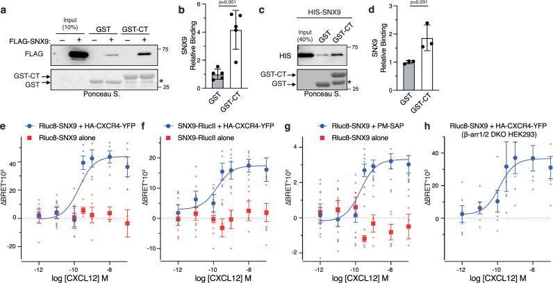
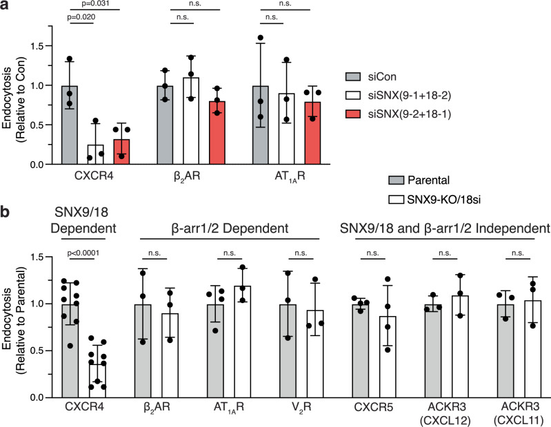
Agonist-stimulated GPCR endocytosis typically occurs via the multi-faceted adaptor proteins known as βarrestins. However, endocytosis of several GPCRs occurs independently of β-arrestins, suggesting an additional mode of GPCR endocytosis, but the mechanisms remain unknown. Here we provide evidence that sorting nexin 9 (SNX9), a previously described endocytic remodeling protein, functions as a novel cargo adaptor that promotes agonist-stimulated GPCR endocytosis. We show that SNX9 and SNX18, but not β-arrestins, are necessary for endocytosis of the chemokine receptor CXCR4. SNX9 is recruited to CXCR4 at the plasma membrane and interacts directly with the carboxyl-terminal tail of the receptor in a phosphorylation-dependent manner. We also provide evidence that some receptors do not require SNX9 and SNX18 nor β-arrestins for endocytosis, suggesting additional modes for GPCR endocytosis. These results provide novel insights into the mechanisms regulating GPCR trafficking and broaden our overall understanding of GPCR regulation.


Conflict of interest statement

The authors declare no competing interests.

Figures

Fig. 1
Fig. 1. Role of β-arrestins in CXCR4 endocytosis.
a CXCR4 endocytosis was examined in parental HeLa cells or HeLa cells deleted of β-arrestin1 (β-arr1 KO), β-arrestin2 (β-arr2 KO), or both β-arr1 and β-arr2 (β-arr1/2 DKO). Cells were stimulated with vehicle or CXCL12 (50 nM) for 30 mins at 37 °C, and endogenous cell surface receptor expression was measured by flow cytometry using an antibody against CXCR4. Endocytosis was calculated as a fractional decrease in cell surface receptors in CXCL12-treated cells compared with vehicle-treated cells after background subtraction, which was assessed in parallel using an isotype control antibody. Data were normalized to endocytosis in parental cells. Data represent the mean ± SD from four independent experiments. b Whole cell lysates from β-arr1 KO, β-arr2 KO, or β-arr1/2 DKO were analyzed by immunoblot for β-arr1 and β-arr2 using an antibody that recognizes both isoforms or for GAPDH, using here as a loading control. c GPCR endocytosis was examined in the β-arr1/2 DKO HEK293 cell line. Parental and β-arr1/2 KO HEK293 cells transiently expressing FLAG-CXCR4, FLAG-β2AR, or FLAG-AT1AR were treated with vehicle or CXCL12 (50 nM), isoproterenol (10 µM), or angiotensin II (10 µM), respectively, for 30 min at 37 °C, and endocytosis was measured by ELISA. Endocytosis was calculated as a percent decrease in cell surface receptors in CXCL12-treated cells compared with vehicle-treated cells after background subtraction from cells not transfected with the receptor. Data were normalized to endocytosis in parental cells. Bars represent the mean ± SD from four (CXCR4 and AT1AR) or three (β2AR) independent experiments. Cell surface levels relative to FLAG-CXCR4 in parental (1 ± 0.1 for CXCR4) and DKO (1 ± 0.1 for CXCR4) cells were 0.9 ± 0.1 and 1.0 ± 0.2 for FLAG-β2AR and 0.9 ± 0.3 and 1.0 ± 0.2 for FLAG-AT1AR, respectively. Data were analyzed by one-way ANOVA (a) or Student’s t-test (c). P values are indicated for each GPCR (c). None of the conditions in panel a were significant. n.s. not significant.
Fig. 2
Fig. 2. SNX9 binding to the carboxy-terminal tail of CXCR4.
ad SNX9 binding to the carboxyl-terminal tail (CT) of CXCR4 was assessed by GST pulldown. Equimolar amounts (~1 µM) of GST-CT or GST immobilized on glutathione-sepharose resin were incubated with HEK293 cells lysates transiently expressing FLAG-SNX9 (a) or with bacterially-purified HIS-SNX9 (0.3 µM) (b), and binding was analyzed by immunoblotting for the FLAG or HIS tag, respectively. Ponceau S-stain shows GST and GST-CT from the binding reactions. Input represents 10% (a) or 40% (b) used in binding reactions. The asterisk (*) indicates degradation products. Binding was quantified by densitometric analysis of FLAG (c) or HIS Immunoblots (d) using Image J software and normalized to GST or GST-CT. Data represent the mean ± SD from five (b) or three (d) independent experiments. Data were analyzed by Student’s t-test. P-values are indicated. e, f SNX9 recruitment to CXCR4 by BRET in HEK293 cells. HEK293 cells transiently co-expressing HA-CXCR4-YFP and Rluc8-SNX9 (e) or SNX9-RlucII (f) were stimulated with increasing concentrations of CXCL12 at 37 °C for 30 min. Coelenterazine-H (1 µM) was added, and BRET was immediately measured in a microplate reader at room temperature. Data represent the mean ± SEM from seven (e) or five (f) independent experiments. For each independent experiment, BRET was determined from the average of 6 consecutive reads (1–6 min) (e) or the average of three reads (4–6 min) (f). The pEC50 was 9.727 (e) and 9.862 (f). No detectable BRET response was measured in cells transfected with Rluc8-SNX9 or SNX9-RlucII alone. g Analysis of SNX9 recruitment to the plasma membrane by BRET. HEK293 cells transiently expressing FLAG-CXCR4, Rluc8-SNX9, and plasma membrane-targeted BRET acceptor Sapphire (PM-SAP) were stimulated for 30 min at 37 °C with increasing concentrations of CXCL12. Coelenterazine-400A (10 µM) was added, and BRET was immediately measured in a microplate reader at room temperature. The pEC50 was determined to be 9.699. No detectable BRET was measured when Rluc8-SNX9 was transfected alone. Data represent the mean ± SEM from five to ten independent experiments. BRET from each independent experiment was determined from the average of 3 consecutive reads (4–6 min). h SNX9 recruitment to CXCR4 by BRET was examined in β-arr1/2 DKO HEK293 cells transiently expressing HA-CXCR4-YFP and Rluc8-SNX9. Cells were stimulated for 30 min at 37 °C with increasing concentrations of CXCL12. Coelenterazine-H (1 µM) was added, and BRET was immediately measured in a microplate reader at room temperature. The pEC50 was 9.904. Data represent the mean ± SEM from six independent experiments. BRET from each independent experiment was determined from the average of three consecutive reads (4–6 mins). All curves were fit by nonlinear regression (log(agonist) vs. response (three-parameters)) using GraphPad Prism.
Fig. 3
Fig. 3. Role of SNX9 in CXCR4 endocytosis.
a CXCR4 endocytosis was examined in wild-type HEK293 cells transfected with combinations of siRNA against SNX9 and SNX18. Cells were treated with vehicle or CXCL12 (50 nM) for 30 min at 37 °C and FLAG-CXCR4 endocytosis was measured by ELISA. Bars represent the mean ± SD from three-four independent experiments. Data were analyzed by one-way ANOVA, followed by Dunnett’s multiple comparison test compared to Con. P values are indicated for each comparison group. b CXCR4 endocytosis was examined in matched parental HEK293 cells or monoclonal CRISPR/Cas9 gene edited SNX9 knock out HEK293 cell line SNX9 KO #2. Parental and SNX9 KO #2 HEK293 cells transfected with FLAG-CXCR4 and control siRNA (Con) or two different siRNA against SNX18 were treated with vehicle or CXCL12 (50 nM) for 30 mins at 37 °C, and FLAG-CXCR4 endocytosis was measured by ELISA. Data were normalized to endocytosis in the parental cells. Bars represent the mean ± SD CXCR4 from three independent experiments. Data were analyzed by one-way ANOVA, followed by Dunnett’s multiple comparison test compared to Con. P values are indicated for each comparison group. c CXCR4 endocytosis was examined in Parental HEK293 cells transfected with FLAG-CXCR4 and control siRNA (Con) or SNX9 KO #2 HEK293 cells transfected with FLAG-CXCR4 and siRNA against SNX18 with increasing amounts of SNX9-RlucII (0, 50, and 500 ng). Cells were treated with vehicle or CXCL12 (50 nM) for 30 min at 37 °C and FLAG-CXCR4 endocytosis was measured by ELISA. Data were normalized to endocytosis in parental cells. Bars represent the mean ± SD from five independent experiments. Data were analyzed by one-way ANOVA, followed by Dunnett’s multiple comparison test compared to SNX9 KO #2 with SNX18 siRNA. P values are indicated for each comparison group.
Fig. 4
Fig. 4. SNX9 knockdown does not have a broad role in GPCR endocytosis.
a Agonist stimulated endocytosis of CXCR4, β2AR, and AT1AR was examined in HEK293 cells transfected with control siRNA (siCon) or two combinations of siRNA against SNX9 and SNX18. Cells were treated with a vehicle or agonist for 30 min at 37 °C, and FLAG-tag receptor endocytosis was measured by ELISA. Agonists used for each respective GPCR: CXCL12 (50 nM; CXCR4; n = 3), isoproterenol (1 µM; β2AR; n = 3), angiontensin II (1 µM; AT1AR; n = 3). b Agonist-stimulated endocytosis of CXCR4, β2AR, AT1AR, V2R, CXCR5, and CXCR7/ACKR3 was examined in parental HEK293 cells transfected with control siRNA or SNX9 KO #2 HEK293 cells transfected with siRNA against SNX18 (18-1) (SNX9-KO/18si). Cells were treated with a vehicle or agonist, and FLAG-tag receptor endocytosis was measured by ELISA. Agonists used for each respective GPCR: CXCL12 (50 nM; CXCR4; n = 11), isoproterenol (1 µM; β2AR; n = 3), angiontensin II (1 µM; AT1AR; n = 4), vasopressin (1 µM; V2R; n = 3), CXCL13 (100 nM; CXCR5; n = 4), CXCL12 and CXCL11 (50 nM and 100 nM, respectively; CXCR7/ACKR3; n = 3). Stimulation for V2R was 1 h, while for all others was for 30 min. Not all receptors were analyzed in parallel, but each experiment was run in parallel to CXCR4 as a positive control. Endocytosis was calculated as a decrease in cell surface receptors in agonist-treated cells compared with vehicle-treated cells after background subtraction from cells not transfected with the receptor. Data were normalized to endocytosis in control cells (siCon). Bars represent the mean ± SD from multiple independent experiments as described above for each receptor. Data were analyzed by one-way ANOVA, followed by Dunnett’s multiple comparison test compared to control (a), or t-test (b). P values for comparison groups are indicated, while others are not significant (n.s).
Fig. 5
Fig. 5. Role of phosphorylation on SNX9 recruitment to CXCR4.
a Schematic representation of the carboxyl-terminal tail of CXCR4. The phosphorylation site clusters are indicated, and the serine residues (S) within each cluster are shown and numbered according to their position in the receptor. Serine residues were substituted with alanine residues within each cluster to create proximal (4A) and distal (5A) receptor variants. b CXCR4 endocytosis was examined in HEK293 cells transfected with FLAG-tag WT, 4A, or 5A receptors. Cells were treated with vehicle or CXCL12 (50 nM) for 30 min at 37 °C and FLAG-tag endocytosis was measured by ELISA. Endocytosis was calculated as a decrease in cell surface receptors in agonist-treated cells compared with vehicle-treated cells after background subtraction from cells not transfected with the receptor. Data were normalized to endocytosis of the WT receptor. Bars represent the mean ± SD in four independent experiments. Data were analyzed by one-way ANOVA, followed by Dunnett’s multiple comparison test. P values are indicated for each comparison group. c, d SNX9 recruitment to receptor (WT, 4A and 5A) (c) or plasma membrane (d) was examined by BRET in HEK293 cells transfected with FLAG-CXCR4-YFP (WT, 4A, or 5A) and Rluc8-SNX9 for receptor BRET (c); or FLAG-receptor (WT, 4A, 5A), Rluc8-SNX9, and PM-Sapphire for plasma membrane BRET (d). Cells were stimulated with increasing concentrations of CXCL12 at 37 °C for 30 min. Colenterazine-H (1 µM) (c) or Colenterazine-400A (10 µM) (b) were added, and BRET was immediately measured in a microplate reader at room temperature. Data represent the mean ± SEM from five (c) or six (d) independent experiments, and BRET from each independent experiment was determined from the average of three consecutive reads (9–11 min for c, or 1–3 min for d). The pEC50 for receptor BRET was 10.03 for WT, 9.726 for 4A variant receptor (in an inverse manner), and not detectable (n.d.) for the 5A variant receptor (c). The pEC50 was 9.524 for WT, 9.639 for the 4A variant receptor (in an inverse manner), and not detectable (n.d.) 5A variant receptor (d). Data were fit by nonlinear regression (log(agonist) vs. response (three-parameters)) using GraphPad Prism.
Fig. 6
Fig. 6. Role of GRKs on SNX9 recruitment to CXCR4.
ac SNX9 recruitment to CXCR4 was examined by BRET in cells transfected with HA-CXCR4-YFP and SNX9-RlucII. BRET was examined in the presence of GRK2/3 inhibitor compound 101 (Cmpd101; 30 µM) in HEK293 cells (a); or in WT or GRK knockout (∆Q-GRK KO) HEK293 cells in which GRK2/3/5/6 have been deleted by CRISPR/Cas9 gene editing (b); or in ∆Q-GRK KO cells transfected with DNA encoding GRK2 and GRK5 (c), as described in “Materials and methods”. BRET was determined from the average of three consecutive reads (a, b) (1–3 min for a, or 9–11 min for b) or six consecutive reads (4–11) (c). Data represent the mean ± SEM from three (a) or four (b, c) independent experiments. The pEC50 was 10.45 for Con and not detectable (n.d.) when treated with Cmpd101 (a). The pEC50 was 9.474 for WT and 9.933 GRK KO (in an inverse manner), and the pEC50 was 9.630 for +GRK2/5, 9.665 for +GRK2 (in an inverse manner) and not detected (n.d.) for +GRK5. The WT response from panel (b) is superimposed in panel (c) (dotted line). BRET curves were fit by nonlinear regression (log(agonist) vs. response (three-parameters)) using GraphPad Prism. d, e SNX9 binding to CXCR4 was examined by microscale thermophoresis (MST). Cy5 labeled HIS-SNX9 [20 nM (d) or 5 nM (e)] was incubated with varying concentrations of carboxyl-terminal (CT) peptides (amino acid residues 321–352). Unphosphorylated (CT-WT), phosphorylated (pCT-WT; pS324/325/338/339) or phosphomimetic (CT-EE; S to E substitution at S324/325/338/339) peptides were used. Samples were prepared at room temperature and incubated for 20–30 min, followed immediately by MST measurements. Data represent the mean ± SEM from three independent experiments. Data were normalized and fit by nonlinear regression (one site-specific binding) using GraphPad Prism.

References

    1. Weinberg, Z. Y. & Puthenveedu, M. A. Regulation of G protein-coupled receptor signaling by plasma membrane organization and endocytosis. Traffic20, 121–129 (2019). - PMC - PubMed
    1. Tian, X., Kang, D. S. & Benovic, J. L. beta-arrestins and G protein-coupled receptor trafficking. Handb. Exp. Pharm.219, 173–186 (2014). - PMC - PubMed
    1. Khoury, E., Nikolajev, L., Simaan, M., Namkung, Y. & Laporte, S. A. Differential regulation of endosomal GPCR/beta-arrestin complexes and trafficking by MAPK. J. Biol. Chem.289, 23302–23317 (2014). - PMC - PubMed
    1. Gurevich, V. V. & Gurevich, E. V. GPCR signaling regulation: the role of GRKs and arrestins. Front Pharm.10, 125 (2019). - PMC - PubMed
    1. Komolov, K. E. & Benovic, J. L. G protein-coupled receptor kinases: past, present and future. Cell Signal.41, 17–24 (2018). - PMC - PubMed

LinkOut - more resources