. 2024 Nov;56(11):2475-2490.
doi: 10.1038/s12276-024-01339-3. Epub 2024 Nov 1.

The inflammasome-activating poxvirus peptide IAMP29 promotes antimicrobial and anticancer responses

Affiliations

The inflammasome-activating poxvirus peptide IAMP29 promotes antimicrobial and anticancer responses

Taylor Roh et al. Exp Mol Med. 2024 Nov.

Abstract

Poxviruses are implicated in a variety of infectious diseases; however, little is known about the molecular mechanisms that underlie the immune response during poxvirus infection. We investigated the function and mechanisms of the monkeypox virus envelope protein (A30L) and its core peptide (IAMP29) during the activation of innate immune responses. The A30L protein and its core peptide, IAMP29 (a 29-amino-acid inflammasome-activating peptide encompassing His40 to Asp69 of A30L), strongly activated the nucleotide-binding oligomerization domain, leucine rich repeat and pyrin domain-containing 3 (NLRP3) inflammasome by inducing the production of mitochondrial reactive oxygen species in human monocytes. Specifically, IAMP29 triggered metabolic reprogramming toward glycolysis and interacted with pyruvate kinase M isoforms (PKM1 and PKM2), thus activating the NLRP3 inflammasome and interleukin (IL)-1β production in human monocytes and murine macrophages. In human primary monocyte-derived macrophages, IAMP29-induced inflammasome activation promoted an antimicrobial response to rapidly growing non-tuberculous mycobacteria. Furthermore, IAMP29 exhibited cytotoxic activity against leukemia cells, which was mediated by pyroptosis and apoptosis. These findings provide insights into the immunological function of the poxvirus envelope peptide and suggest its therapeutic potential.


Conflict of interest statement

Competing interests: The authors declare no competing interests.

Figures
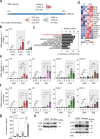
Fig. 1
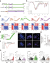
Fig. 1. The A30L protein enhances the inflammatory response in human PBMCs and monocytes.
a Schematic overview and domain organization of the MPXV E8L, A35R, and A30L proteins used in this study. b PBMCs were treated with A35R, A30L, or E8L (0.2 or 1 μg/mL) for 6 h. Total RNA extracted from the cells was then subjected to qRT-PCR analysis to measure the IL1B mRNA expression level. c Gene Ontology analysis of genes whose expression was upregulated in A30L-treated PBMCs. The list was ranked by the number of overlapping genes. d Heatmap showing the expression of inflammatory response-related genes in untreated and A30L-treated human PBMC samples. IL1Β, IL15, CX3CL1, and CXCL10 mRNA expression levels in human PBMCs (e) or monocytes (f) treated with A35R or A30L (0.2, 1, or 5 μg/mL for 6 h). g IL-1β concentrations in the supernatants of the indicated cells (human PBMCs, human monocytes, PMA-differentiated THP-1 cells, and murine BMDMs) treated with A30L (5 μg/ml for 18 h) were measured via ELISA. Western blots showing the levels of phosphorylated NF-κB (p-p65) (h), phosphorylated ERK (p-ERK), phosphorylated JNK (p-JNK), and phosphorylated p38 (p-p38) (i) in A30L-treated PBMCs or monocytes (5 μg/ml for 18 h). Statistical analysis was performed via one-way ANOVA with Tukey’s multiple comparison test or an unpaired t-test with the Mann‒Whitney test, and the results are presented as the means ± SDs. *p < 0.05; **p < 0.01. a.u. arbitrary unit, ns not significant.
Fig. 2
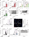
Fig. 2. A30L activates the NLRP3 inflammasome in human monocytes.
a Boxplot of IL1B expression levels in uninfected and MPXV-infected HeLa cells (from the GSE36854 cohort). b IL-1β and TNF concentrations in the supernatants of the indicated cells (human PBMCs and monocytes) stimulated with LPS (10 ng/mL for 4 h) followed by A30L (5 μg/mL for 18 h) were measured via ELISA. c IL-1β concentrations in the supernatants of PMA-differentiated THP-1 cells stimulated with LPS (10 ng/mL for 4 h) followed by A30L (5 μg/mL for 18 h), as measured by ELISA. d IL-1β concentrations in the supernatants of murine BMDMs and PMs stimulated with A30L (with the indicated concentration for 18 h), as measured by ELISA. e Western blot analysis of mature IL-1β and caspase-1 levels in culture supernatants (Sup.) and pro-IL-1β, pro-caspase-1, NLRP3, and ASC levels in cell lysates (Lys.) from human primary monocytes treated with A30L (5 μg/mL for 18 h) after LPS priming. f Western blot analysis of NLRP3 and mature IL-1β levels in human primary monocytes treated with A30L (5 μg/mL for 18 h) after NLRP3 knockdown and LPS priming. g IL-1β concentrations in the supernatants of monocytes stimulated with LPS (10 ng/mL for 4 h) followed by A30L (5 μg/mL for 18 h) after NLRP3 knockdown, as measured by ELISA. Representative images of immunofluorescence staining (scale bar = 20 μm; h) and quantitative analysis of the relative fluorescence intensities of DAPI and mtROS (i) in A30L-treated human primary monocytes (5 μg/mL for 4 h) after pretreatment with MitoTEMPO (100 μM for 2 h). The mtROS and nuclei were stained with MitoSOX Red (red) and DAPI (blue), respectively. IL-1β concentrations in the culture supernatants of cells incubated in the presence or absence of MitoTEMPO (j) or KCl (k). Statistical analysis was performed with an unpaired t-test with the Mann‒Whitney test or one-way ANOVA with Tukey’s multiple comparison test, and the results are presented as the means ± SDs. *p < 0.05; **p < 0.01; ***p < 0.001. ns not significant.
Fig. 3
Fig. 3. IAMP29, a core peptide of A30L, induces inflammatory responses in human monocytes.
a Schematic overview and sequence of the studied IAMP29 peptide. b Models of the top 5 ranked prediction results, featuring predicted aligned error diagrams showing two distinct domains: the N-terminal helical region and the C-terminal core region. c Predicted IDDT per position, indicating the confidence of the model at each residue. All five models exhibited similar values. d Structural prediction of the MPXV A30L protein using AlphaFold2. e CD spectrum of the IAMP29 peptide, displaying a typical curve of a random coil. These findings indicate that residues 40–69 of the MPXV A30L protein are mostly unstructured in solution, which is consistent with the AlphaFold2 model. Representative images of immunofluorescence staining (scale bar = 10 μm) (f) and quantitative analysis of the relative fluorescence intensities of DAPI and anti-FLAG staining (g). Human primary monocytes were treated with IAMP29 (25 μg/mL for 1 or 18 h). FLAG-IAMP29 and nuclei were stained with an anti-FLAG antibody (red) and DAPI (blue), respectively. h IL1Β, IL15, CX3CL1, and CXCL10 mRNA expression levels in IAMP29-treated human PBMCs or monocytes. Statistical analysis was conducted with an unpaired t-test with the Mann‒Whitney test or one-way ANOVA with Tukey’s multiple comparison test, and the results are presented as the means ± SDs. *p < 0.05; ****p < 0.0001. a.u. arbitrary unit, ns not significant.
Fig. 4
Fig. 4. IAMP29 activates the NLRP3 inflammasome in human monocytes and murine BMDMs.
Human IL-1β (a), human IL-18 (b), murine IL-1β (c), and human TNF (d) concentrations in the supernatants of the indicated IAMP29-treated cells after LPS priming, as measured by ELISA. e Western blots showing the levels of mature IL-1β and mature caspase-1 in culture supernatants (Sup.) and pro-IL-1β, pro-caspase-1, and β-actin in cell lysates (Lys.) from IAMP29-treated human primary monocytes (1, 5, or 25 μg/mL for 18 h) after LPS priming. f Western blots showing the levels of mature IL-1β in culture supernatants and pro-IL-1β and β-actin in lysates from IAMP29-treated murine BMDMs (40 or 80 μg/mL for 18 h) after LPS priming. g IL-1β concentrations in the supernatants of NLRP3-knockdown monocytes treated with IAMP29 (5 μg/mL for 18 h) after LPS priming, as measured with ELISA. h Representative images of immunofluorescence staining (scale bar = 10 μm) and quantitative analysis of the relative fluorescence intensities of DAPI and mtROS. Human primary monocytes were treated with IAMP29 (25 μg/mL for 4 h) after pretreatment with MitoTEMPO (100 μM for 2 h). The mtROS and nuclei were stained with MitoSOX Red (red) and DAPI (blue), respectively. IL-1β concentrations in the culture supernatants of cells incubated in the presence or absence of MitoTEMPO (i) or KCl (j). k Western blots showing the levels of mature IL-1β in culture supernatants and pro-IL-1β in the lysates of LPS-primed human primary monocytes treated with IAMP29 (5 μg/mL for 18 h) after KCl pretreatment (5 mM for 2 h). Statistical analysis was conducted with an unpaired t-test with the Mann‒Whitney test or one-way ANOVA with Tukey’s multiple comparison test, and the results are presented as the means ± SDs. *p < 0.05; **p < 0.01; ***p < 0.001; ****p < 0.0001. ns not significant.
Fig. 5
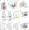
Fig. 5. IAMP29 activates the NLRP3 inflammasome via an interaction with PKM2.
a Heatmap showing the top protein hits identified by the LC‒MS analysis in PMA-differentiated THP-1 cells with FLAG or FLAG-IAMP29. b Volcano plot depicting the significant increase in PKM gene expression in A30L-treated human primary PBMCs. c Venn diagram illustrating the overlap of PKM2 in the three indicated groups. d, e Validation using PKM1 and PKM2 antibodies to detect immunoprecipitates from FLAG- or FLAG-IAMP29-treated cells (PMA-differentiated THP-1 and RAW 264.7 cells). f IL-1β concentrations in the supernatants of the indicated cells treated with IAMP29 (25 μg/mL for 18 h) after pretreatment with shikonin (5 μM for 2 h), as measured by ELISA. g Western blots showing the levels of mature IL-1β and mature caspase-1 in culture supernatants and pro-IL-1β, caspase-1, and β-actin in lysates of IAMP29-treated monocytes cultured in the absence or presence of shikonin (5 μM for 2 h). h IL-1β concentrations in the supernatants of IAMP29-treated monocytes (25 μg/mL for 18 h) with PKM2 knockdown, as measured by ELISA. IL-1β concentrations in the supernatants of shikonin-treated (i) or PKM2-knockdown monocytes (j) treated with the indicated NLRP3 inflammasome activators were measured by ELISA. k Western blots showing the levels of mature IL-1β and mature caspase-1 in culture supernatants and PKM2, pro-IL-1β, pro-caspase-1, and β-actin in the lysates of IAMP29-treated monocytes after PKM2 knockdown. Statistical analysis was conducted with an unpaired t-test with the Mann‒Whitney test or one-way ANOVA with Tukey’s multiple comparison test, and the results are presented as the means ± SDs. *p < 0.05; **p < 0.01; ***p < 0.001; ****p < 0.0001.
Fig. 6
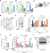
Fig. 6. IAMP29 treatment increases the PKM2 mRNA expression level and activates the NLRP3 inflammasome via PKR signaling.
Dose (a)- or time (b)-dependent expression of PKM1 and PKM2 in IAMP29-treated human monocytes. c Western blots showing the levels of PKM2 in the lysates of IAMP29-treated human monocytes or PMA-differentiated THP-1 cells. d, e The ECAR in PMA-differentiated THP-1 cells treated with IAMP29 (25 μg/mL for 18 h) after LPS stimulation. f Hif1a mRNA expression in IAMP29-treated murine BMDMs (25 μg/mL for 3, 6, and 18 h). g Pkm1 and Pkm2 mRNA expression in IAMP29-treated murine BMDMs after pretreatment with the indicated inhibitors. h IL1B and IL18 mRNA expression in A30L- or IAMP29-treated human monocytes after pretreatment with the indicated inhibitors. i Western blots showing the levels of mature IL-1β in culture supernatants and pro-IL-1β, PKR, and phospho-PKR in the lysates of IAMP29-treated monocytes cultured in the presence of the indicated inhibitors. Statistical analysis was conducted with an unpaired t-test with the Mann‒Whitney test or one-way ANOVA with Tukey’s multiple comparison test, and the results are presented as the means ± SDs. *p < 0.05; **p < 0.01; ***p < 0.001; ****p < 0.0001. a.u. arbitrary unit, ns not significant.
Fig. 7
Fig. 7. IAMP29 treatment restricts the intracellular growth of NTMs in human and murine macrophages.
a The cytotoxicity of IAMP29 in PBMCs and monocytes was assessed via a CCK-8 assay. The cells were treated with IAMP29 (0.2, 1, 5, or 25 μg/mL for 24 h) after LPS or BCG pretreatment. b Intracellular survival of smooth strains of Mboll and Mabc in IAMP29-treated human primary MDMs after Mboll or Mabc infection (MOI = 1 for 2 h), as determined by a CFU assay. IL-1β (c) and TNF (d) concentrations in the supernatants of IAMP29-treated human MDMs. The cells were treated with IAMP29 (25 μg/mL for 18 h) after Mboll or Mabc infection (MOI = 1 for 2 h). e Western blots showing the levels of mature IL-1β and mature caspase-1 in culture supernatants (Sup.) and pro-IL-1β, pro-caspase-1, NLRP3, and ASC in cell lysates (Lys.) of Mboll- or Mabc-infected human MDMs treated with IAMP29 (25 μg/mL for 18 h). Intracellular survival of Mboll (f) and Mabc (g) in human MDMs with PKM2 knockdown after Mboll or Mabc infection (MOI = 1 for 2 h), as determined by a CFU assay. Intracellular survival of Mboll (h) and Mabc (i) in Mboll- or Mabc-infected murine BMDMs treated with IAMP29 (40 μg/mL for 18 h) after pretreatment with MitoTEMPO (1 and 10 μM for 2 h) or 2-DG (10 and 100 μM for 2 h), as determined by a CFU assay. Statistical analysis was conducted with an unpaired t-test with the Mann‒Whitney test or one-way ANOVA with Tukey’s multiple comparison test, and the results are presented as the means ± SDs. *p < 0.05; **p < 0.01; ***p < 0.001; ****p < 0.0001. ns not significant.
Fig. 8
Fig. 8. IAMP29 treatment activates apoptosis and pyroptosis in human leukemic cells.
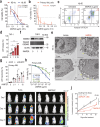
a, b The cytotoxic effect of IAMP29 on human KG-1α, HL-60 (48 h), and patient-derived AML cells (72 h) was assessed using a CCK-8 assay. c Flow cytometry analysis of IAMP29-treated HL-60 cells (2 μg/mL for 24 h) using annexin V/PI staining. d LDH release from IAMP29-treated THP-1 cells. The cells were treated with IAMP29 (1, 5, or 25 μg/mL for 24 h) and assessed with an LDH assay kit. e Transmission electron micrographs of HL-60 and KG-1α cells treated with IAMP29 (5 μg/mL for 24 h). f Western blots showing the levels of cleaved GSDMD in culture supernatants and GSDMD in the lysates of THP-1 cells treated with IAMP29 (1 and 5 μg/mL for 36 h). g LDH release from IAMP29-treated primary AML cells with NLRP3 knockdown. The cells were treated with IAMP29 (0.1, 1, or 10 μg/mL for 24 h) and assessed with an LDH assay kit. h The cytotoxic effect of IAMP29 in the presence or absence of z-VAD was assessed using a CCK-8 assay. i Luminescence intensity in NSG mice treated with PLGA or IAMP29-P (36 mg/kg) for 7 days after confirming the engraftment of K562-luc cells. NSG mice were orthotopically implanted with K562-luc cells (5 × 106 cells/mouse) and injected with IAMP29-P through the tail vein twice a week for 2 weeks. The indicated dates represent “days” following the first IAMP29-P treatment. j Quantification of the luminescence intensity of photons released from each tumor in NSG mice treated with PLGA or IAMP29-P. Statistical analysis was conducted with an unpaired t-test with the Mann‒Whitney test or one-way ANOVA with Tukey’s multiple comparison test, and the results are presented as the means ± SDs. *p < 0.05; **p < 0.01; ***p < 0.001; ****p < 0.0001. ns not significant.

References

    1. De Clercq, E., Jiang, Y. & Li, G. Therapeutic strategies for human poxvirus infections: monkeypox (mpox), smallpox, molluscipox, and orf. Travel Med. Infect. Dis.52, 102528 (2023). - PMC - PubMed
    1. McFadden, G. Poxvirus tropism. Nat. Rev. Microbiol.3, 201–213 (2005). - PMC - PubMed
    1. Hung, Y. P. et al. A brief on new waves of monkeypox and vaccines and antiviral drugs for monkeypox. J. Microbiol. Immunol. Infect.55, 795–802 (2022). - PMC - PubMed
    1. Lum, F. M. et al. Monkeypox: disease epidemiology, host immunity and clinical interventions. Nat. Rev. Immunol.22, 597–613 (2022). - PMC - PubMed
    1. Rahman, M. M. & McFadden, G. Role of cytokines in poxvirus host tropism and adaptation. Curr. Opin. Virol.57, 101286 (2022). - PMC - PubMed

LinkOut - more resources