. 2024 Nov 7;22(1):521.
doi: 10.1186/s12916-024-03724-1.

ANXA4 restricts HBV replication by inhibiting autophagic degradation of MCM2 in chronic hepatitis B

Affiliations

ANXA4 restricts HBV replication by inhibiting autophagic degradation of MCM2 in chronic hepatitis B

Luo Yang et al. BMC Med. .

Abstract

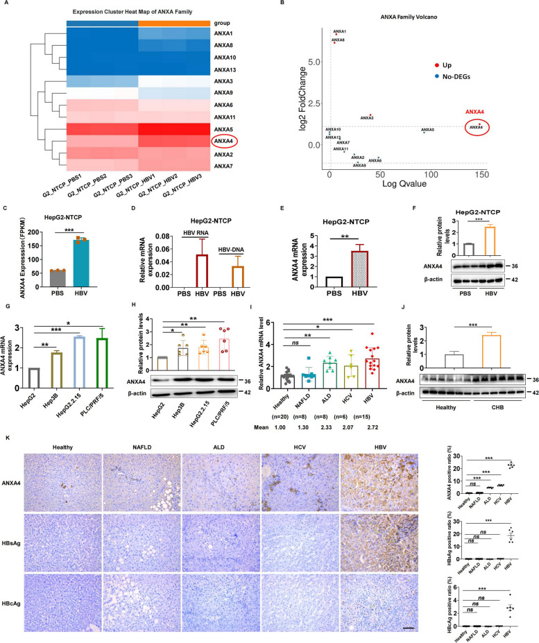
Background: Hepatitis B virus (HBV) is an enveloped DNA virus that causes chronic hepatitis B (CHB) infection. Annexin, a Ca2+-activated protein, is widely expressed in various organs and tissues and has potential utility in disease diagnosis and treatment. However, the relationship between the annexin family and CHB remains unclear.

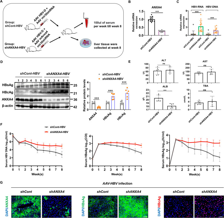
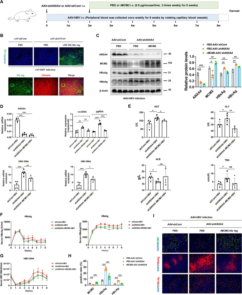
Methods: Clinical samples from hepatitis patients and donors or healthy individuals were collected. Transcriptome sequencing in CHB liver tissues and HBV-infected cells were performed. HepG2.2.15 cells with the full-length HBV genome and HBV-infected HepG2-NTCP cell models were established. HBV-infected mouse model was constructed and adeno-associated virus was utilized.

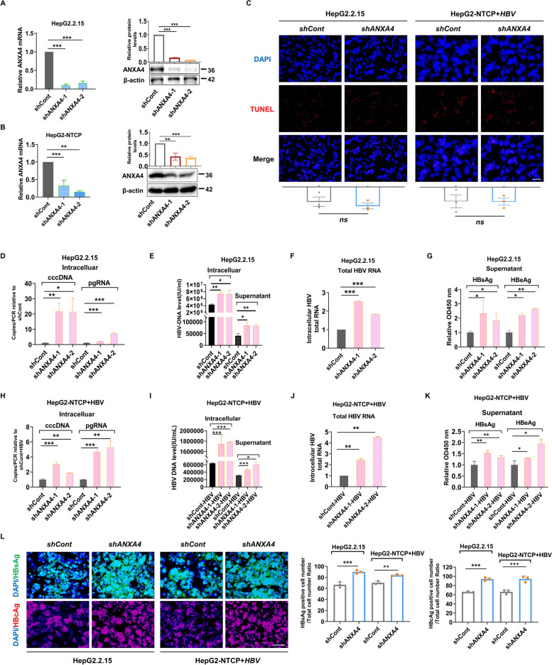
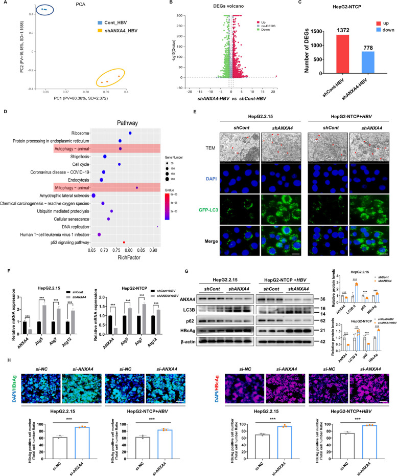
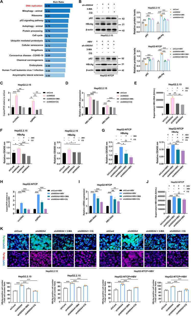
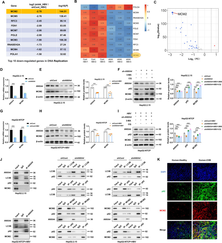
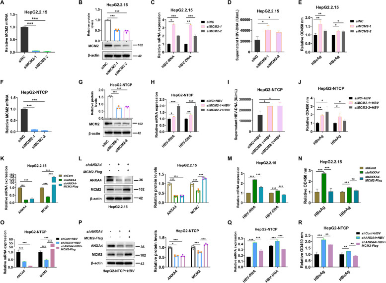
Results: ANXA4 expression was elevated during CHB infection. ANXA4 knockdown promoted HBV replication and aggravated liver injury, while ANXA4 overexpression alleviated that. Mechanistically, autophagy pathway was activated by ANXA4 deficiency, promoting autophagic degradation of minichromosome maintenance complex component 2 (MCM2). MCM2 inhibition activated HBV replication, while MCM2 overexpression attenuated ANXA4 deficiency-induced HBV replication and liver injury. Clinically, the expression of hepatitis B viral protein was negatively correlated with the ANXA4 levels, and CHB patients with high ANXA4 levels (> 8 ng/ml) showed higher sensitivity to interferon therapy.

Conclusions: ANXA4 functions as a protective factor during HBV infection. ANXA4 expression is elevated under HBV attack to restrict HBV replication by inhibiting autophagic degradation of MCM2, thereby alleviating liver injury and suppressing the CHB infection process. ANXA4 also enhances the sensitivity of CHB patients to interferon therapy. Therefore, ANXA4 is expected to be a new target for CHB treatment and prognostic evaluation.

Keywords: Annexin A4 (ANXA4); Autophagy; Chronic hepatitis B (CHB); HBV replication; Minichromosome maintenance complex component 2 (MCM2).


Conflict of interest statement

The authors declare no competing interests.

Figures

Fig. 1
Fig. 1
ANXA4 expression is elevated during CHB infection. AB Gene expression heatmap and volcano plot of ANXA family in HBV-infected HepG2-NTCP cells and non-HBV-infected HepG2-NTCP cells. C ANXA4 FPKM values in the sequencing group. D HBV RNA and HBV DNA levels in the HBV-infected and control groups. E ANXA4 mRNA levels in the HBV-infected and control groups. F ANXA4 protein expression in the infected group versus the control group. G ANXA4 mRNA levels in HBV-related cells. H ANXA4 protein expression levels in HBV-related cells. I The levels of ANXA4 mRNA expression in liver tissues of healthy individuals and hepatitis patients with different etiologies were determined by RT-qPCR. J ANXA4 protein expression in CHB and healthy liver tissues. K Immunohistochemistry results of ANXA4 expression in liver tissues (bar = 100 µm). The values are presented as the means ± SEMs. * p < 0.05, ** p < 0.01, *** p < 0.001
Fig. 2
Fig. 2
ANXA4 inhibits HBV replication and viral protein expression. A, B ANXA4 knockdown was validated at the mRNA and protein level. C TUNEL staining in HepG2.2.15 and HepG2-NTCP + HBV cells with shCont or shANXA4 transfection (bar = 50 µm). D, H Intracellular ccDNA and pgRNA levels. E, I Intracellular and supernatant levels of HBV DNA. F, J Total HBV RNA levels. G, K Levels of HBsAg and HBeAg in the cell supernatant. L HBsAg and HBcAg immunofluorescence in HepG2.2.15 cells and HBV-infected HepG2-NTCP cells transfected with shCont or shANXA4 (bar = 50 µm). The values are presented as the means ± SEMs. * p < 0.05, ** p < 0.01, *** p < 0.001
Fig. 3
Fig. 3
ANXA4 attenuates HBV replication and liver injury in mice. A Flow chart of the experimental setup. B ANXA4 mRNA expression in liver tissues from HBV-infected mice treated with AAV-shCont or AAV-shANXA4. C Levels of HBV RNA and HBV DNA in mouse liver tissues. D The expression levels of HBsAg, HBcAg, and ANXA4 in the above groups were measured by WB. E Measurement of serum biochemical indexes in the above mice. F Serum HBV genomic DNA was quantified by real-time PCR. Serum HBeAg and HBsAg were quantified by a chemiluminescent microparticle immunoassay. G ANXA4, HBcAg, and HBsAg levels were determined by IF analysis (bar = 50 µm). The values are presented as the means ± SEMs. * p < 0.05, ** p < 0.01, *** p < 0.001
Fig. 4
Fig. 4
The autophagy pathway is activated when ANXA4 is inhibited. A PCA of sequencing samples. B Volcano plot of DEGs. C Histogram of DEGs. A total of 1372 DEGs were upregulated and 778 were downregulated in HBV-infected HepG2-NTCP cells transduced with shANXA4 compared to those transduced with shCont. D Top 15 enriched KEGG pathways. E Transmission electron microscopy (bar = 2 µm) and fluorescence microscopy (bar = 10 µm) indicated that ANXA4 downregulation increased autophagosome formation. The red arrows indicate autophagosomes. F The mRNA levels of autophagy markers were measured by RT-qPCR. G ANXA4, LC3B, p62, and HBcAg expression was analyzed by WB. H HBsAg and HBcAg immunofluorescence in HepG2 2.2.15 cells and HBV-infected HepG2-NTCP cells treated with si-NC or si-ANXA4 (bar = 50 µm). The values are presented as the means ± SEMs. * p < 0.05, ** p < 0.01, *** p < 0.001
Fig. 5
Fig. 5
Inhibition of ANXA4 promotes HBV replication via activating autophagy. A Enrichment ratios of KEGG pathways in HBV-infected HepG2-NTCP cells. B p62 and HBcAg levels in shCont- and shANXA4-transduced HepG2 2.2.15 cells and HBV-infected HepG2-NTCP cells treated with CQ or 3-MA. C cccDNA and pgRNA levels in HepG2.2.15 cells with above treatments. D HBV DNA and HBV RNA were detected in HepG2.2.15 cells. EF HBV DNA, HBsAg, and HBeAg secreted by HepG2.2.15 cells were detected. G HBV DNA and HBV RNA levels in HBV-infected HepG2-NTCP cells were measured by RT-qPCR. H cccDNA and pgRNA levels in HBV-infected HepG2-NTCP cells with above treatments. IJ HBV DNA and HBV RNA in HBV-infected HepG2-NTCP cells were detected. K HBsAg and HBcAg expression was analyzed by immunofluorescence staining (bar = 50 µm). The values are presented as the means ± SEMs. * p < 0.05, ** p < 0.01, *** p < 0.001
Fig. 6
Fig. 6
ANXA4 silencing enhances HBV replication by promoting autophagic degradation of MCM2. A Top DEGs in the DNA replication pathway. B, C Heatmap and volcano plot of the top DEGs in the DNA replication pathway. D, E ANXA4 and MCM2 levels in shCont- and shANXA4-transduced HepG2.2.15 cells. F ANXA4, p62, and MCM2 expression in HepG2.2.15 cells transduced with shANXA4 and treated with an autophagy inhibitor. G, H ANXA4 and MCM2 levels in shCont- and shANXA4-transduced HBV-infected HepG2-NTCP cells. I ANXA4, p62, and MCM2 expression in HBV-infected HepG2-NTCP cells transduced with shANXA4 and treated with an autophagy inhibitor. J Protein interactions were detected by immunoprecipitation in HepG2.2.15 cells and HBV-infected HepG2-NTCP cells. K p62 and MCM2 co-localization analysis in healthy individuals and CHB patients (bar = 50 µm). The values are presented as the means ± SEMs. * p < 0.05, ** p < 0.01, *** p < 0.001
Fig. 7
Fig. 7
MCM2 inhibits HBV replication and viral protein expression in vitro. AB, FG MCM2 mRNA and protein levels were reduced in HepG2.2.15 cells and HBV-infected HepG2-NTCP cells treated with si-MCM2. C, H HBV DNA and HBV RNA levels in HepG2.2.15 cells and HBV-infected HepG2-NTCP cells were measured after MCM2 knockdown. D, I HBV DNA levels in the cell supernatant were measured. E, G The levels of HBsAg and HBeAg in the supernatant were measured by ELISA. KL, OP mRNA and protein levels of ANXA4 and MCM2 in HepG2.2.15 cells and HBV-infected HepG2-NTCP cells treated with shANXA4 and MCM2-Flag. M, Q HBV DNA and HBV RNA levels in HepG2.2.15 cells and HBV-infected HepG2-NTCP cells after shANXA4 and MCM2-Flag treatment. N, R The levels of HBsAg and HBeAg in the supernatant were evaluated. The values are presented as the means ± SEMs. * p < 0.05, ** p < 0.01, *** p < 0.001
Fig. 8
Fig. 8
MCM2 overexpression attenuates HBV replication activity and liver injury in ANXA4-deficient mice. A Schematic overview of the experimental setup. B His tag labeled rMCM2 proteins can be engulfed by the liver and hepatocytes (bar = 50 µm). C Protein expression analysis of ANXA4, MCM2, HBsAg, and HBcAg in HBV-infected mice with AAV-shCont or AAV-shANXA4 treatment together with PBS or rMCM2 injection. D Relative ANXA4 mRNA expression, HBV RNA, HBV DNA, cccDNA, and pgRNA levels in above groups. E Measurement of serum AST, ALT, ALB, and TBA in the above mice. F Serum HBeAg and HBsAg were quantified by a chemiluminescent microparticle immunoassay in “shCont + HBV,” “shANXA4 + HBV,” and “shANXA4 + rMCM2 + HBV” group. G Serum HBV genomic DNA was also quantified in “shCont + HBV,” “shANXA4 + HBV,” and “shANXA4 + rMCM2 + HBV” mice. H, I IF of MCM2, HBcAg, and HBsAg expression (bar = 50 µm). The values are presented as the means ± SEMs. * p < 0.05, ** p < 0.01, *** p < 0.001

Similar articles

Cited by

References

    1. Mohsen N, Haidong W, Rafael L, et al. Global, regional, and national age-sex specific all-cause and cause-specific mortality for 240 causes of death, 1990–2013: a systematic analysis for the Global Burden of Disease Study 2013. Lancet. 2015;385(9963):117–71. - PMC - PubMed
    1. Trépo C, Chan HLY, Lok A. Hepatitis B virus infection. Lancet. 2014;384(9959):2053–63. - PubMed
    1. Asandem DA, Segbefia SP, Kusi KA, Bonney JHK. Hepatitis B virus infection: a mini review. Viruses. 2024;16(5):724. - PMC - PubMed
    1. Liu Z, Lin C, Mao X, Guo C, Suo C, Zhu D, et al. Changing prevalence of chronic hepatitis B virus infection in China between 1973 and 2021: a systematic literature review and meta-analysis of 3740 studies and 231 million people. Gut. 2023;72(12):2354–63. - PMC - PubMed
    1. Jose-Abrego A, Roman S, Laguna-Meraz S, Panduro A. Host and HBV interactions and their potential impact on clinical outcomes. Pathogens (Basel, Switzerland). 2023;12(9):1146. - PMC - PubMed

Publication types

MeSH terms

LinkOut - more resources