. 2024 Nov 7;24(1):1365.
doi: 10.1186/s12885-024-13074-z.

Targeting LLT1 as a potential immunotherapy option for cancer patients non-responsive to existing checkpoint therapies in multiple solid tumors

Affiliations

Targeting LLT1 as a potential immunotherapy option for cancer patients non-responsive to existing checkpoint therapies in multiple solid tumors

Tirtha Mandal et al. BMC Cancer. .

Abstract

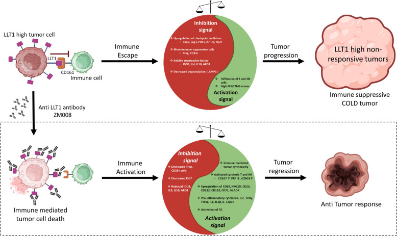
Background: High levels of LLT1 expression have been found in several cancers, where it interacts with CD161 on NK cells to facilitate tumor immune escape. Targeting LLT1 could potentially relieve this inhibitory signal and enhance anti-tumor responses mediated through NK cells. Using the 'The Cancer Genome Atlas' (TCGA) database, we investigated the role of LLT1 in the tumor microenvironment (TME) across various cancers. Identifying such biomarkers could create new therapeutic options for patients in addition to complementing existing immunotherapies.

Methods: LLT1 expression was evaluated in 33 cancers using TCGA transcriptome data. Univariate Cox regression analysis was employed to assess the correlation of LLT1 expression with patient survival. The relationship between LLT1 expression with immune infiltrates, immune gene signatures, and cancer genomic biomarkers (TMB, MSI, and MMR) was also investigated. Immunofluorescence studies were conducted to validate LLT1 expression in tumors. Furthermore, using the CRI iAtlas data, we evaluated LLT1 distribution and its correlation with other immune checkpoint genes in patients non-responsive to existing immune checkpoint therapies across multiple solid cancers.

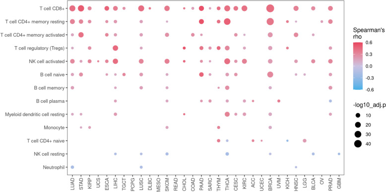
Results: High expression of LLT1 was observed in 12 cancers, including BRCA, CHOL, ESCA, GBM, HNSC, KIRC, KIRP, LIHC, LUAD, STAD, SARC, and PCPG. In certain cancers like COAD, KICH, and KIRC, high LLT1 expression was associated with poor prognosis. Further analysis revealed that upregulated LLT1 was associated with an abundance of NK and T cell infiltrates in the TME, as well as exhaustive immune biomarkers, and inversely associated with pro-inflammatory and tumor suppressor signatures. High LLT1 expression is also positively correlated with genomic biomarkers in certain cancers. Immunofluorescence studies confirmed moderate to high LLT1 expression in immune-resistant prostate cancer, glioma, ovarian cancer, and immune-sensitive liver cancer cell lines. An independent assessment of clinical cohorts from CRI iAtlas showed a correlation of upregulated LLT1 with multiple immunosuppressive genes in patients non-responsive to current ICIs.

Conclusions: The biomarker analysis revealed a clear association between elevated LLT1 expression and an immunosuppressive TME in patient cohorts from TCGA and clinical databases. Therefore, this study provides a foundation for utilizing LLT1 as a potential target to improve clinical responses in ICI non-responsive patients with upregulated LLT1.

Keywords: Immune Checkpoint Inhibitors; Immunotherapy; LLT1; Natural Killer Cells; Tumor Biomarkers; Tumor Microenvironment.


Conflict of interest statement

TM, SK, AT, AD, SB, YM, and SK are employees of Zumutor Biologics. SG is a former employee of Zumutor Biologics. MG, AT, AD, SB, and YM have stock options in Zumutor Biologics, the company that owns the anti-LLT1 antibody, ZM008. MSM and GR declared no competing interests.

Figures

Fig. 1
Fig. 1
LLT1 expression across tumors and normal adjacent tissues. A LLT1 expression in the indicated cancer types. The Mann-Whitney test was used to compare the difference in LLT1 expression between the tumor and its adjacent normal. Cancers that have significantly higher LLT1 expression in the tumor tissue compared to the normal are shown on the left. B Cancer types that did not contain data for adjacent normal tissue in the GDC database accessed through the UCSC Xena browser. C The expression levels of LLT1 transcripts across different AJCC pathologic stages compared to the adjacent normal expression levels through the Mann-Whitney Test. LLT1 expression tends to be higher in higher stages compared to normals. *P < 0.05, ** P < 0.01, *** P < 0.001. D Plot showing the correlation between the expression of LLT1 and methylation across different cancer types. The size of the dots represents the adjusted p-values, and the color represents the correlation coefficient. Unfaded dots represent statistically significant data points, while the faded dots represent statistically insignificant data points. E CNV profile showing percentage of heterozygous or homozygous CNV, including amplification or deletion for the LLT1 gene in each cancer. F Plot showing the correlation between the expression of LLT1 and CNV across different cancer types. The size of the dots represents the adjusted p-values, and the color represents the correlation coefficient. Unfaded dots represent statistically significant data points, while the faded dots represent statistically insignificant data points
Fig. 1
Fig. 1
LLT1 expression across tumors and normal adjacent tissues. A LLT1 expression in the indicated cancer types. The Mann-Whitney test was used to compare the difference in LLT1 expression between the tumor and its adjacent normal. Cancers that have significantly higher LLT1 expression in the tumor tissue compared to the normal are shown on the left. B Cancer types that did not contain data for adjacent normal tissue in the GDC database accessed through the UCSC Xena browser. C The expression levels of LLT1 transcripts across different AJCC pathologic stages compared to the adjacent normal expression levels through the Mann-Whitney Test. LLT1 expression tends to be higher in higher stages compared to normals. *P < 0.05, ** P < 0.01, *** P < 0.001. D Plot showing the correlation between the expression of LLT1 and methylation across different cancer types. The size of the dots represents the adjusted p-values, and the color represents the correlation coefficient. Unfaded dots represent statistically significant data points, while the faded dots represent statistically insignificant data points. E CNV profile showing percentage of heterozygous or homozygous CNV, including amplification or deletion for the LLT1 gene in each cancer. F Plot showing the correlation between the expression of LLT1 and CNV across different cancer types. The size of the dots represents the adjusted p-values, and the color represents the correlation coefficient. Unfaded dots represent statistically significant data points, while the faded dots represent statistically insignificant data points
Fig. 2
Fig. 2
Association of LLT1 expression with patient prognosis. A Univariate Cox-Proportional Hazards regression was performed with LLT1 expression as the independent variable, and overall survival information was obtained through the clinical files associated with each of the cancers. LLT1 was found to be a prognostic risk factor in KIRC, KICH, and COAD with Hazard ratios significantly more than 1. (KIRC-HR = 1.067, KICH-HR = 2.114, and COAD-HR = 1.114) B The empirically estimated Kaplan Meier Curves for these three cancers. The ‘High’ group shown in red is the sub-population of patients whose LLT1 expression is higher than its median as measured in TPM, while the ‘Low’ group shown in blue is the population of patients who have LLT1 expression lower than its median as measured in TPM
Fig. 2
Fig. 2
Association of LLT1 expression with patient prognosis. A Univariate Cox-Proportional Hazards regression was performed with LLT1 expression as the independent variable, and overall survival information was obtained through the clinical files associated with each of the cancers. LLT1 was found to be a prognostic risk factor in KIRC, KICH, and COAD with Hazard ratios significantly more than 1. (KIRC-HR = 1.067, KICH-HR = 2.114, and COAD-HR = 1.114) B The empirically estimated Kaplan Meier Curves for these three cancers. The ‘High’ group shown in red is the sub-population of patients whose LLT1 expression is higher than its median as measured in TPM, while the ‘Low’ group shown in blue is the population of patients who have LLT1 expression lower than its median as measured in TPM
Fig. 3
Fig. 3
LLT1-related gene set enrichment analysis. The Normalized Enrichment Score (NES) for important cancer-relevant pathways from the Hallmark Pathways gene sets was estimated across 33 cancers. Positive NES indicates an enrichment of that pathway in the ‘High LLT1 expression’ group while a negative NES score indicates an enrichment of that pathway in the ‘Low LLT1 expression’ group in a given cancer. The sizes of the dots are indicative of the level of enrichment
Fig. 4
Fig. 4
Relationship between LLT1 expression and immune infiltrates in the TME. Spearman’s purity-adjusted correlation coefficient between the infiltration score (estimated by CIBERSORT-ABS from TCGA transcriptomics data) and LLT1 expression is shown in this figure across cancers. The size of the dots represents the adjusted p-values, and the color represents the correlation coefficient
Fig. 5
Fig. 5
Correlation of LLT1 expression with expression of immunoregulatory genes. A Pearson correlation between relevant immune checkpoint genes and LLT1 expression measured in TPM is shown in this heat map. The thatched tiles represent cases where the correlation coefficient could not be reliably estimated owing to poor data. P-values are coded as follows: * < 0.05, ** < 0.01, *** < 0.001. B Individual correlation maps of select immune checkpoint genes are shown. The correlation coefficient (r) values for each gene plot are shown as an inset in each plot. C The gene ontology of the positively correlated gene set is shown. The size of the dots represents the level of abundance and the color represents the adjusted p-values
Fig. 5
Fig. 5
Correlation of LLT1 expression with expression of immunoregulatory genes. A Pearson correlation between relevant immune checkpoint genes and LLT1 expression measured in TPM is shown in this heat map. The thatched tiles represent cases where the correlation coefficient could not be reliably estimated owing to poor data. P-values are coded as follows: * < 0.05, ** < 0.01, *** < 0.001. B Individual correlation maps of select immune checkpoint genes are shown. The correlation coefficient (r) values for each gene plot are shown as an inset in each plot. C The gene ontology of the positively correlated gene set is shown. The size of the dots represents the level of abundance and the color represents the adjusted p-values
Fig. 6
Fig. 6
Correlation between LLT1 expression and tumor genomic markers. A Pearson correlation between LLT1 expression in TPM and TMB score is shown in this radar plot. Cancers where the correlation is significant (p < 0.05) are underlined in the plot (KICH, COAD, OV, TGCT, UCEC). B Pearson correlation between LLT1 expression in TPM and MSI score is shown in this radar plot. Cancers where the correlation is significant (p < 0.05) are underlined in the plot (PRAD, TGCT, LAML, BRCA, COAD, LUAD, LUSC, OV). C Pearson correlation between mismatch repair genes and LLT1 expression is shown in this figure. P-values are coded as follows: * < 0.05, ** < 0.01, *** < 0.001
Fig. 6
Fig. 6
Correlation between LLT1 expression and tumor genomic markers. A Pearson correlation between LLT1 expression in TPM and TMB score is shown in this radar plot. Cancers where the correlation is significant (p < 0.05) are underlined in the plot (KICH, COAD, OV, TGCT, UCEC). B Pearson correlation between LLT1 expression in TPM and MSI score is shown in this radar plot. Cancers where the correlation is significant (p < 0.05) are underlined in the plot (PRAD, TGCT, LAML, BRCA, COAD, LUAD, LUSC, OV). C Pearson correlation between mismatch repair genes and LLT1 expression is shown in this figure. P-values are coded as follows: * < 0.05, ** < 0.01, *** < 0.001
Fig. 7
Fig. 7
LLT1 expression and correlation with immune regulation in ICI non-responders. A The expression of LLT1 in each non-responsive patient is divided by the drug used to treat the patient and the tissue type. The horizontal dotted lines indicate the median LLT1 value of the pooled dataset as described in the method’s section. B The topmost axis indicates the cancer under consideration. The second from the top axis indicates the drug to which the patients were unresponsive. The tiles are colored according to the hypergeometric overlap test’s p-value. The y-axis indicates the immune genes used in the overlap analysis along with LLT1. P-values are coded as follows: * < 0.05, ** < 0.01, *** < 0.001
Fig. 7
Fig. 7
LLT1 expression and correlation with immune regulation in ICI non-responders. A The expression of LLT1 in each non-responsive patient is divided by the drug used to treat the patient and the tissue type. The horizontal dotted lines indicate the median LLT1 value of the pooled dataset as described in the method’s section. B The topmost axis indicates the cancer under consideration. The second from the top axis indicates the drug to which the patients were unresponsive. The tiles are colored according to the hypergeometric overlap test’s p-value. The y-axis indicates the immune genes used in the overlap analysis along with LLT1. P-values are coded as follows: * < 0.05, ** < 0.01, *** < 0.001
Fig. 8
Fig. 8
The expression of LLT1 in different cell lines by confocal microscopy. A The surface expression of LLT1 was examined using confocal microscopy. An anti-LLT1 antibody (4C7) was used to probe the cells. DAPI was used for counterstaining the nucleus. The left panel shows cells treated with a secondary antibody attached to a fluorescent probe (Alexa Fluor 488 goat anti-mouse IgG), while the right panel shows cells treated with the anti-LLT1 antibody (4C7) followed by the secondary antibody attached to a fluorescent probe. The green color indicates the binding of the anti-LLT1 antibody to the target antigen on respective cell lines. The binding signal of the anti-LLT1 antibody (green color) was observed only in LLT1 transfected CHO-K1 cells (right panel), while no binding was detected in untransfected CHO-K1 (control) cells (left panel). The scale bar for both panels is 10 μm. B The surface expression of LLT1 on different tumor cell lines, including prostate cancer (hormone-refractory PC3, hormone-sensitive DU145, and 22Rv1), liver cancer (Hep G2), glioma (LN229), and ovarian cancer (SK-O-V3), was also investigated. DAPI was used for counterstaining the nucleus, while the same anti-LLT1 antibody (4C7) followed by secondary antibody with the fluorescent probe (Alexa Fluor 488 goat anti-mouse IgG) were used to probe the cells. The green color indicates the binding of the anti-LLT1 antibody to the target antigen on respective tumor cell lines. The scale bar for both panels is 10 μm
Fig. 8
Fig. 8
The expression of LLT1 in different cell lines by confocal microscopy. A The surface expression of LLT1 was examined using confocal microscopy. An anti-LLT1 antibody (4C7) was used to probe the cells. DAPI was used for counterstaining the nucleus. The left panel shows cells treated with a secondary antibody attached to a fluorescent probe (Alexa Fluor 488 goat anti-mouse IgG), while the right panel shows cells treated with the anti-LLT1 antibody (4C7) followed by the secondary antibody attached to a fluorescent probe. The green color indicates the binding of the anti-LLT1 antibody to the target antigen on respective cell lines. The binding signal of the anti-LLT1 antibody (green color) was observed only in LLT1 transfected CHO-K1 cells (right panel), while no binding was detected in untransfected CHO-K1 (control) cells (left panel). The scale bar for both panels is 10 μm. B The surface expression of LLT1 on different tumor cell lines, including prostate cancer (hormone-refractory PC3, hormone-sensitive DU145, and 22Rv1), liver cancer (Hep G2), glioma (LN229), and ovarian cancer (SK-O-V3), was also investigated. DAPI was used for counterstaining the nucleus, while the same anti-LLT1 antibody (4C7) followed by secondary antibody with the fluorescent probe (Alexa Fluor 488 goat anti-mouse IgG) were used to probe the cells. The green color indicates the binding of the anti-LLT1 antibody to the target antigen on respective tumor cell lines. The scale bar for both panels is 10 μm
Fig. 9
Fig. 9
Summary of LLT1’s role in TME of cancers and possible implications for therapy. Interaction of LLT1 with CD161 on immune cells like T cells and NK cells impairs their anti-tumor activity, leading to immune escape and tumor progression. As our study suggests, high LLT1 expression is associated with various inhibitory signals in the TME, as shown in the yin-yang-like representation in the top panel. These inhibitory signals induce immune dysfunction, thereby creating a TME conducive to immune-resistant “COLD” tumors. Blocking the inhibitory LLT1-CD161 axis could shift the balance towards immune activation, mediating immune cell-mediated killing of tumors and prompting tumor regression. Anti-LLT1 antibody ZM008 could provide a potential treatment option for these immune-resistant tumors. This antibody drug could potentially transform the TME into an immune-responsive “HOT” tumor. The boxed panel in the figure indicates the proposed hypothesis of this prospective anti-LLT1 antibody-based immunotherapy approach

References

    1. Braud VM, Meghraoui-Kheddar A, Elaldi R, Petti L, Germain C, Anjuere F. LLT1-CD161 interaction in cancer: promises and challenges. Front Immunol. 2022;13: 847576. - PMC - PubMed
    1. Germain C, Bihl F, Zahn S, Poupon G, Dumaurier MJ, Rampanarivo HH, et al. Characterization of alternatively spliced transcript variants of CLEC2D gene. J Biol Chem. 2010;285(46):36207–15. - PMC - PubMed
    1. Boles KS, Barten R, Kumaresan PR, Trowsdale J, Mathew PA. Cloning of a new lectin-like receptor expressed on human NK cells. Immunogenetics. 1999;50(1–2):1–7. - PubMed
    1. Skalova T, Blaha J, Harlos K, Duskova J, Koval T, Stransky J, et al. Four crystal structures of human LLT1, a ligand of human NKR-P1, in varied glycosylation and oligomerization states. Acta Crystallogr D Biol Crystallogr. 2015;71(Pt 3):578–91. - PMC - PubMed
    1. Germain C, Meier A, Jensen T, Knapnougel P, Poupon G, Lazzari A, et al. Induction of lectin-like transcript 1 (LLT1) protein cell surface expression by pathogens and interferon-gamma contributes to modulate immune responses. J Biol Chem. 2011;286(44):37964–75. - PMC - PubMed

MeSH terms

LinkOut - more resources