

Development of a deep learning model for automated detection of calcium pyrophosphate deposition in hand radiographs
- PMID: 39512610
- PMCID: PMC11540928
- DOI: 10.3389/fmed.2024.1431333
Development of a deep learning model for automated detection of calcium pyrophosphate deposition in hand radiographs
Abstract
Background: Calcium pyrophosphate deposition (CPPD) disease is a leading cause of arthritis, which can mimic or strongly interfere with other rheumatic diseases such as gout, osteoarthritis (OA) or rheumatoid arthritis (RA). In the recently established ACR/EULAR CPPD classification criteria, calcification and OA features of the wrist and hand joints are substantial features.
Objectives: To develop and test a deep-learning algorithm for automatically and reliably detecting CPPD features in hand radiographs, focusing on calcification of the triangular fibrocartilage complex (TFCC) and metacarpophalangeal (MCP)-2 and -3 joints, in separate or combined models.
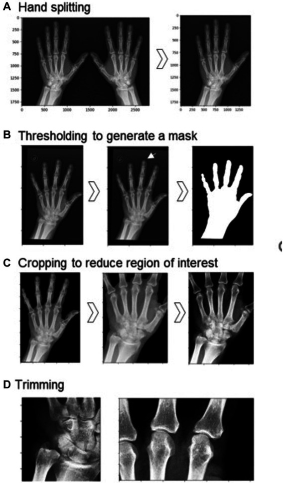
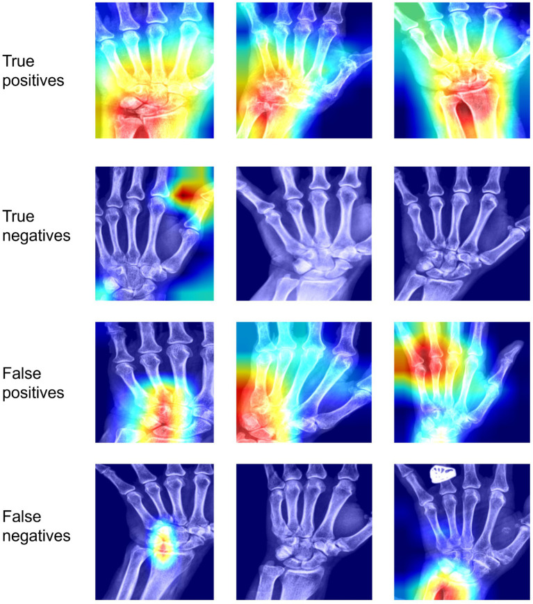
Methods: Two radiologists independently labeled a dataset of 926 hand radiographs, yielding 319 CPPD positive and 607 CPPD negative cases across the three sites of interest after adjudicating discrepant cases. CPPD presence was then predicted using a convolutional neural network. We tested seven CPPD models, each with a different combination of sites out of TFCC, MCP-2 and MCP-3. The model performance was assessed using the area under the receiver operating characteristic (AUROC) and area under the precision-recall (AUPR) curves, with heatmaps (Grad-CAM) aiding in case discrimination.
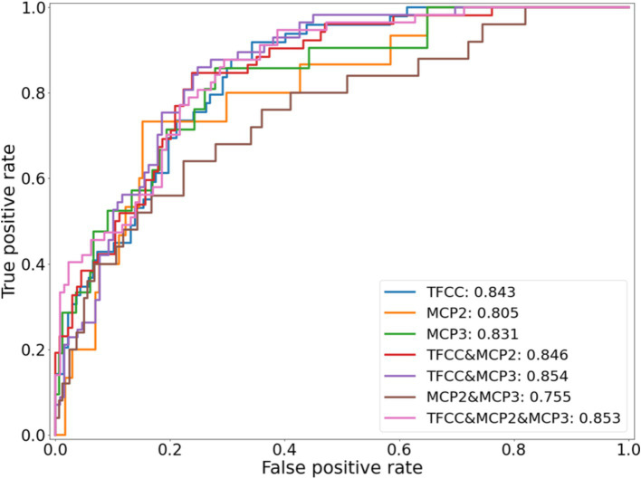
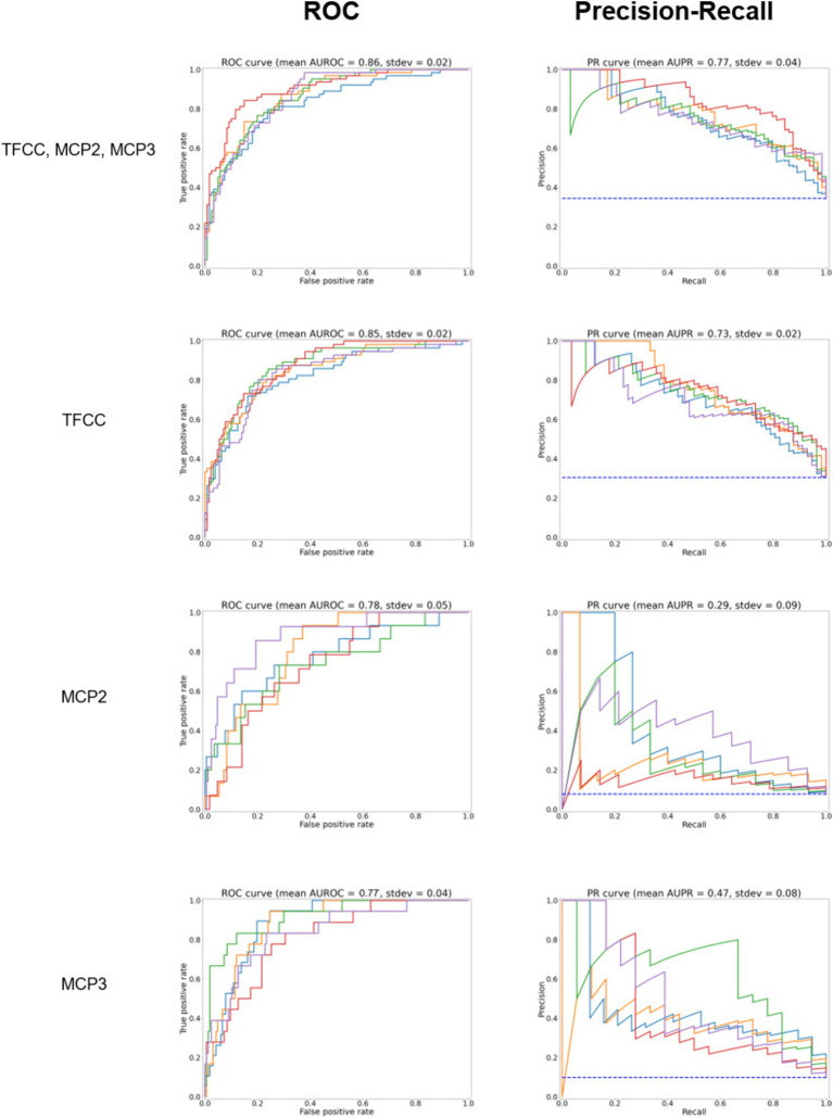
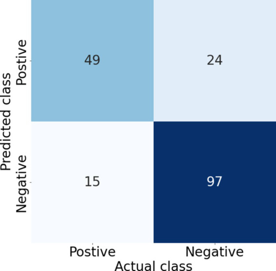
Results: All models trialed gave good class separation, with the combined TFCC, MCP-2 and MCP-3 model showing the most robust performance with a mean AUROC of 0.86, mean AUPR of 0.77, sensitivity of 0.77, specificity of 0.80, and precision of 0.67. The TFCC-alone model had a slightly lower mean AUROC of 0.85 with a mean AUPR of 0.73. The MCP-2-alone and MCP-3-alone models exhibited mean AUROCs of 0.78-0.87, but lower mean AUPRs of 0.29-0.47. Heatmap analysis revealed activation in the regions of interest for positive cases (true and false positives), but unexpected highlights were encountered possibly due to correlated features in different hand regions.
Conclusion: A combined deep-learning model detecting CPPD at the TFCC and MCP-2/3 joints in hand radiographs provides the highest diagnostic performance. The algorithm could be used to screen larger OA or RA databases or electronic medical records for CPPD cases. Future work includes dataset expansion and validation with external datasets.
Keywords: CPPD; automated; chondrocalcinosis; detection; image recoginiton; machine learning; radiograph (X-ray).
Copyright © 2024 Hügle, Rosoux, Fahrni, Markham, Manigold and Becce.
Conflict of interest statement
The authors declare that the research was conducted in the absence of any commercial or financial relationships that could be construed as a potential conflict of interest.
Figures
References
-
- FDA: artificial intelligence and machine learning (AI/ML)-enabled medical devices. (2023). Available at:https://www.fda.gov/medical-devices/software-medical-device-samd/artific...
LinkOut - more resources
Full Text Sources
Miscellaneous