

A semantic segmentation method to analyze retinal vascular parameters of diabetic nephropathy
- PMID: 39512612
- PMCID: PMC11540694
- DOI: 10.3389/fmed.2024.1494659
A semantic segmentation method to analyze retinal vascular parameters of diabetic nephropathy
Abstract
Introduction: By using spectral domain optical coherence tomography (SD-OCT) to measure retinal blood vessels. The correlation between the changes of retinal vascular structure and the degree of diabetic nephropathy is analyzed with a full-pixel Semantic segmentation method.
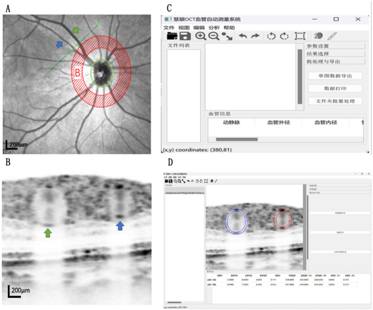
Methods: A total of 120 patients with diabetic nephropathy who were treated in the nephrology department of Quzhou People's Hospital from March 2023 to March 2024 were selected and divided into three groups according to the urinary albumin creatinine ratio (UACR). The groups included simple diabetes group (UACR < 30 mg/g), microalbuminuria group (30 mg/g ≤ UACR <300 mg/g) and macroalbuminuria group (UACR ≥300 mg/g). SD-OCT was used to scan the arteries and veins in the superior temporal area B of the retina. The semantic segmentation method built into the SD-eye software was used to automatically identify the morphology and structure of the vessels and calculate the parameters of arteriovenous vessels. The parameters of arteriovenous vessels are as follows: outer diameter of the retinal artery (RAOD); inner diameter of the retinal artery (RALD); arterial wall thickness (AWT); arterial wall to lumen ratio (AWLR); cross sectional area of arterial wall (AWCSA); retinal vein outer diameter (RVOD); retinal vein inner diameter (RVLD); vein wall thickness (VWT); vein wall to lumen ratio (VWLR); cross sectional area of vein wall (VWCSA). Statistical analysis software was used to compare and analyze the parameters of retinal arteriovenous vessels of the three groups.
Results: The study revealed statistically significant differences in RAOD and RALD among the three groups (p < 0.05) with the RAOD and RALD of the macroalbuminuria group and microalbuminuria group being lower than those of the simple diabetes group. Conversely, there were no significant differences in AWT, AWLR and AWCSA among the three groups (p > 0.05). Additionally, the differences in RVOD and RVLD among the three groups were found to be statistically significant (p < 0.05) with the RVOD and RVLD of the simple diabetes group being lower than those of the microalbuminuria group and macroalbuminuria group. No significant differences were observed in VWT and VWL among the groups. Additionally, RVOD and RVLD were weakly associated with UACR (R = 0.247, p = 0.007; R = 0.210, p = 0.021). Full-pixel semantic segmentation method combined with OCT images is a new retinal vascular scanning technology, which can be used as a new method for early diagnosis of diabetic nephropathy. The structural changes of retinal vessels can be used to predict the severity of diabetic nephropathy during the development of diabetic nephropathy.
Keywords: diabetic nephropathy; full pixel semantic segmentation method; optical coherence tomography; urinary protein creatinine ratio; vascular structure.
Copyright © 2024 Lu, Fang, Xu, Feng, Zhu, Yu and Tong.
Conflict of interest statement
The authors declare that the research was conducted in the absence of any commercial or financial relationships that could be construed as a potential conflict of interest.
Figures
Similar articles
-
Exploring the effect of gestational diabetes mellitus on retinal vascular morphology by PKSEA-Net.Front Cell Dev Biol. 2025 Jan 8;12:1532939. doi: 10.3389/fcell.2024.1532939. eCollection 2024. Front Cell Dev Biol. 2025. PMID: 39845084 Free PMC article.
-
Application of the full-width-at-half-maximum image segmentation method to analyse retinal vascular changes in patients with internal carotid artery stenosis.Front Cell Dev Biol. 2024 Aug 19;12:1467374. doi: 10.3389/fcell.2024.1467374. eCollection 2024. Front Cell Dev Biol. 2024. PMID: 39224436 Free PMC article.
-
Application of the Full-Width-at-Half-Maximum Image Segmentation Method to Analyse Retinal Vascular Changes in Patients with Diabetic Retinopathy.J Healthc Eng. 2022 Aug 8;2022:6726499. doi: 10.1155/2022/6726499. eCollection 2022. J Healthc Eng. 2022. PMID: 39296953 Free PMC article. Review.
-
Update on retinal vessel structure measurement with spectral-domain optical coherence tomography.Microvasc Res. 2014 Sep;95:7-14. doi: 10.1016/j.mvr.2014.06.007. Epub 2014 Jun 27. Microvasc Res. 2014. PMID: 24976361
-
[A new approach for studying the retinal and choroidal circulation].Nippon Ganka Gakkai Zasshi. 2004 Dec;108(12):836-61; discussion 862. Nippon Ganka Gakkai Zasshi. 2004. PMID: 15656089 Review. Japanese.
References
LinkOut - more resources
Full Text Sources