. 2024 Oct 27;17(5):401-421.
doi: 10.1007/s12195-024-00818-x. eCollection 2024 Oct.

Remote-Controlled Gene Delivery in Coaxial 3D-Bioprinted Constructs using Ultrasound-Responsive Bioinks

Affiliations

Remote-Controlled Gene Delivery in Coaxial 3D-Bioprinted Constructs using Ultrasound-Responsive Bioinks

Mary K Lowrey et al. Cell Mol Bioeng. .

Abstract

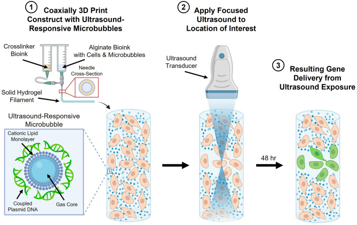
Introduction: Coaxial 3D bioprinting has advanced the formation of tissue constructs that recapitulate key architectures and biophysical parameters for in-vitro disease modeling and tissue-engineered therapies. Controlling gene expression within these structures is critical for modulating cell signaling and probing cell behavior. However, current transfection strategies are limited in spatiotemporal control because dense 3D scaffolds hinder diffusion of traditional vectors. To address this, we developed a coaxial extrusion 3D bioprinting technique using ultrasound-responsive gene delivery bioinks. These bioink materials incorporate echogenic microbubble gene delivery particles that upon ultrasound exposure can sonoporate cells within the construct, facilitating controllable transfection.

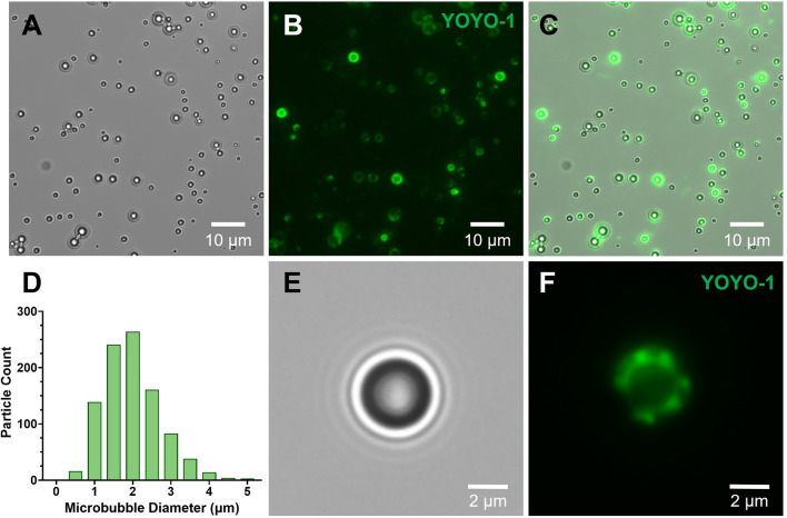
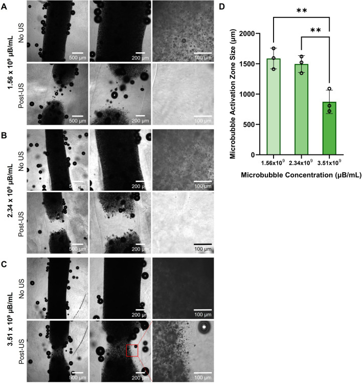
Methods: Phospholipid-coated gas-core microbubbles were electrostatically coupled to reporter transgene plasmid payloads and incorporated into cell-laden alginate bioinks at varying particle concentrations. These bioinks were loaded into the coaxial nozzle core for extrusion bioprinting with CaCl2 crosslinker in the outer sheath. Resulting bioprints were exposed to 2.25 MHz focused ultrasound and evaluated for microbubble activation and subsequent DNA delivery and transgene expression.

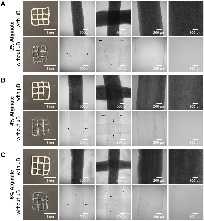
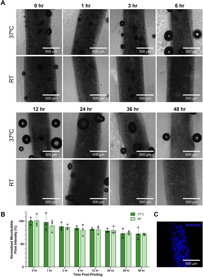
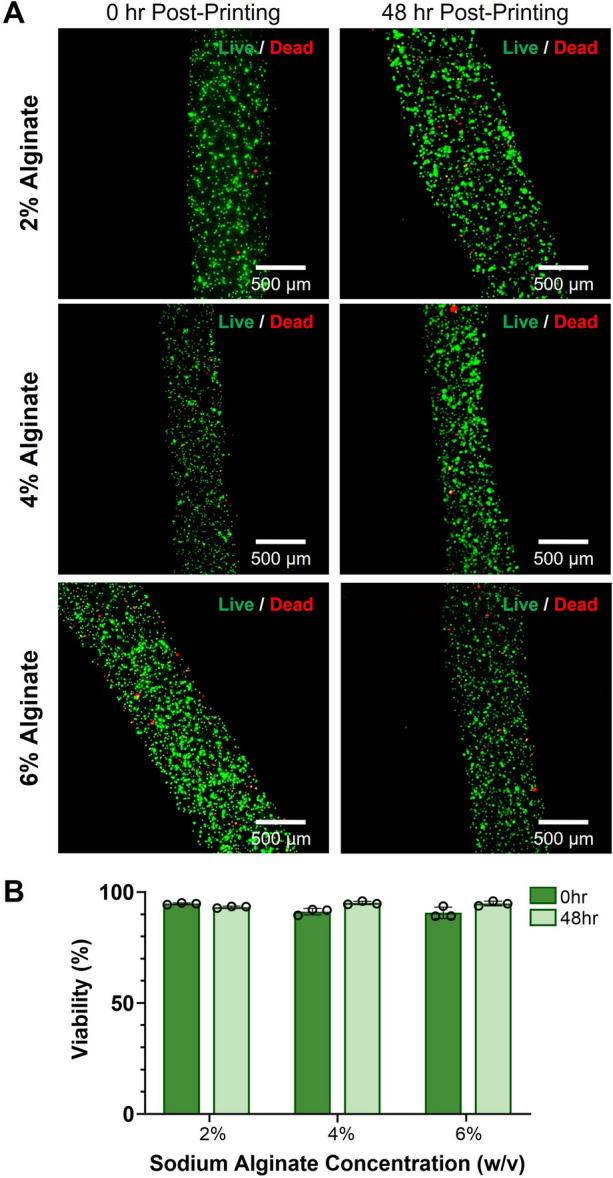
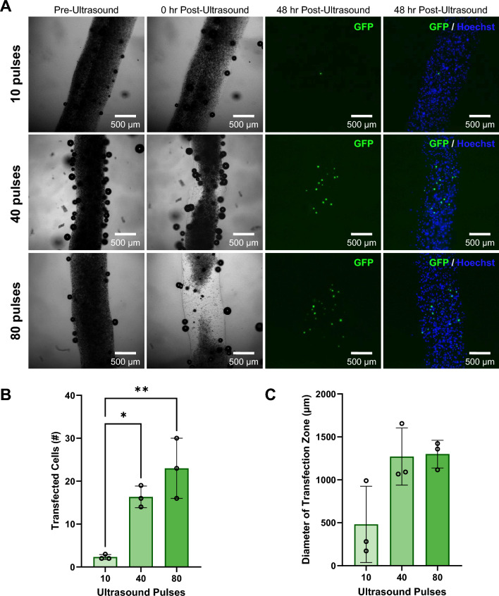
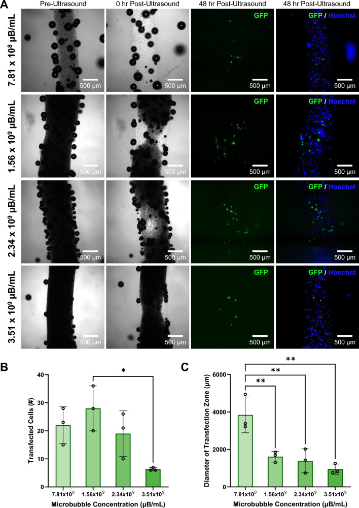
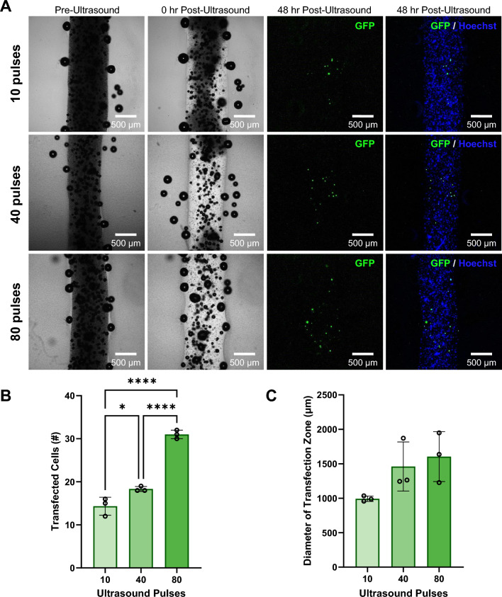
Results: Coaxial printing parameters were established that preserved the stability of ultrasound-responsive gene delivery particles for at least 48 h in bioprinted alginate filaments while maintaining high cell viability. Successful sonoporation of embedded cells resulted in DNA delivery and robust ultrasound-controlled transgene expression. The number of transfected cells was modulated by varying the number of focused ultrasound pulses applied. The size region over which DNA was delivered was modulated by varying the concentration of microbubbles in the printed filaments.

Conclusions: Our results present a successful coaxial 3D bioprinting technique designed to facilitate ultrasound-controlled gene delivery. This platform enables remote, spatiotemporally-defined genetic manipulation in coaxially bioprinted tissue constructs with important applications for disease modeling and regenerative medicine.

Supplementary information: The online version contains supplementary material available at 10.1007/s12195-024-00818-x.

Keywords: Bioink; Biomaterials; Coaxial 3D bioprinting; Controlled delivery; Focused ultrasound; Gene delivery; Microbubbles; Sonoporation; Ultrasound.


Conflict of interest statement

Conflict of interestThe authors MKL, HD, KJS, KTH, CMF and CES declare no conflicts of interest.

Figures

Fig. 1
Fig. 1
Coaxial 3D bioprinting of microbubble gene delivery vehicles. The schematic depicts coaxial bioprinting of a 3D construct containing cells and DNA-coupled ultrasound-responsive microbubbles. The construct is printed with a coaxial needle where the needle core contains sodium alginate bioink precursor solution, microbubble gene delivery vehicles, and cells. The sheath compartment contains calcium crosslinking agent thereby enabling printing of microbubble-containing crosslinked hydrogel filaments. Post-printing, focused ultrasound is applied to user-defined areas of the construct resulting in spatiotemporally controlled regions of gene delivery
Fig. 2
Fig. 2
Ultrasound-responsive microbubble gene delivery vehicles. A-C Representative microscopy images of DNA-coupled ultrasound-responsive microbubbles in A brightfield, B fluorescence (green; plasmid DNA visualized via YOYO-1 stain), and C brightfield/fluorescence overlay (green; plasmid DNA visualized via YOYO-1 stain). D Representative size distribution of DNA-coupled ultrasound-responsive microbubbles. E-F Enlarged microscopy image of DNA-coupled ultrasound-responsive microbubble in E brightfield, showing the dark microbubble and characteristic ring distortions surrounding it due to the lensing effect of the gas bubble core, and F fluorescence (green; plasmid DNA visualized via YOYO-1 stain)
Fig. 3
Fig. 3
Coaxial bioprinting of microbubble gene delivery vehicles. AC Representative macroscale and microscopy images of coaxially-bioprinted alginate at A 2% w/v, B 4% w/v, C 6% w/v, with and without ultrasound-responsive microbubble (µB) gene delivery vehicles. Black arrows indicate filament boundaries
Fig. 4
Fig. 4
Stability of coaxially-printed microbubble gene delivery vehicles. A Representative microscopy images of coaxially-bioprinted HEK293T-laden 4% w/v alginate filaments containing microbubbles from 0-48 hr, in samples kept incubated at 37 °C (top row) and at room temperature (RT, bottom row). B Quantification of microbubble stability over time in 4% w/v alginate in samples incubated at 37 °C and at room temperature. C Representative fluorescence image of bioprinted filament containing microbubbles and Hoechst stained HEK293T cells
Fig. 5
Fig. 5
Effect of microbubble concentration on the size of the ultrasound induced activation zone in 4% w/v alginate bioprinted constructs. A–C Brightfield images of 4% w/v alginate pre- and post-ultrasound (US) exposure containing A 1.56 × 109 µB/mL B 2.34 × 109 µB/mL and C 3.51 × 109 µB/mL. D Quantified size of microbubble activation zone in 4% alginate with varying microbubble concentrations (**p < 0.01, n = 3, one-way ANOVA, Tukey’s multiple comparisons test, error bars denote standard deviation)
Fig. 6
Fig. 6
Viability of bioprinted cells with ultrasound-responsive microbubbles in alginate bioinks. A Representative fluorescence microscopy images of 2%, 4%, and 6% w/v alginate bioprints containing HEK293T cells and microbubbles at 0 hr and 48 hr post-printing. Constructs were stained with calcein-AM (green, live cells) and ethidium homodimer-1 (red, dead cells). B Viability of cells printed in 2%, 4%, and 6% w/v alginate at 0 hr and 48 hr post-printing (n = 3, error bars denote standard deviation)
Fig. 7
Fig. 7
Effect of varying the number of ultrasound pulses on ultrasound-controlled transfection of HEK293T cells in coaxially-bioprinted constructs. A Coaxially-bioprinted HEK293T-laden 4% alginate bioink containing GFP-coupled microbubbles before ultrasound, 0 hr post-ultrasound, and 48 hr post-ultrasound with varying ultrasound exposure (10, 40, or 80 pulses). B Number of transfected cells in bioprinted constructs at 48 hr post-ultrasound for varying numbers of ultrasound pulses (*p < 0.05, **p < 0.01, one-way ANOVA, Tukey’s multiple comparisons test, error bars denote standard deviation). C Diameter of zone containing transfected cells at 48 hr post-ultrasound for varying numbers of ultrasound pulses (one-way ANOVA, Tukey’s multiple comparisons test, error bars denote standard deviation)
Fig. 8
Fig. 8
Effect of increasing microbubble concentration on ultrasound-controlled HEK293T cellular transfection in coaxially-bioprinted constructs. A Increasing concentrations of GFP-coupled microbubbles in coaxially-bioprinted HEK293T-laden 4% alginate bioink, before ultrasound, 0 hr post-ultrasound, and 48 hr post-ultrasound. B Number of transfected cells in bioprinted constructs at 48 hr post-ultrasound for varying microbubble concentrations (*p < 0.05, one-way ANOVA, Tukey’s multiple comparisons test, error bars denote standard deviation). C Diameter of zone containing transfected cells at 48 hr post-ultrasound for varying microbubble concentrations (**p < 0.01, one-way ANOVA, Tukey’s multiple comparisons test, error bars denote standard deviation)
Fig. 9
Fig. 9
Effect of varying the number of ultrasound pulses on ultrasound-controlled osteoblast cellular transfection in coaxially-bioprinted constructs. A Coaxially-bioprinted osteoblast-laden (hFOB 1.19) 4% alginate bioink containing GFP-coupled microbubbles before ultrasound, 0 hr post-ultrasound, and 48 hr post-ultrasound with varying ultrasound exposure (10, 40, or 80 pulses). B Number of transfected cells in bioprinted constructs at 48 hr post-ultrasound for varying numbers of ultrasound pulses (*p < 0.05, ****p < 0.0001, one-way ANOVA, Tukey’s multiple comparisons test, error bars denote standard deviation). C Diameter of zone containing transfected cells at 48 hr post-ultrasound for varying numbers of ultrasound pulses (one-way ANOVA, Tukey’s multiple comparisons test, error bars denote standard deviation)

References

    1. Debnath, J., S. K. Muthuswamy, and J. S. Brugge. Morphogenesis and oncogenesis of MCF-10A mammary epithelial acini grown in three-dimensional basement membrane cultures. Methods. 30:256–268, 2003. - PubMed
    1. Alemany-Ribes, M., and C. E. Semino. Bioengineering 3D environments for cancer models. Adv. Drug Deliv. Rev. 79–80:40–49, 2014. - PubMed
    1. Pal, A., and C. G. Kleer. Three dimensional cultures: a tool to study normal acinar architecture vs. malignant transformation of breast cells. J. Vis. Exp. 86:51311, 2014. - PMC - PubMed
    1. Langley, R. R., and I. J. Fidler. Tumor cell-organ microenvironment interactions in the pathogenesis of cancer metastasis. Endocr. Rev. 28:297–321, 2007. - PubMed
    1. Gupta, M. K., et al. 3D printed programmable release capsules. Nano Lett. 15:5321–5329, 2015. - PMC - PubMed

LinkOut - more resources