Case Reports
. 2024 Oct 31;14(5):974-981.
doi: 10.21037/cdt-24-216. Epub 2024 Oct 22.

Covered stent implantation for calcified nodule to physically hinder its protrusion causing restenosis: a case report

Affiliations
Case Reports

Covered stent implantation for calcified nodule to physically hinder its protrusion causing restenosis: a case report

Satoshi Kitahara et al. Cardiovasc Diagn Ther. .

Abstract

Background: Calcified nodule (CN) is a phenotypic feature of calcified plaques which causes acute coronary syndrome (ACS). Recent studies reported that culprit lesions harboring CN has been shown to increase a risk of repeat revascularization after percutaneous coronary intervention (PCI) with the implantation of newer-generation drug-eluting stent (DES) or debulking device. Mechanistically, a re-protrusion of CN into the lumen has been considered as an important cause associated with repeat revascularization after PCI. These observations suggest the need for additional therapeutic approach to mitigate a risk of repeat revascularization at CN lesions. Here we report a case who received the implantation of one covered stent due to coronary artery perforation after stent implantation at coronary lesion exhibiting CN. This case is unique in terms of preventing restenosis by using covered stent which could physically hinder protrusion of CN through the stent strut.

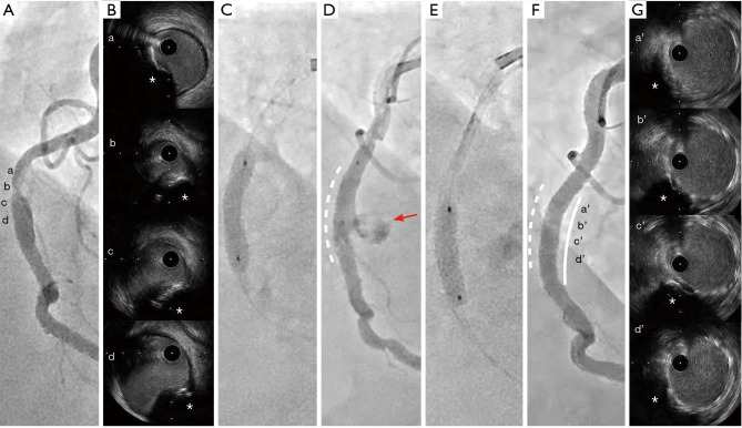
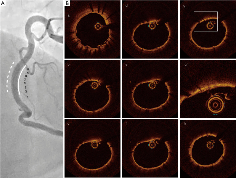
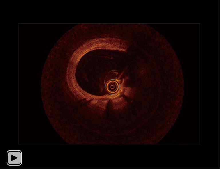
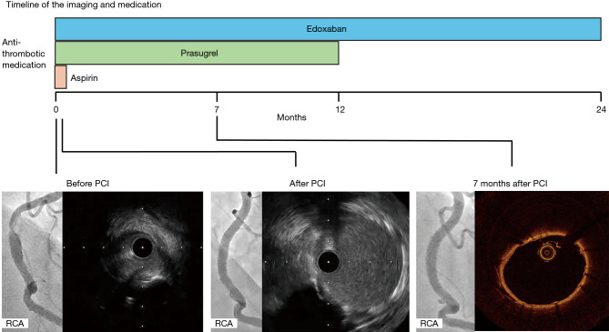
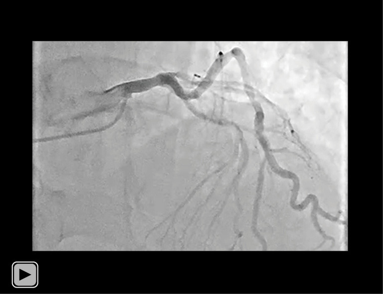
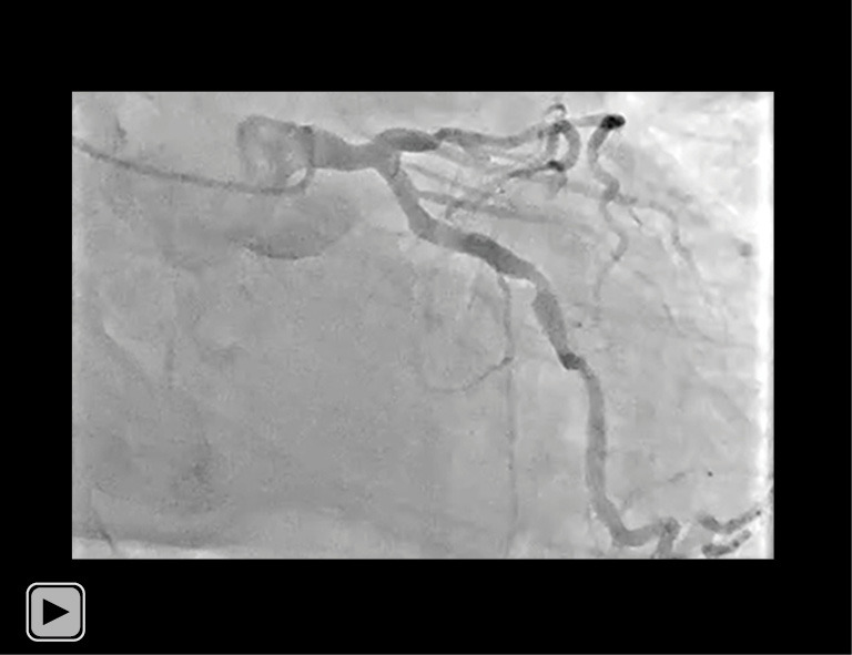
Case description: A 79-year-old man presented to the emergency department with his prolonged chest pain. Although he had a history of hypertension and adrenal hypertrophy, he was not taking any medication prior to his admission. He was diagnosed as ST-segment elevation myocardial infarction. Emergent coronary angiography revealed one severe stenosis in the middle segment of his right coronary artery (RCA). Primary PCI was performed under the guidance of intravascular ultrasound (IVUS) imaging. IVUS imaging prior to PCI revealed a protruding shape of calcification and its irregular surface at his culprit lesion, suggesting the presence of a CN. Following one DES implantation, coronary artery perforation occurred at the segment receiving DES implantation. We implanted one covered stent for the coronary artery perforation. This procedure resulted in successfully sealing coronary artery perforation. Seven months later, follow-up coronary angiography and optical coherence tomography (OCT) imaging were conducted to evaluate his RCA. Any in-stent restenosis (ISR) was not observed. Furthermore, OCT imaging elucidated a small amount of neointimal proliferation without any re-protruding feature of CN through the segment receiving a covered stent. Of note, he did not experience any clinically-driven target lesion revascularization (TLR) for 2 years after PCI.

Conclusions: Our case indicates the use of covered stent as an effective approach to physically hinder the re-protrusion of calcification tissues into the lumen, potentially mitigating a risk of ISR.

Keywords: Calcified nodule (CN); acute coronary syndrome (ACS); case report; covered stent; in-stent restenosis (ISR).


Conflict of interest statement

Conflicts of Interest: All authors have completed the ICMJE uniform disclosure form (available at https://cdt.amegroups.com/article/view/10.21037/cdt-24-216/coif). Y.K. serves as an unpaid editorial board member of Cardiovascular Diagnosis and Therapy from September 2023 to August 2025. Y.K. has received research support from Kowa, Nipro and Abbott, and honoraria from Nipro, Abbott, Kowa, Amgen, Sanofi, Astellas, Takeda and Daiichi-Sankyo. The other authors have no conflicts of interest to declare.

Figures

Figure 1
Figure 1
Coronary angiography and IVUS images before and after PCI. (A) Severe stenosis at the middle segment of his RCA [a-d correspond to IVUS images in (B)]. (B) IVUS images of CN (asterisks). (C) Implantation of drug-eluting stent. (D) Coronary artery perforation (red arrow) at segment receiving drug-eluting stent (white dotted line). (E) Covered stent implantation. (F) Final coronary angiography (white line = implanted covered stent; white dotted line = implanted drug eluting stent) [a’-d’ correspond to IVUS images in (G)]. (G) IVUS images at segment receiving covered stent. CNs (asterisks) did not erupt into the stent. IVUS, intravascular ultrasound; PCI, percutaneous coronary intervention; RCA, right coronary artery; CN, calcified nodule.
Video 1
Video 1
IVUS imaging of his RCA prior to PCI. IVUS imaging revealed a convex shape of the luminal surface, convex shape of the luminal side of calcium, an irregular luminal surface, and an irregular leading edge of calcium at his culprit lesion, suggesting the presence of type 1 eccentric CN. IVUS, intravascular ultrasound; RCA, right coronary artery; PCI, percutaneous coronary intervention; CN, calcified nodule.
Video 2
Video 2
IVUS imaging of his RCA after PCI. The IVUS imaging after post dilatation of covered stent, demonstrated optimal stent apposition and stent expansion. IVUS, intravascular ultrasound; RCA, right coronary artery; PCI, percutaneous coronary intervention.
Figure 2
Figure 2
Follow-up coronary angiography and OCT imaging at 7 months after PCI. (A) In-stent restenosis did not occur at segment receiving covered stent (white line) [a-h correspond to OCT images in (B)]. Dotted white line indicates the implanted drug-eluting stent. (B) Protruding of CN was not observed at segment receiving covered stent by OCT imaging (g’ is an enlargement of the frame of g). OCT, optical coherence tomography; PCI, percutaneous coronary intervention; CN, calcified nodule.
Video 3
Video 3
OCT imaging 7 months after PCI. OCT imaging 7 months after PCI elucidated a small amount of neointimal proliferation without any protruding feature of CN through the segment receiving a covered stent. OCT, optical coherence tomography; PCI, percutaneous coronary intervention; CN, calcified nodule.
Figure 3
Figure 3
The timeline of the imaging and treatment. The x-axis displays the clinical course, with coronary angiogram, intravascular imaging, and medications. One covered stent was implanted to the coronary perforation in lesion with calcified nodule of his RCA. He started taking 100 mg of aspirin and 3.75 mg prasugrel and 30 mg edoxaban once daily. Aspirin was stopped when he discharged, the others had been kept one year after the PCI. In-stent restenosis did not occur 7 months after the PCI. One year after the PCI, he discontinued taking 3.75 mg of prasugrel. He did not experience any clinically-driven target lesion revascularization or no adverse events for 2 years after PCI. PCI, percutaneous coronary intervention; RCA, right coronary artery.
Video S1
Video S1
Coronary angiography of his LCA (cranial view). Coronary angiogram revealed one moderate stenosis at the middle segment of his left anterior descending artery. LCA, left coronary artery.
Video S2
Video S2
Coronary angiography of his LCA (caudal view). Coronary angiogram revealed one moderate stenosis at the middle segment of his left circumflex artery. LCA, left coronary artery.

References

    1. Nakamura N, Torii S, Tsuchiya H, et al. Formation of Calcified Nodule as a Cause of Early In-Stent Restenosis in Patients Undergoing Dialysis. J Am Heart Assoc 2020;9:e016595. 10.1161/JAHA.120.016595 - DOI - PMC - PubMed
    1. Watanabe Y, Sakakura K, Taniguchi Y, et al. Comparison of clinical outcomes of intravascular ultrasound-calcified nodule between percutaneous coronary intervention with versus without rotational atherectomy in a propensity-score matched analysis. PLoS One 2020;15:e0241836. 10.1371/journal.pone.0241836 - DOI - PMC - PubMed
    1. Sugane H, Kataoka Y, Otsuka F, et al. Cardiac outcomes in patients with acute coronary syndrome attributable to calcified nodule. Atherosclerosis 2021;318:70-5. 10.1016/j.atherosclerosis.2020.11.005 - DOI - PubMed
    1. Tada T, Miura K, Ikuta A, et al. Prevalence, predictors, and outcomes of in-stent restenosis with calcified nodules. EuroIntervention 2022;17:1352-61. 10.4244/EIJ-D-21-00504 - DOI - PMC - PubMed
    1. Takahashi Y, Otake H, Kuramitsu S, et al. Prevalence and outcomes of stent thrombosis with in-stent calcified nodules: substudy from the REAL-ST registry. EuroIntervention 2022;18:749-58. 10.4244/EIJ-D-21-00976 - DOI - PMC - PubMed

Publication types

LinkOut - more resources