

Voltage-Gated Ion Channel Compensatory Effect in DEE: Implications for Future Therapies
- PMID: 39513870
- PMCID: PMC11544952
- DOI: 10.3390/cells13211763
Voltage-Gated Ion Channel Compensatory Effect in DEE: Implications for Future Therapies
Abstract
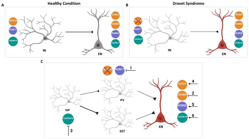
Developmental and Epileptic Encephalopathies (DEEs) represent a clinically and genetically heterogeneous group of rare and severe epilepsies. DEEs commonly begin early in infancy with frequent seizures of various types associated with intellectual disability and leading to a neurodevelopmental delay or regression. Disease-causing genomic variants have been identified in numerous genes and are implicated in over 100 types of DEEs. In this context, genes encoding voltage-gated ion channels (VGCs) play a significant role, and part of the large phenotypic variability observed in DEE patients carrying VGC mutations could be explained by the presence of genetic modifier alleles that can compensate for these mutations. This review will focus on the current knowledge of the compensatory effect of DEE-associated voltage-gated ion channels and their therapeutic implications in DEE. We will enter into detailed considerations regarding the sodium channels SCN1A, SCN2A, and SCN8A; the potassium channels KCNA1, KCNQ2, and KCNT1; and the calcium channels CACNA1A and CACNA1G.
Keywords: Developmental and Epileptic Encephalopathy (DEE); compensatory effect; ion channels.
Conflict of interest statement
The authors declare no conflict of interest. All authors are full employee of Servier.
Figures


Similar articles
-
Reduction of Kcnt1 is therapeutic in mouse models of SCN1A and SCN8A epilepsy.Front Neurosci. 2023 Oct 13;17:1282201. doi: 10.3389/fnins.2023.1282201. eCollection 2023. Front Neurosci. 2023. PMID: 37901435 Free PMC article.
-
Rates of Status Epilepticus and Sudden Unexplained Death in Epilepsy in People With Genetic Developmental and Epileptic Encephalopathies.Neurology. 2023 Apr 18;100(16):e1712-e1722. doi: 10.1212/WNL.0000000000207080. Epub 2023 Feb 7. Neurology. 2023. PMID: 36750385 Free PMC article.
-
Phenotypic and genetic spectrum of SCN8A-related disorders, treatment options, and outcomes.Epilepsia. 2019 Dec;60 Suppl 3:S77-S85. doi: 10.1111/epi.16319. Epilepsia. 2019. PMID: 31904124
-
Developmental and Epileptic Encephalopathy: Pathogenesis of Intellectual Disability Beyond Channelopathies.Biomolecules. 2025 Jan 15;15(1):133. doi: 10.3390/biom15010133. Biomolecules. 2025. PMID: 39858526 Free PMC article. Review.
-
SCN1A-related phenotypes: Epilepsy and beyond.Epilepsia. 2019 Dec;60 Suppl 3:S17-S24. doi: 10.1111/epi.16386. Epilepsia. 2019. PMID: 31904117 Review.
References
-
- Howell K.B., Eggers S., Dalziel K., Riseley J., Mandelstam S., Myers C.T., McMahon J.M., Schneider A., Carvill G.L., Mefford H.C., et al. A Population-Based Cost-Effectiveness Study of Early Genetic Testing in Severe Epilepsies of Infancy. Epilepsia. 2018;59:1177–1187. doi: 10.1111/epi.14087. - DOI - PMC - PubMed
-
- Symonds J.D., Zuberi S.M., Stewart K., McLellan A., O’Regan M., MacLeod S., Jollands A., Joss S., Kirkpatrick M., Brunklaus A., et al. Incidence and Phenotypes of Childhood-Onset Genetic Epilepsies: A Prospective Population-Based National Cohort. Brain. 2019;142:2303–2318. doi: 10.1093/brain/awz195. - DOI - PMC - PubMed
-
- De Lange I.M., Koudijs M.J., Van’T Slot R., Sonsma A.C.M., Mulder F., Carbo E.C., Van Kempen M.J.A., Nijman I.J., Ernst R.F., Savelberg S.M.C., et al. Assessment of Parental Mosaicism in SCN1A-Related Epilepsy by Single-Molecule Molecular Inversion Probes and next-Generation Sequencing. J. Med. Genet. 2019;56:75–80. doi: 10.1136/jmedgenet-2018-105672. - DOI - PubMed
Publication types
MeSH terms
Substances
LinkOut - more resources
Full Text Sources
Medical
